Glioma Stem Cells Enable the Production of 3D Human “Mini Brain” to Improve Glioblastoma Treatment

Discover how 3D models of glioblastoma using patient-derived stem cells could enable more effective treatment

24 Jul 2018
Aidan Barry
Administrator / Office Personnel

Editorial article

Glioblastoma (GBM) is an extremely aggressive, difficult to treat, malignant brain tumor. Being resistant to conventional therapies, research into the development and proliferation of these tumors is crucial to develop a greater understanding of how to provide more effective treatment. Current clinical research involves producing 3D models of GBM using animal-derived cells, which have a number of limiting factors as they are not a true representation of the human brain due to interspecies differences.   Dr. Amanda Linkous, of Weill Cornell Medicine, is a pioneering scientist at the forefront of a project providing ex vivo models of GBM using human-derived stem cells in order to closely mimic the microenvironment of the human brain in vivo, thus enabling more effective treatment. Here, SelectScience speaks to Linkous to hear more about her fascinating and potentially revolutionary research.   An image of the glioma cerebral organoid model developed by Dr. Amanda Linkous

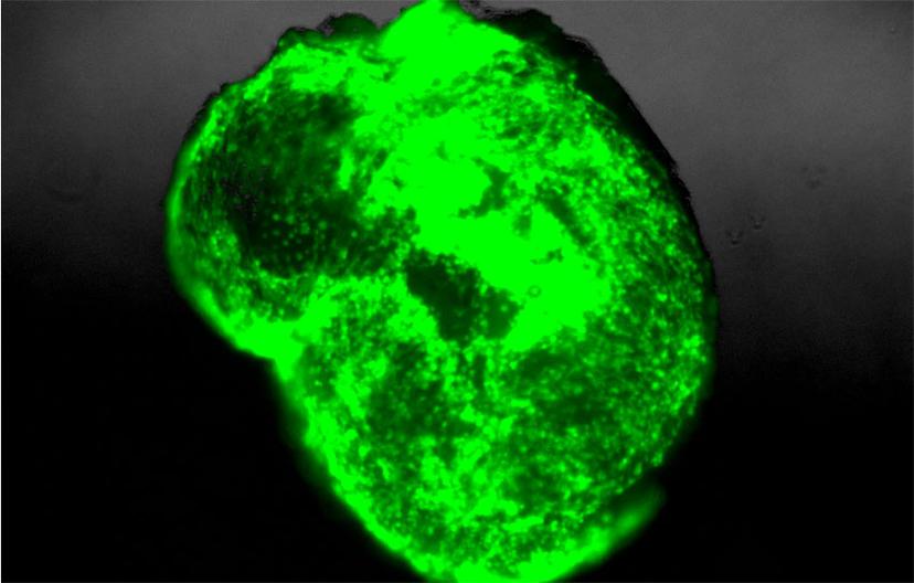
SS: Tell us a little about your background and your role at Weill Cornell Medicine (WCM)

AL: I have over 13 years of scientific training in a combination of government institutes and multiple premier academic medical centers including the National Institutes of Health, Vanderbilt University Medical Center and, most recently, New York-Presbyterian Hospital/Weill Cornell Medicine (WCM). I currently serve as the Director of the Starr Foundation Cerebral Organoid Translational Core here at WCM. In addition to establishing a “mini-brain” ex vivo model of glioblastoma (GBM) for our core, I manage the derivation of glioma stem cells (GSCs) from patient tumor tissue and oversee the compliance and key operations of our core facility.

SS: Why is there a need for organoids? What benefits do organoids offer over regular cell culture or even animal models?

AL: Current preclinical models of GBM are significantly limited by the lack of a normal human microenvironment. For example, animal models of GBM are restricted by the unknown interspecies differences between human GSCs and murine brain cells. Coupled with variably long tumor latencies and the inability to genetically manipulate and image tumor progression in real time, mouse models present a multitude of obstacles when trying to accurately reproduce the biology of human GBM. Tumor organoids overcome some of the logistical constraints of animal models, but they fail to address the important issue of cell-cell interactions between tumor cells and a normal host microenvironment.

SS: Tell us about your glioma cerebral organoid model

AL: To address the limitations of current preclinical GBM models, we have retro-engineered patient-specific GBMs using patient-derived glioma stem cells and human embryonic stem cell (hESC)-derived cerebral organoids. Our cerebral organoid glioma (GLICO) model demonstrates a clear infiltration of the human cerebral organoid by the patient-derived GSCs. Following the invasion into the organoid, the GSCs then proliferate and migrate throughout the host tissue, ultimately forming tumors that closely phenocopy patient glioblastomas.

Glioma tumor cells grow inside the organoid. Corning® Matrigel® matrix forms an integral component of every stage of organoid development. Image courtesy of Dr. Amanda Linkous.

SS: Please briefly describe your project using the glioma cerebral organoid model and share your findings

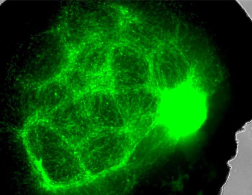
AL: Our GLICO model addresses a number of the limitations of current preclinical GBM models, as it allows us to study patient-specific GBMs within a human microenvironment similar to that of a normal human brain. Additionally, our cerebral organoids cultured and differentiated in Corning Matrigel matrix, allows us to recapitulate the same biological behavior and histopathological features of patient GBMs. Our findings also show that GLICO tumors are supported by a network of interconnecting microtubes that facilitate the invasion and proliferation of the glioma stem cells within the organoid.

SS: How important is it to use the right labware, surfaces and reagents to obtain an organoid culture?

AL: Selecting the appropriate labware is vital to generating a successful batch of cerebral organoids. As with any biomedical research involving human samples, it is critical that our data is consistent and reproducible each and every time. Failure to use the correct culture vessels, matrix or reagents can drastically alter the morphological structure and viability of the organoids. Corning® Matrigel® matrix is an integral component of every stage of organoid development in our lab. From culturing the hESCs and generating neuroectoderm/neural rosettes, to ultimately providing a three-dimensional matrix that promotes the growth of a fully differentiated cerebral organoid, Matrigel matrix is an indispensable component of our organoid research.

SS: What’s the future of organoid research? What projects do you have in the pipeline?

AL: The future of organoid research is incredibly exciting! Along with our collaborators, we are continuing to expand the GLICO model and make it an ever-closer representative of the human clinical disease. Furthermore, by pairing patient-specific glioma stem cells with these miniature brains, we are able to use our unique model to test multiple therapeutic combinations against these deadly tumors. It is our sincere hope that the GLICO model will allow us to develop new, effective treatment options for patients suffering from this horrible disease.

Learn more about Corning’s Matrigel technology here.

For the latest in cell culture technology, visit our 3D Cell Culture special feature.

Links

Tags

Fluorescence SpectroscopyFluorometers and spectrofluorometers (also called fluorescence spectrometers) are used to measure the intensity and wavelength of fluorescent light emitted from a sample after excitation by illumination. Spectrofluorometers utilize monochromators to select the desired wavelengths, whereas filter fluorometers employ a set of filters. Spectrofluorometers for measuring steady-state fluorescence and lifetime fluorescence (or time-resolved fluorescence) are available, as well as fluorescence microscopes and microplate readers. Find the best fluorescence spectroscopy products in our peer-reviewed product directory: compare products, check customer reviews and receive pricing direct from manufacturers.Cell / Tissue CultureCell culture or tissue culture is used to study the biology of cells or tissues and to isolate cellular products in an environment which can be manipulated and well defined. Accurately control your culture environment with bioreactors or culture incubators, bind your cells to a surface or together with an extracellular matrix. Distinguish cell types with differential media or proliferate cells with certain characteristics using selective media. Enrich your media with supplements such as growth factors, sera and vitamins. Find the best cell and tissue culture products, kits and equipment in our peer-reviewed product directory: compare products, check customer reviews and receive pricing direct from manufacturers.Cell Lines Stem Cells and Primary CellsPrimary cell cultures, established cell lines and stem cells are vital for <i>in vitro</i> and <i>ex vivo</i> experimentation. High-quality cells, optimized for your applications, alongside optimized cell substrates, growth medium and supplements, are critical for experimental success. Explore a range of cells suitable for your applications, including isogenic cell lines, competent cells, induced pluripotent stem cell (iPSC)-derived cell lines, fungal/bacterial/mammalian cell lines, stem cells and cancer cell lines. Find the best cells for your research in our peer-reviewed product directory: compare products, check customer reviews and receive pricing direct from manufacturers.hESCCancer CellsCancer cells are abnormal cells that divide uncontrollably, leading to the formation of tumors and the spread of cancer. Studying cancer cells is crucial for developing new treatments and understanding tumor biology. Explore cancer cell research products in our peer-reviewed product directory; compare products, check reviews, and get pricing directly from manufacturers.Fluorescence MicroscopyFluorescence microscopy has become an essential tool in biology, as well as in materials science. The application of many fluorochromes has made it possible to identify cells and sub-microscopic cellular components with a high degree of specificity. Using multiple fluorescence labels, different probes can simultaneously identify several target molecules.TumorsTumor research focuses on understanding abnormal cell growth that leads to cancer. Identifying biomarkers, studying tumor microenvironments, and developing targeted therapies are critical for advancing cancer treatment. Early detection and personalized treatment options are key to improving outcomes for patients. Browse our peer-reviewed product directory to explore tools for tumor research, diagnostics, and cancer therapies; compare products, read customer reviews, and get pricing directly from manufacturers.Cancer ResearchAlthough cancer is often referred to as a single condition, it actually consists of more than 100 different diseases. Microscopy, mass spectrometry, high throughput sequencing and flow cytometry are some of the most common techniques employed in cancer research labs.