Ten Plasma Proteins Possibly Predict Alzheimer’s Disease
written by Luminex
25 Jan 2016
Editorial article
written by Luminex
The World Alzheimer Report 2009 estimated that 36 million people worldwide are living with dementia, with numbers doubling every 20 years to 66 million by 2030, and 115 million by 2050. The majority of those individuals will have Alzheimer’s disease (AD).1
The rapid growth of AD cases worldwide, estimated by the National Institutes of Health (NIH) to be more than five million in the US alone, is approaching epidemic proportions and is threatening to overwhelm healthcare systems.
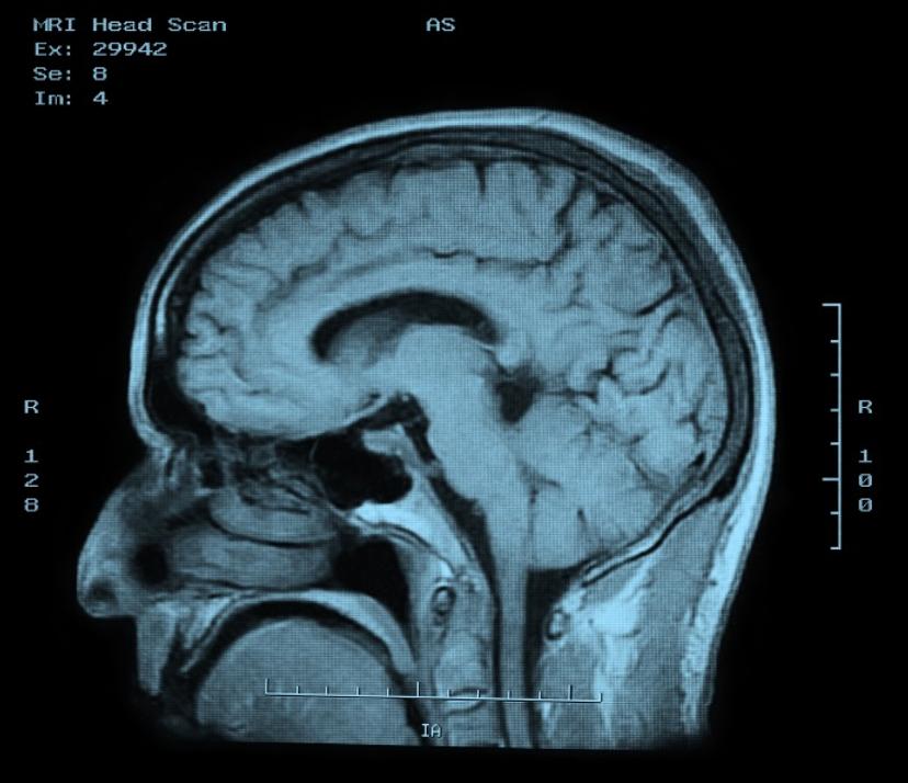
Definitive diagnosis of AD has traditionally relied upon post-mortem neuropathologic tissue analysis, requiring histological examination of brain tissue at autopsy, or in a rare case, following brain biopsy.3 Biomarkers are measurable proteins or genes in the body that relate to normal and abnormal processes within the body. In the case of AD, two biomarkers within the cerebrospinal fluid (CSF) can be measured to monitor Alzheimer’s disease progression—amyloid ß and tau proteins. It has been shown that the level of amyloid ß protein decreases during AD pathology.2
Conversely, the level of the tau protein increases as neuroaxonal degeneration progresses, and the level of hyper-phosphorylated tau protein goes up as tangle pathology occurs. While CSF biomarkers may provide accurate diagnostic information concerning the progression of the disease, the availability of samples for research studies and the practicality of their use for screening are limited by the invasiveness of the procedure required to collect the sample. Sample volume is also limited, and multiple spinal punctures are not an option. The most obvious alternative sample source is blood. However, Aß42 levels in CSF and plasma do not correlate, and tau is apparently present at concentrations too low to be useful.4 As a result, several efforts are under way to identify biomarkers in blood that can potentially be used to reliably diagnose AD.
The top ten
A recent study published in Alzheimer’s & Dementia has identified a set of ten proteins in the blood that can potentially predict the onset of Alzheimer’s Disease. This test could be used for clinical trials of experimental treatments being developed to counter the development of AD. The research team used samples from three international studies: AddNeuroMed (ANM)—a multicenter European study, Kings Health Partners-Dementia Case Register (KHP-DCR)—a UK clinic and population based study, and Genetics AD Association (GenADA)—a multisite case-control study based in Canada. Samples from 1148 subjects, 476 with AD, 220 with MCI (mild cognitive impairment), and 452 elderly controls with no dementia were used in the study.
Seven Milliplex® MAP multiplex panels (96-well plate format; EMD Millipore), based on Luminex® xMAP® Technology, were utilized, and screened a total of 27 proteins that had been previously indicated as potential biomarkers of AD.
Ten proteins (TTR, Clusterin, cystatin C, A1AcidG, ICAM1, CC4, pigment epithelium-derived factor [PEDF], A1AT, RANTES, ApoC3) plus APOE were strongly associated with disease severity and disease progression.
The original article was posted by Luminex here.
Related products
Request Quote for All Products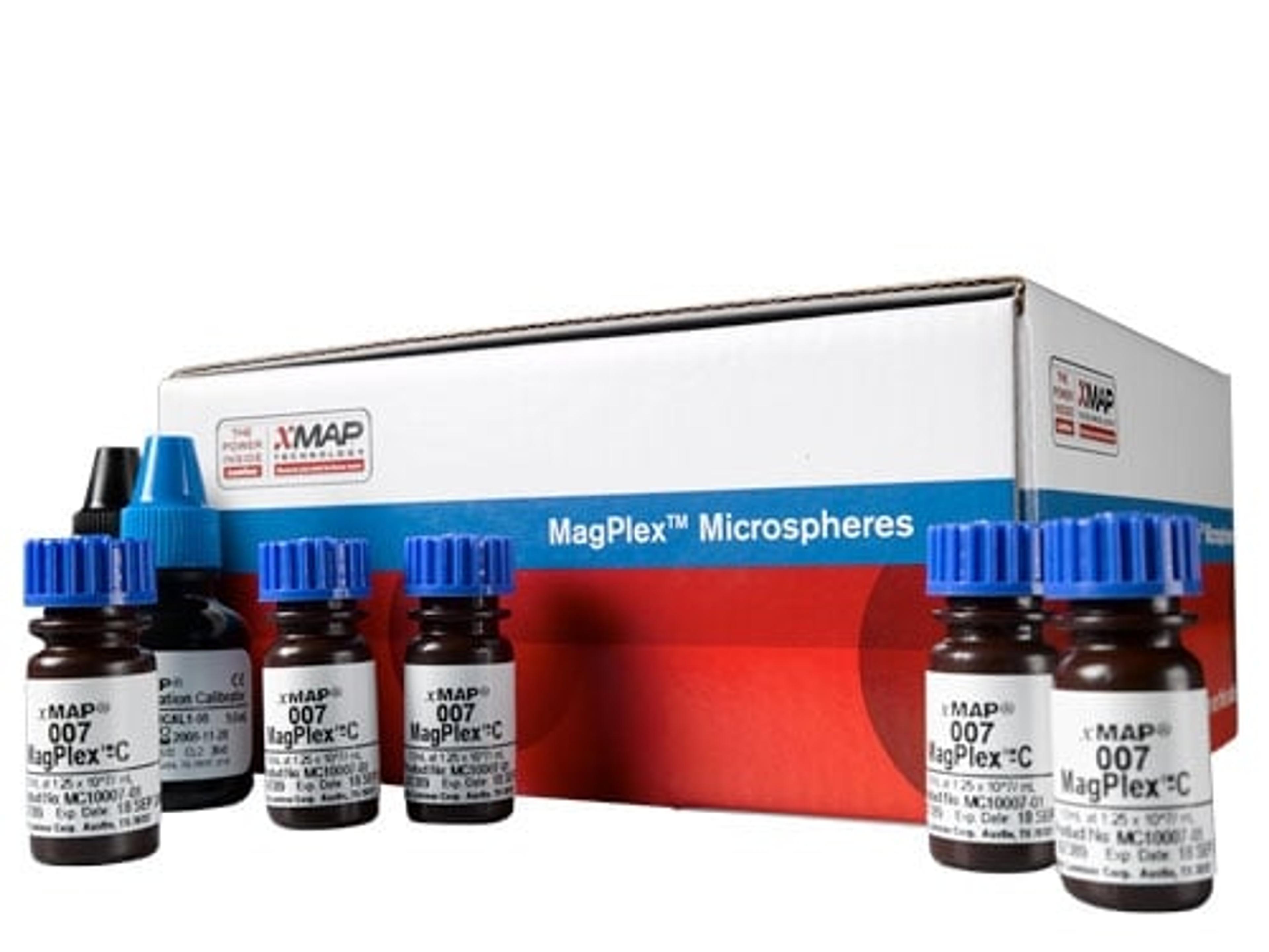
MagPlex® Multiplexing Microspheres
LuminexxMAP® Technology uses color-coded microspheres to detect up to 500 protien or oligonucleotide analytes from a single aliquot of sample in a single reaction well, enabling you to generate more data while saving sample, time, and reagents. Depending on your targets and the application, there are five different microsphere types to select from to produce the best experimental results. MagPlex® are magnetic carboxylated polystyrene microspheres that have been dyed into 500 spectrally distinct sets or “regions”, allowing them to be individually identified by an xMAP instrument. In the multiplex assay environment, the microspheres act as both the surface for the solution phase assay and the spectral identifier that the instrument detects. The open architecture of xMAP Technology enables users to build custom multiplex assays or select from a broad menu commercially available kits. MagPlex are the most versatile xMAP microspheres and are: Compatible with both protein and nucleic acid applications Maximum xMAP microsphere regions available: 500 Superparamagnetic, eliminating the need for filter plates and offering superior washing efficiency Ideal for automated high throughput applications Available in two concentrations: 2.5 (10 regions only) & 12.5 (millions/mL) Available in multiple vial sizes: 1 mL, 4 mL, Custom Compatible with all xMAP instruments: FLEXMAP 3D® (500 regions), Luminex® 100/200™ (80 regions), MAGPIX® (50 regions) Additional xMAP microspheres: MagPlex-TAG™, MicroPlex®, MagPlex-Avidin®, SeroMAP™
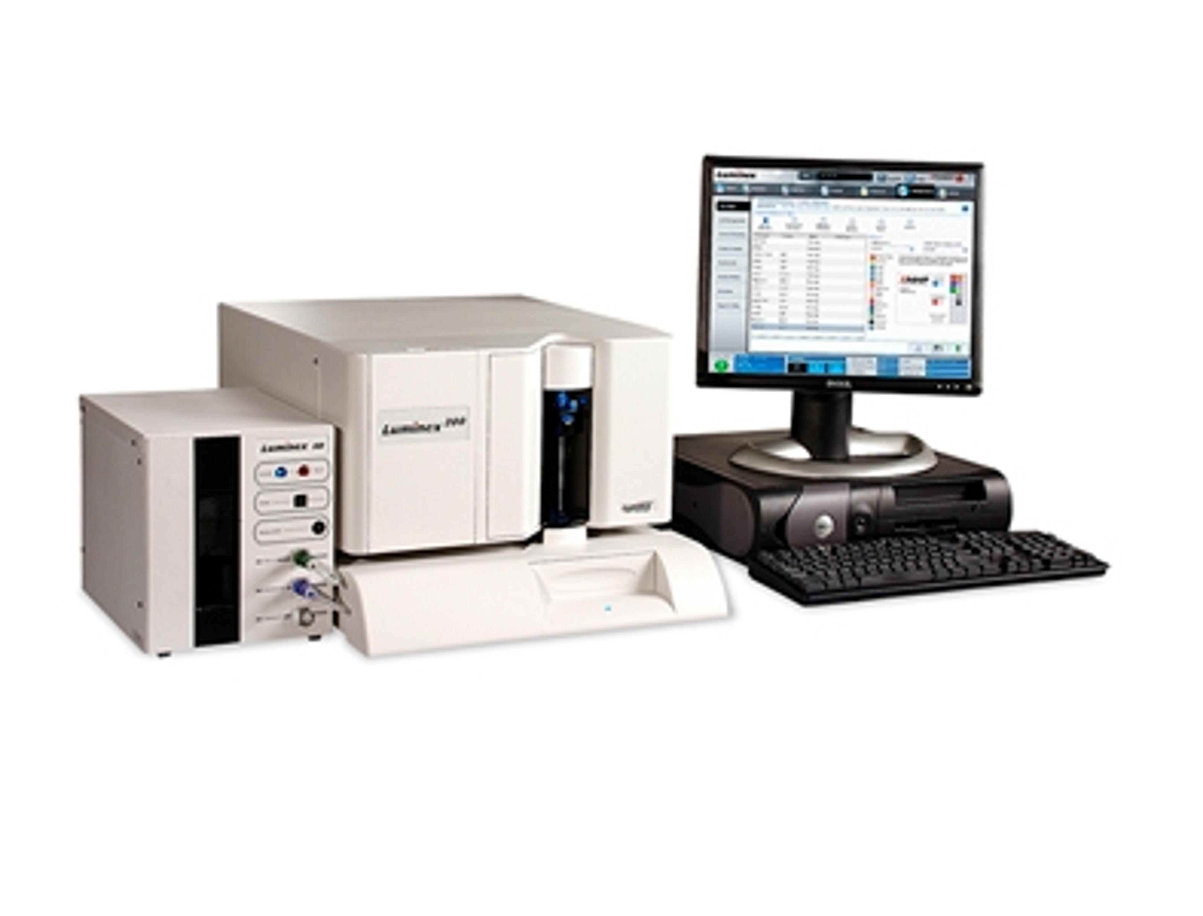
Luminex® 200™Multiplexing Instrument
LuminexThe Luminex® 200™ detects up to 100 protein or nucleic acid analytes from a single aliquot of sample in a single reaction well, enabling you to generate more data while saving sample, time, and reagents. With more than 11,000 units sold globally to date, the Luminex 100/200 is the established standard in multiplexing instrumentation. The xMAP® multiplexing platform is composed of an instrument and associated reagents: The Luminex 200 instrument is based on the principles of flow cytometry, which integrates key xMAP detection components such as lasers, optics, fluidics, and high-speed digital signal processors. xMAP microspheres are a family of fluorescently dyed carboxylated polystyrene microspheres that act as both the surface for the solution phase assay and the spectral identifier that the instrument detects. The open architecture of xMAP Technology enables users to build custom multiplex assays or select from a broad menu commercially available kits. Instrument Details: Multiplex Capacity: Up to 100 non-magnetic; 80 magnetic Microtiter Plate: 96 well Throughput: ~40 min/96-well plate (up to 12,800 tests per hour) Dynamic Range: 3.5 logs Sensitivity: Detects a minimum of 1,000 fluorochromes of phycoerythrin (PE) per xMAP microsphere Optics: Lasers/APDs/PMTs Hardware: Flow Cytometry based Software: xPONENT® Software supports protocol-based data acquisition with robust data regression analysis Dimensions: All components combined (Analyzer, XY Platform and Sheath Delivery System) 26.5" W x 23.5" D x 12.5" H (67.3 cm W x 59.7 cm D x 31.8 cm H) Weight: All components combined (Analyzer, XY Platform and Sheath Delivery System) 113 lbs (49 kg) xMAP Reagent Compatibility: Magnetic and nonmagnetic microspheres