New advanced imaging center powered by two MuVi and LCS SPIM microscopes
25 Mar 2021
Industry news
Bruker has announced that two Luxendo MuVi™and LCS SPIM™ light-sheet microscopes have been installed by Memorial Sloan Kettering Cancer Center (MSK). The funding for the two light-sheet fluorescence microscopes was supported by Cycle for Survival. The new SPIM microscopes will help researchers visualize the cellular and tissue hallmarks of cancer and translate those findings into better cancer treatment methods.
“By understanding how cells mobilize to build organs, researchers can glean insights into why some cells become cancerous and lead to organ destruction,” said Dr. Anna-Katerina Hadjantonakis, MSK Chair of the Developmental Biology Program. “Instruments such as these are useful for imaging across differing length scales — from subcellular to single cells to tissue-level processes — allowing researchers to study cellular dynamics and cellular motion, processes that enable cells to metastasize.”
“Light-sheet fluorescence microscopy has emerged as a uniquely powerful method for high-resolution, cleared-sample and dynamic biological imaging,” added Dr. Lars Hufnagel, Vice President and General Manager of Bruker’s Luxendo light-sheet microscopy business. “We couldn’t be more pleased that our technology will be assisting the great MSK researchers and programs in such important work.”
Want the latest science news straight to your inbox? Become a SelectScience member for free today>>
Related products
Request Quote for All Products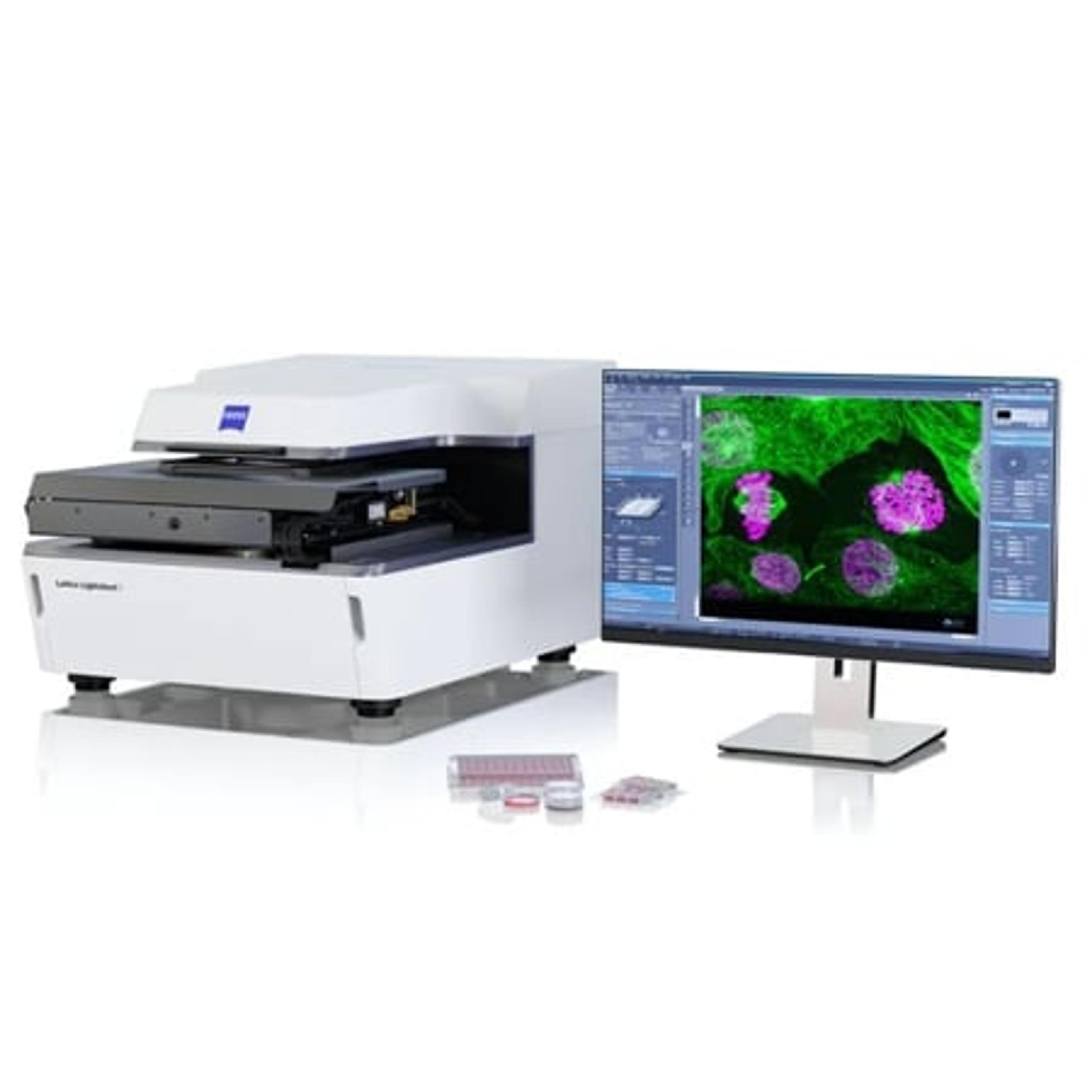
ZEISS Lattice Lightsheet 7
ZEISS Research Microscopy SolutionsLong-term volumetric imaging of living cells