Leica, Indica Labs, and Lunit partner to advance AI-driven digital pathology
Collaboration integrates AI image analysis with digital pathology workflows to support consistent biomarker scoring
5 Jun 2026
Leica Biosystems, Indica Labs, and Lunit have entered into a strategic collaboration to develop AI-powered image analysis algorithms for PD-L1 and emerging immunohistochemistry (IHC) biomarkers. The partnership aims to deliver scalable, consistent biomarker quantification through computational pathology tools, enhancing cancer research workflows worldwide.
Expanding a market-defining computational pathology ecosystem
Building on the market-defining computational pathology partnership between Leica Biosystems and Indica Labs, this new collaboration integrates Lunit’s advanced AI capabilities into a best-in-class IHC staining and digital pathology ecosystem. The first product from this three-way partnership is an AI-powered Lunit SCOPE algorithm designed specifically for Leica Biosystems’ PD-L1 primary antibody (CAL10).
Addressing variability in biomarker scoring with AI
Leica Biosystems, Indica Labs, and Lunit will work together to support pharmaceutical partners in integrating AI-enabled digital pathology into routine clinical research. Biomarker scoring for PD-L1 and other IHC biomarkers remains highly variable, as disease state assessments and treatment decisions often rely on manual evaluation of stained tissue under a microscope. This manual process can be time-consuming and susceptible to human variability.
By combining Leica Biosystems’ expertise in IHC staining and digital pathology, Indica Labs’ image management platform, and Lunit’s advanced AI analysis, the collaboration is designed to support consistent scoring approaches, enable robust biomarker analysis, and accelerate oncology research workflows.
Seamless, AI-powered digital pathology workflow
The partnership unites Leica Biosystems’ PD-L1 IHC assay platform¹, the Aperio GT 450 digital pathology scanner¹, the Aperio HALO AP® image management solution², and the Lunit SCOPE biomarker scoring algorithm¹ to deliver a seamless, AI-powered pathology workflow.
“As a first step, we’re integrating Lunit’s algorithm directly into our image management software powered by Indica Labs, which enables a continuous workflow for users of the Aperio GT 450 scanner,” said Karan Arora, SVP of Advanced Assays, AI, and Pharma Partnerships at Leica Biosystems. “This launch aligns with our long-term vision to deliver scalable, seamlessly integrated research workflows to the market.”
Commitment to open pathology and interoperability
This three-way integration reflects a shared commitment to open pathology and interoperability across the digital pathology ecosystem. Indica Labs CEO Steven Hashagen emphasized that, “robust end-to-end solutions like these are a critical step toward realizing the promise of AI in digital pathology.”
Brandon Suh, CEO of Lunit, added, “We are encouraged by the strong interest from pharmaceutical partners and excited about the potential this collaboration holds. Together, we’re applying AI to pathology image analysis at unprecedented scale, with a shared goal of delivering precision diagnostics that translate into tangible improvements in patient care.”
References
1. For research use only. Not for use in diagnostic procedures.
2. Aperio HALO AP® is CE-IVDR marked for in vitro diagnostic use in Europe, the UK, and Switzerland. Aperio HALO AP® is for research use only in the US and is not FDA cleared for clinical diagnostic use.
This news contains forward-looking statements.
Want the latest science news straight to your inbox? Become a SelectScience member for free today>>
Related products
Request Quote for All Products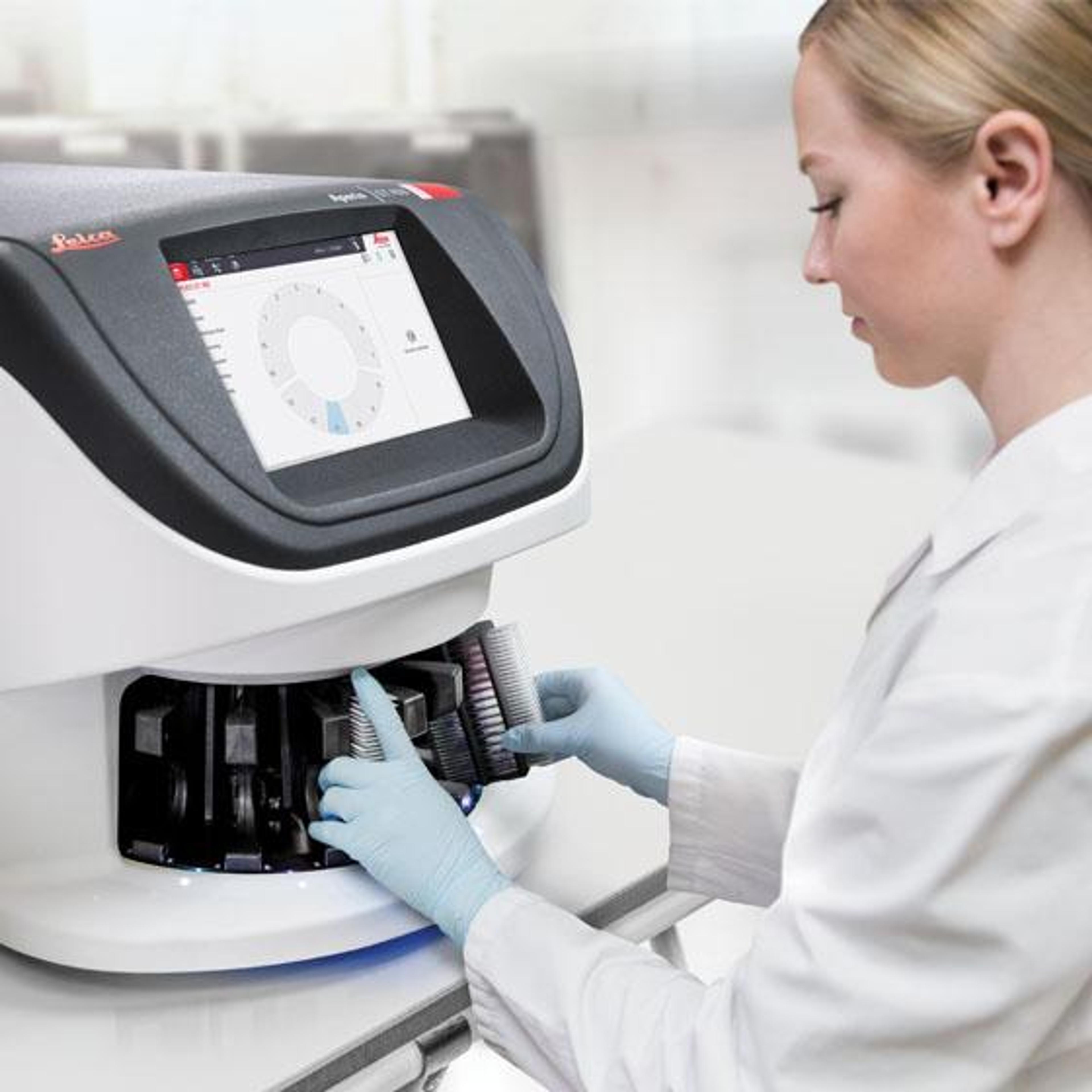
Aperio GT 450 DX Digital Pathology Scanner
Leica BiosystemsAutomated, High Capacity Digital Pathology Slide Scanner
Frequently asked questions
How does the Leica Biosystems, Indica Labs, and Lunit collaboration advance AI-powered PD-L1 and IHC biomarker analysis in digital pathology?
The collaboration between Leica Biosystems, Indica Labs, and Lunit focuses on developing AI-powered image analysis algorithms for PD-L1 and emerging immunohistochemistry (IHC) biomarkers. By combining Leica Biosystems’ expertise in IHC staining and digital pathology, Indica Labs’ image management platform, and Lunit’s advanced AI analysis, the partnership aims to deliver scalable, consistent biomarker quantification.
The first product of this three-way collaboration is an AI-powered Lunit SCOPE algorithm designed specifically for Leica Biosystems’ PD-L1 primary antibody (CAL10), supporting robust biomarker analysis and enhancing cancer research workflows worldwide.
What digital pathology ecosystem components are integrated in the Leica Biosystems, Indica Labs, and Lunit AI workflow for oncology research?
The AI-powered digital pathology workflow created by Leica Biosystems, Indica Labs, and Lunit integrates several key components into a market-defining computational pathology ecosystem. These include Leica Biosystems’ PD-L1 IHC assay platform, the Aperio GT 450 digital pathology scanner, the Aperio HALO AP® image management solution from Indica Labs, and the Lunit SCOPE biomarker scoring algorithm.
Lunit’s algorithm is integrated directly into the image management software powered by Indica Labs, enabling a continuous workflow for users of the Aperio GT 450 scanner and supporting scalable, seamlessly integrated oncology research workflows.
How does AI-enabled digital pathology from Leica Biosystems, Indica Labs, and Lunit address variability in PD-L1 and IHC biomarker scoring for cancer research?
Leica Biosystems, Indica Labs, and Lunit are collaborating to help pharmaceutical partners integrate AI-enabled digital pathology into routine clinical research, specifically to address variability in PD-L1 and other IHC biomarker scoring.
Traditional biomarker assessment relies on manual evaluation of stained tissue under a microscope, which can be time-consuming and susceptible to human variability. By uniting Leica Biosystems’ IHC staining and digital pathology expertise, Indica Labs’ image management platform, and Lunit’s AI analysis, the partnership is designed to support consistent scoring approaches, reduce variability, enable robust biomarker analysis, and accelerate oncology research workflows, while maintaining a commitment to open pathology and interoperability.