How slide choice helps improve confidence across histology workflows
As laboratories embrace digital pathology, one histology team discovered that improving workflow quality began with re-evaluating one of the most fundamental components of the entire process: the microscope slide
7 Sept 2026

Chandra Garrett, lab supervisor at Atrium Health
For most people outside a histology lab, a microscope slide is a flat piece of glass holding a tissue section in place. But for the histology professionals, supervisors and pathologists who depend on it every day, the slide is a critical part of the diagnostic workflow. Adhesion strength, optical clarity, and chemical resistance all determine whether a specimen survives staining, cover slipping and, increasingly, digital scanning intact.
That is the reality Chandra Garrett has spent her career navigating. Garrett is a lab supervisor at Atrium Health's Charlotte, North Carolina, campus, part of the Atrium Health and Advocate Health system. She described her role as working closely between administrators and bench staff, "I'm the one that speaks both languages, where you have the people in the office versus the people that are doing the science, and I try to meet them in the middle," Garrett says. That dual perspective recently put her at the center of a decision that would support how her lab prepares, stains and images tissue: a switch to KT Premium Slides from StatLab.
Managing diverse tissue types across histology workflows
Atrium Health's Charlotte lab is not a specialty operation. "It has been an honor to work here at this hospital because we see every organ from the body," Garrett says. "A lot of labs are specified, but we see every organ from every organ system." That includes gastrointestinal, genitourinary, neurological, and gynecological specimens, along with ENT, hematopathology cases, and placentas.
That diversity runs through a wide instrument fleet. "Our LIS that we're using is Epic," Garrett says. "We also have an H&E 600, which is by Roche Ventana. We have the Sakura Tissue-Tek, which we use for our auto embedder. We also have a couple more items by Roche, their Benchmark Special Stainer and Benchmark Ultra. Another Leica item that we have is our Leica Bond that we do stains on also in our IHC area." A Dako stainer is coming online next, she says, alongside the lab's newest instruments: Leica GT450 whole-slide scanners, which recently went live enterprise-wide across Atrium Health's Southeast and Midwest regions.
Garrett says that rollout matters because slide quality, image quality, and turnaround are closely linked. "The faster those pathologists get those slides, the faster they can turn around a diagnosis and get those patients either released or treated," she says.
How slide performance can affect tissue adhesion and staining quality
Once digital scanning entered the workflow, Garrett's attention turned to the one variable that touches every step before it. "I'm thinking about the tissue, but I'm also thinking about training the techs to make sure they're cutting the tissue correctly," she says. "But then, it's a slide that the pathologists see. So, I want to make sure I have the best slide to put the tissue on when they do cut it."
The slides her lab had been using were creating several workflow challenges. "I wanted to prevent some background staining," Garrett says. "Some of the slides we were using, they weren't a universal slide. You could use it for this, but it's so specific, you can only put them on this platform. And as you see, we have a wide range of platforms, so I needed something that could be dual, or sometimes triple purpose."
Adhesion was the most urgent concern. "The tissue washing off the slides," she says. "You can't diagnose if you don't see it. We're looking at cells at a microscopic level, so we got to get that tissue to adhere to that slide. I needed a super adhesive slide for that."
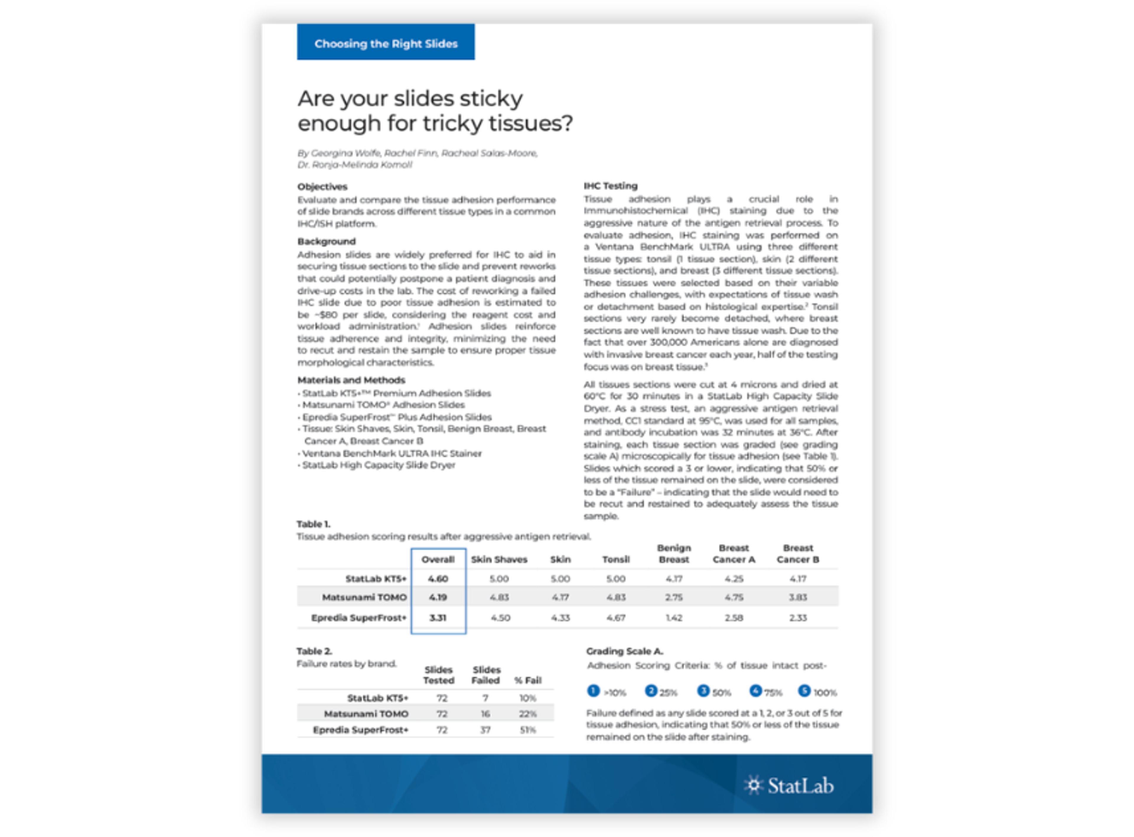
This problem is well-documented in IHC workflows. The StatLab whitepaper, Are your slides sticky enough for tricky tissues?, found that tissue loss under aggressive antigen retrieval protocols remains a leading cause of failed slides and delayed diagnoses, particularly with breast and skin tissue.
Versatility across applications mattered just as much as adhesion strength. "I have routine tissue processing, advanced staining processing, and IHC processing," Garrett says. "So, I needed that slide, any slide that a teammate grabbed, I wanted that first slide to be the best slide that they could use, because the first cut counts. We're talking about irretrievable specimens here."
Choosing adhesion slides for IHC, H&E, and special stains
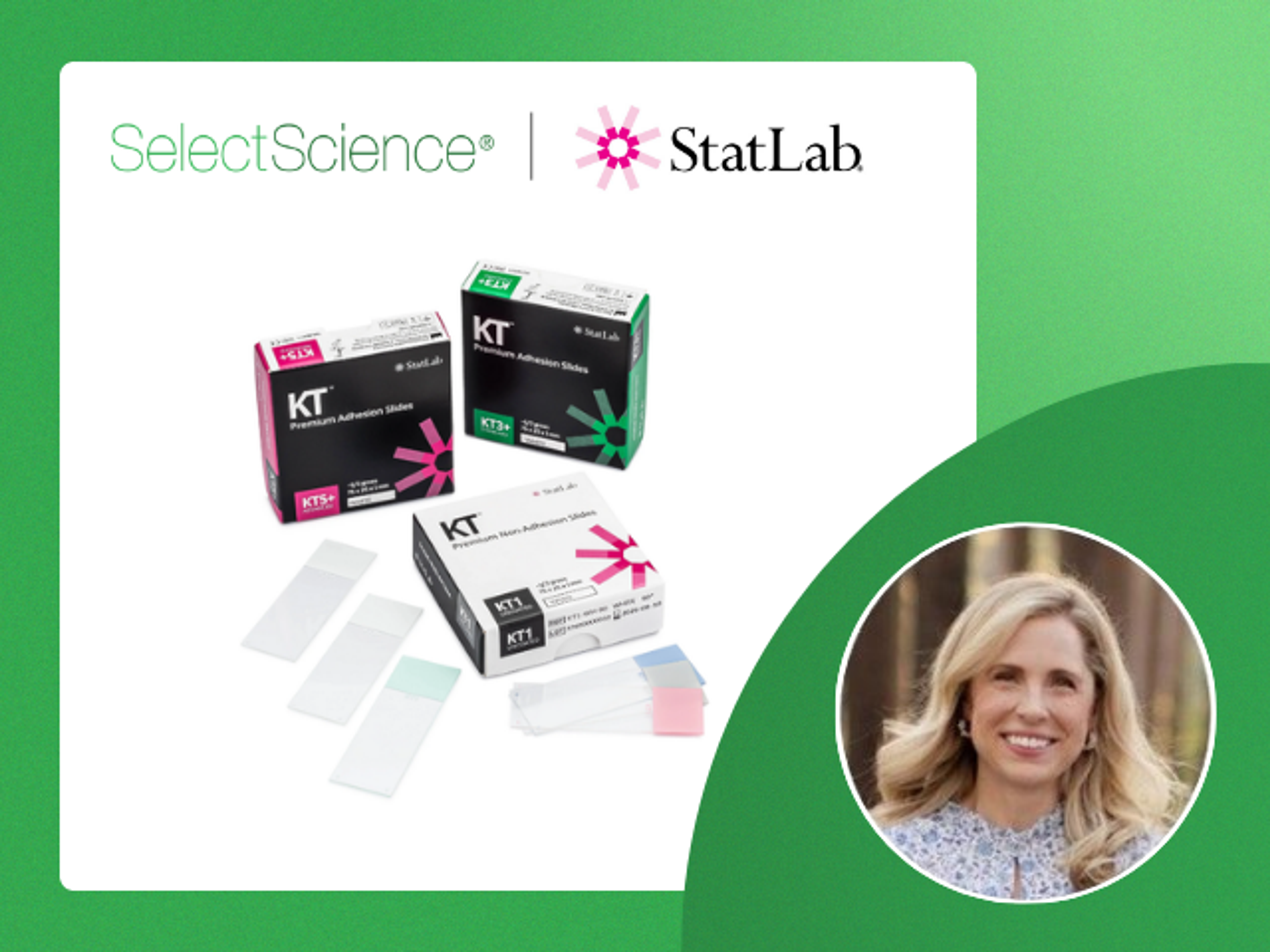
That search led Garrett to StatLab's KT Premium Slides, a line built around three formulas for different laboratory applications: KT5+ for high adhesion on difficult tissue types, including breast and fatty tissue prone to detachment during aggressive chemical processing; KT3+, hydrophobic with strong adhesion for routine staining and immunohistochemistry; and KT1 for routine H&E, special stains and frozen sections.
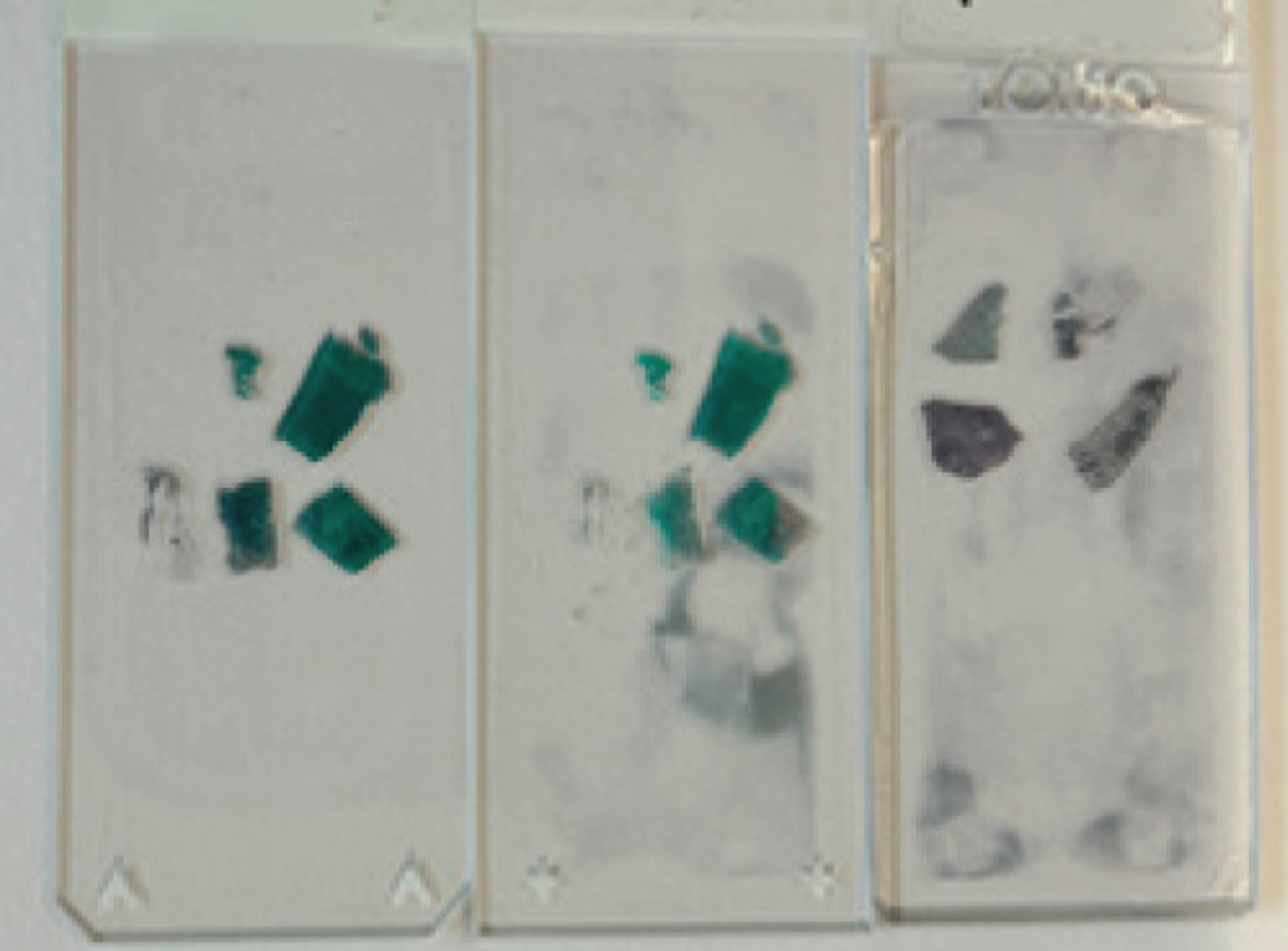
A comparative poster assessing adhesion slide performance across histology applications found significant differences in adhesion strength and background staining between slide brands, especially on breast tissue, highlighting why labs may need to assess slide performance against their own tissue types and workflows.
Availability also influenced the decision. "We could think of all these places like, oh yeah, let's get this slide, but then it's not available to us in our market," Garrett says. "I love that this was available in our market." Color coding factored in too, "We use several different color slides, and that was important to me because they mean different things," she says. "So, the availability of the multiple colors, I thought, this feels right."
Garrett also credited her StatLab representative for shaping the decision. "Oh my gosh, I don't know what I would do without him," she says of the rep, Jermaine. "He listens. He strives to understand what we're doing in this lab, and he's not just pushing out a product. It's like, how can I do what you're doing better?" She says Jermaine often grasped the lab's needs before she had fully articulated them, "A lot of times he heard what I was saying without me actually saying it. I don't speak your language, but I know what I need and I know what's happening. So, I appreciated that, definitely."
Improving histology workflow efficiency with KT Premium Slides
The changeover itself, Garrett says, was uneventful on the supply side. "It was smooth. We didn't have any issues on the StatLab side." The friction that did surface was internal, "The biggest hump that we had to get over was getting those items into Workday and getting a Workday order number," she says. "Once we had those numbers, our materials management department orders everything seamlessly, and it just works on a continuous system."
The workflow benefits followed quickly. "We've had better quality in the slides," Garrett says. "We've had the tissue, of course, stay on the slide so you can actually get a diagnosis. That's huge for our pathologists because they're looking at hundreds of slides a day, and to have to recut and waste tissue or reuse reagents, we don't get any money from that."
Her team also found they no longer needed to reserve specific slides for specific applications. "Why can't we just use KT5 across the board?" She recalled a colleague asking. "It works no matter what tissue we're putting on that slide. Grab any of them; they all work." That consistency simplified a decision their scientists had been making dozens of times a day: "You don't have to think. Choose the right color and just go to work."
Supporting digital pathology performance with low-background slides
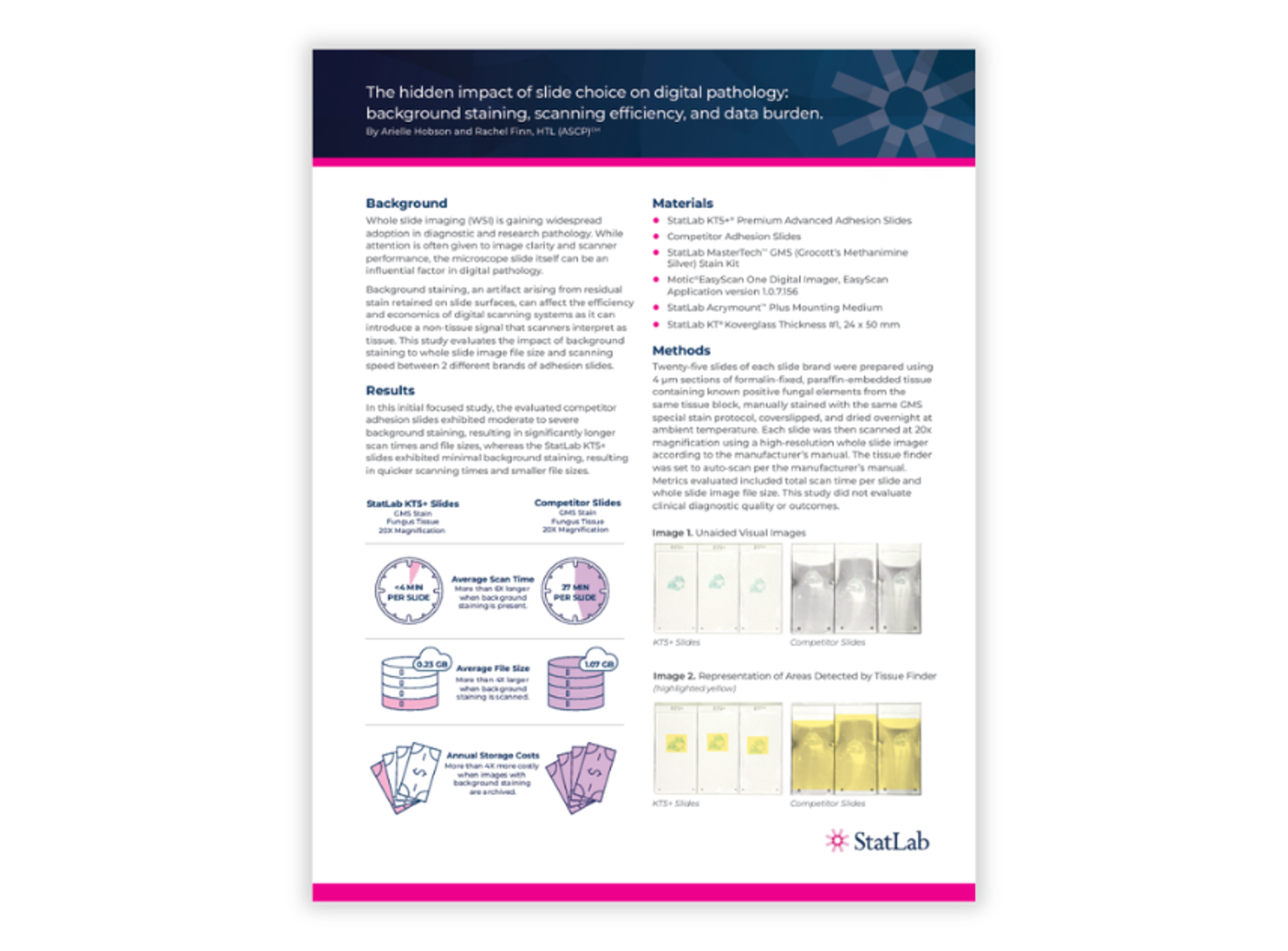
The clearest payoff surfaced once digital scanning entered the picture. "We validated our digital pathology with the KT5 slides, and that's the only slide that we've used," Garrett says. "Across the enterprise, our slides were better with the images with digital pathology." That aligns with StatLab's own data: a whitepaper on the hidden impact of slide choice on digital pathology found that KT5+ slides' minimal background staining produced six times faster scan times and four times smaller file sizes than competitor slides in a whole-slide imaging comparison.
StatLab's slide line is built alongside a companion product, KT Premium Koverglass, a borosilicate glass engineered for chemical resistance and precision-cut edges suited to automated cover slippers. "That's how we're able to get that crisp quality that our pathologists need to diagnose those tissues”, she says. Click here to learn how this Koverglass technology can enhance digital pathology and automated microscopy workflows.
The upgrade also eased skepticism among veteran pathologists still adjusting to screen-based diagnosis. "I think it has kind of calmed some of the fears of some of our more seasoned pathologists," Garrett says. She says the feedback loop with pathologists is now immediate: "Pathologists will text you immediately what they don't like. It was nice to get that text message that's like, hey, what are you doing differently? I like this slide. Keep this slide."
Why slide choice matters for scalable histology and digital pathology workflows
Atrium Health's Charlotte campus is expanding. "We are growing every day. We're taking on new clients, new hospitals," Garrett says. "The most recent thing that will be happening is next September, we will open a new bed tower, and we'll be able to service more patients." Six more GT450 scanners are also on the way, adding to a digital pathology program the slide switch now underpins.
Her advice to other lab managers evaluating slide performance is direct: run a side-by-side trial before assuming the problem lies elsewhere. "It's undeniable what you're looking at and how crisp and how clean it is, especially with those digital pathology images," she says.
Learn more about the KT Premium Slides, and hear expert insights into how slide quality supports consistency and confidence in the lab in the SelectScience® TechTalk, Slide-level precision in histology workflows.
Related products
Request Quote for All Products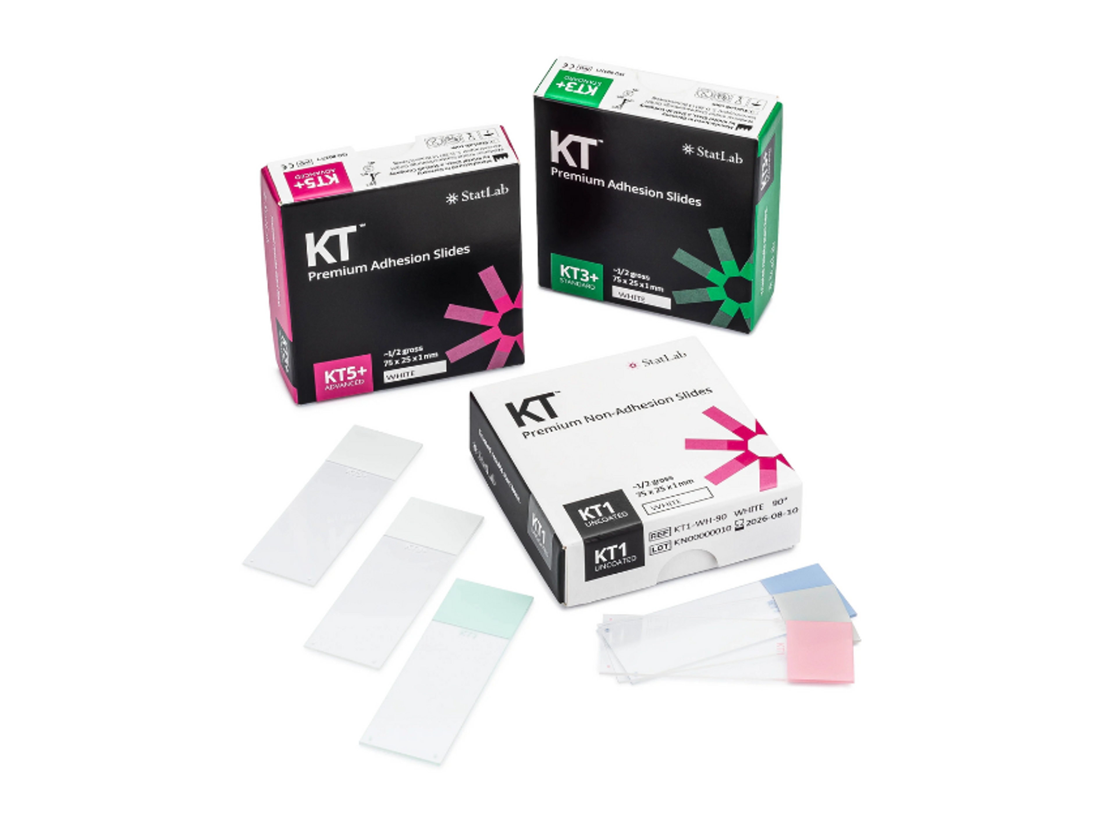
KT™ Premium Slides
StatLabResults you can trust. High performance slides to empower the best possible patient care.
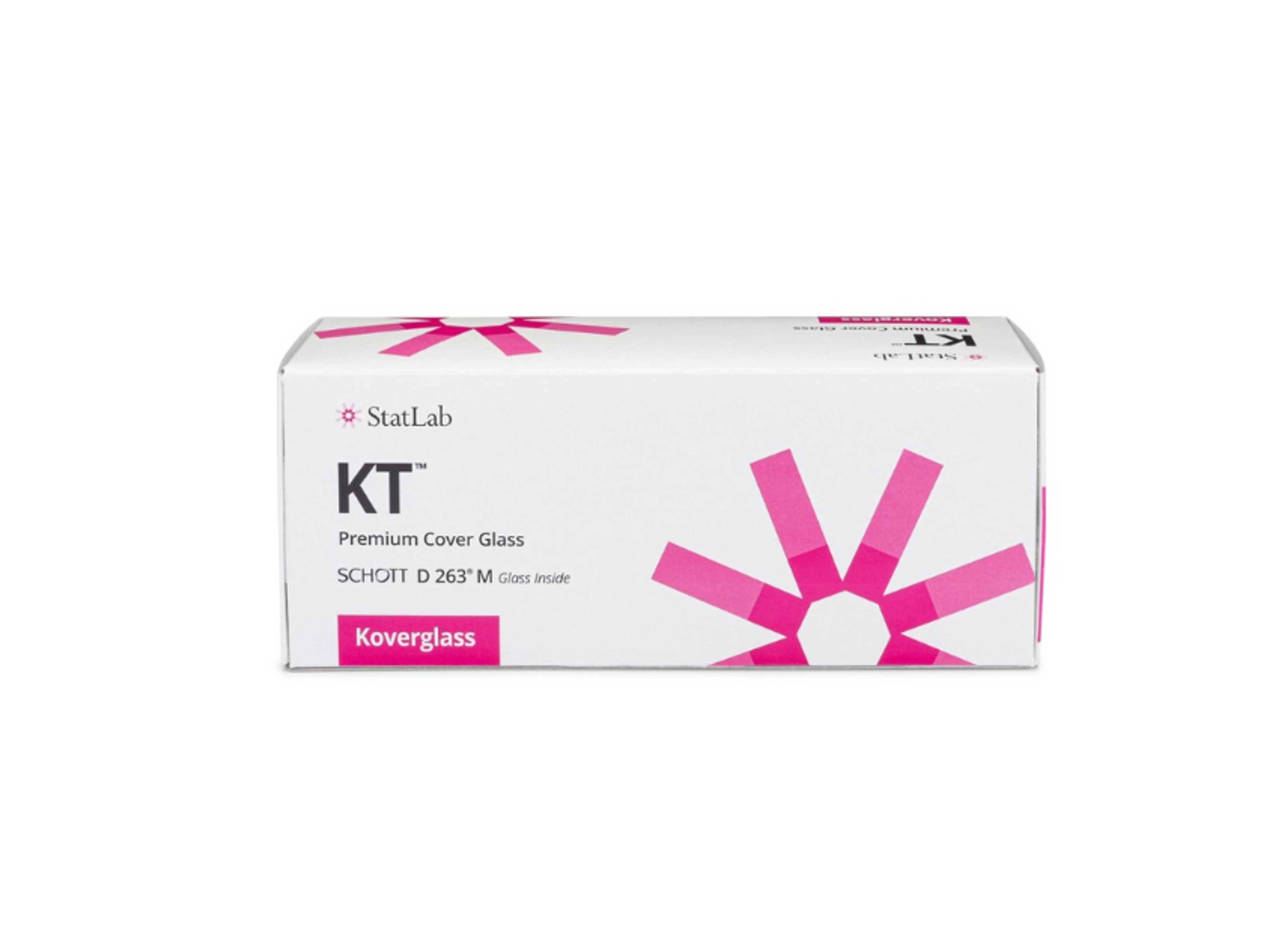
KT™ Premium Koverglass
StatLabHigh quality coverglass to protect and preserve patient samples with clarity.
Links
Tags
Frequently asked questions
Show frequently asked questions
How did Atrium Health’s Charlotte histology lab benefit from switching to StatLab KT Premium Slides?
Atrium Health’s Charlotte lab saw stronger tissue adhesion, reduced background staining, and consistent performance across H&E, IHC, and special stains after adopting StatLab KT Premium Slides, especially KT5+. The upgrade improved diagnostic reliability, minimized recuts and reagent waste, and supported efficient use of Leica GT450 whole-slide scanners, helping pathologists deliver faster, higher-quality digital diagnoses across the Atrium Health and Advocate Health system.
Why is slide adhesion strength critical for IHC, H&E, and digital pathology workflows at Atrium Health?
Chandra Garrett reports that weak adhesion caused tissue to wash off slides, preventing diagnosis, particularly in IHC and difficult tissues like breast and skin. By using KT Premium Slides, especially KT5+, the lab improved tissue retention under aggressive antigen retrieval, reduced failed slides and delays, and ensured high-quality images for digital pathology, directly impacting turnaround times and patient care at Atrium Health’s Charlotte campus.
What features of StatLab KT Premium Slides and KT Premium Koverglass support scalable digital pathology at Atrium Health?
StatLab KT Premium Slides (KT5+, KT3+, KT1) offer high adhesion, low background staining, and compatibility with multiple platforms, while KT Premium Koverglass provides chemically resistant borosilicate glass with precision-cut edges for automated cover slippers. Together, they enabled faster scan times, smaller file sizes, and crisp whole-slide images on Leica GT450 scanners, supporting Atrium Health’s expanding digital pathology program and new bed tower in Charlotte.