ResourceClinical Diagnostics
Assessing adhesion slide performance across histology applications
30 Jan 2025Adhesion slides are essential for securing tissue sections during IHC, H&E, and special stains, helping to prevent costly rework and delays in patient diagnosis. In this poster, StatLab evaluates multiple slide brands, revealing significant differences in tissue adhesion and background staining—especially with difficult tissue types like breast.
With variations in performance, it's crucial for labs to assess their specific staining needs and choose the right slide. Strong adhesion can reduce failure rates, while minimizing background staining is key for digital pathology and pathologist review.
Related products
Request Quote for All Products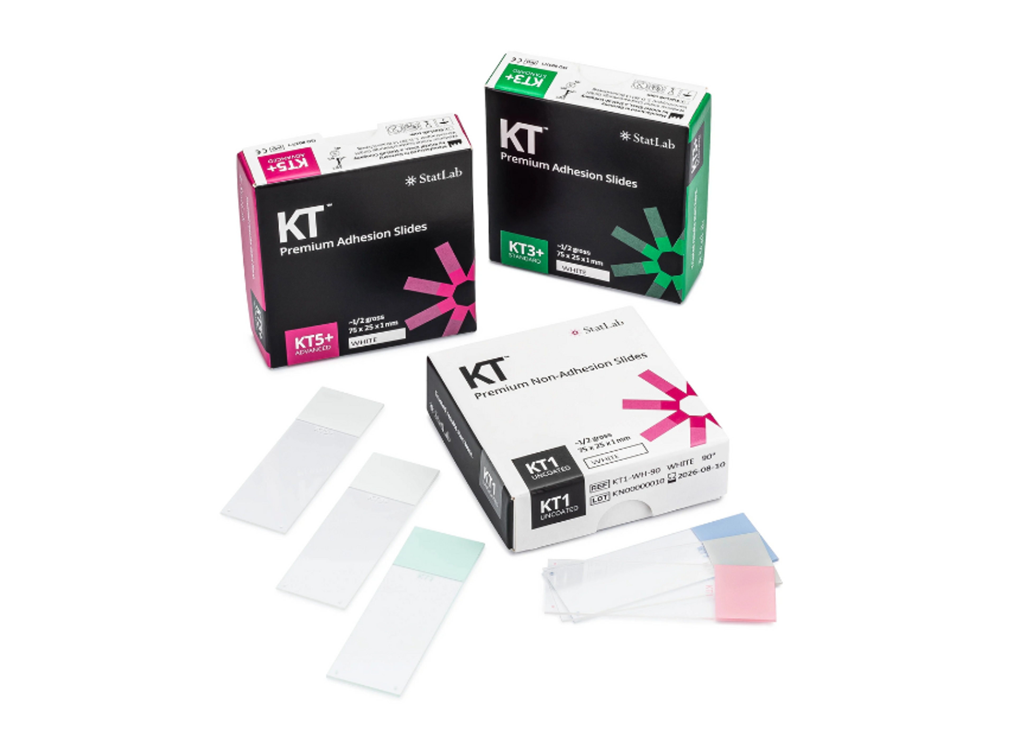
KT™ Premium Slides
StatLabResults you can trust. High performance slides to empower the best possible patient care.
Links
Tags
Cellular PathologyCellular Pathology deals with the microscopic analysis of tissue samples and cells. Sample preparation and processing includes fixation, staining, sectioning and slide mounting, using equipment such microtomes and cryostats. In choosing immunohistochemistry and immunocytochemistry kits, consider chromogens, staining method, antibodies, microscopes and imaging.HistologyImmunohistochemistryImmunohistochemistry (IHC) is a technique used to detect specific proteins in tissue samples by using antibodies that bind to target antigens. IHC is widely applied in pathology, immunology, hematology and cancer diagnostics. Explore IHC tools in our peer-reviewed product directory; compare products, check reviews, and get pricing directly from manufacturers.