How multimodal biomarker innovation is driving precision oncology
Quanterix is bridging spatial biology and ultra-sensitive detection to transform cancer research and diagnostics
9 Jun 2026
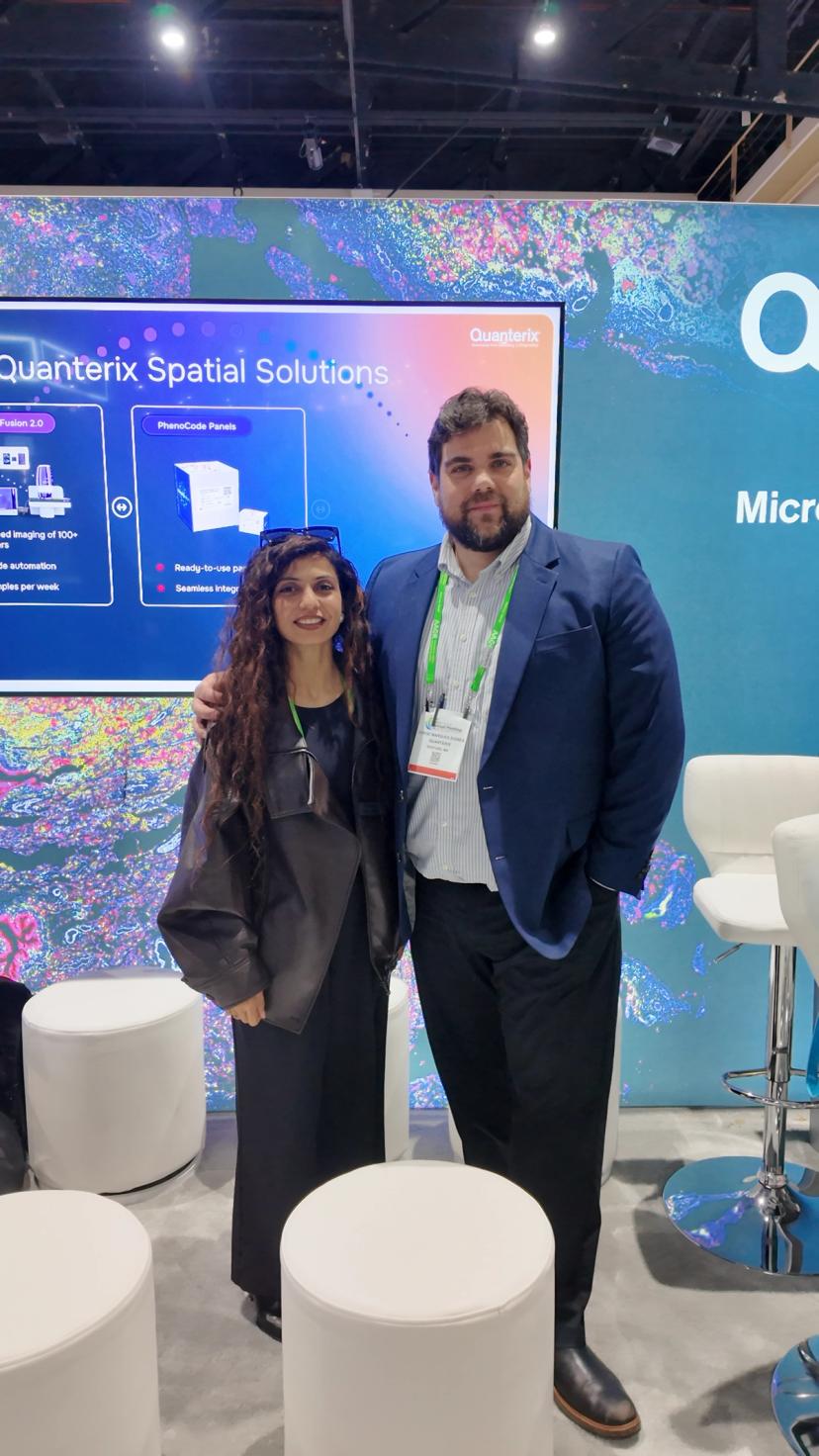
Ritu Mihani and Jorge Marques-Signes from Quanterix at AACR 2026
Cancer research is increasingly moving beyond single technologies toward integrated, multimodal approaches that better reflect the complexity of the disease. While innovations such as spatial biology and ultra-sensitive biomarker detection continue to advance individually, their convergence is opening new opportunities to connect discovery with clinical impact.
In a recent conversation, Ritu Mihani and Jorge Marques-Signes from Quanterix shared how combining complementary technologies is helping researchers build a more complete picture of cancer biology, and why this shift is key to improving patient outcomes.
Moving beyond single-modality analysis
Cancer cannot be fully understood through a single lens. As Ritu explains, context plays a critical role in determining how cells behave: “It’s the same cell type, but the behavior of the cell will change depending on the neighborhood of cells it’s surrounded by.”
This is where spatial biology has emerged as a powerful tool, enabling researchers to study protein expression directly within tissue architecture. By preserving the spatial context of cells, these approaches reveal insights into tumor heterogeneity and microenvironmental interactions-factors that are central to treatment response, particularly for immunotherapies and antibody–drug conjugates (ADCs).
Importantly, this evolution is also shaping how assays are designed. Rather than focusing solely on technical performance metrics such as plex or sensitivity, there is a growing emphasis on biological relevance. At Quanterix, this includes aligning biomarker panels with established frameworks such as the hallmarks of cancer, ensuring that generated data is not only high-quality, but meaningful for downstream interpretation and clinical translation.
Adding sensitivity through blood-based biomarkers
While tissue-based insights are essential, they provide only part of the picture. Complementing spatial analysis with ultra-sensitive blood-based detection introduces another critical dimension.
“Simoa is a high-sensitivity, ultra-high-sensitivity digital ELISA platform,” says Jorge. “We are able to measure molecule by molecule(…)at very low levels of proteins in the bloodstream.”
This level of sensitivity enables the detection of biomarkers that may otherwise go unnoticed, supporting earlier insights into disease progression and treatment response.
Connecting tissue and blood for a fuller picture
Bringing these modalities together represents a fundamental shift toward more holistic cancer research. By linking tissue-based spatial data with biomarkers detected in biofluids, researchers can better understand how molecular changes within tumors relate to signals circulating throughout the body.
This multimodal strategy also supports longitudinal monitoring, offering the potential to track how a patient responds to treatment over time rather than relying on single-timepoint measurements. As Jorge explains, integrating these approaches “opens huge possibilities” for both research and diagnostics.
Looking ahead, this convergence could help bridge discovery and clinical care, enabling more dynamic and personalized treatment strategies.
Bridging discovery and clinical application
Despite rapid technological progress, translating research findings into clinical practice remains a significant challenge. Quanterix is addressing this through its Accelerator laboratory, which focuses on validating biomarker-driven assays in regulated environments and supporting their use in clinical trials.
“We take innovation from the biomarker space and bring this innovation into a validated regulatory environment(…)to enter into the clinical,” says Jorge.
A key part of this process is adopting a fit-for-purpose approach. While discovery platforms often prioritize depth and multiplexing, clinical diagnostics require simplicity, robustness, and reproducibility. As a result, researchers are increasingly working to identify focused biomarker signatures that can be translated into scalable and clinically viable tests.
As Ritu notes, “in research, more is better(…)but in clinic it’s about getting the right information.”
Advancing spatial biology toward clinical use
Although spatial biology continues to evolve rapidly, adoption in clinical settings presents challenges. Traditional pathology workflows remain heavily reliant on immunohistochemistry (IHC), and transitioning to multiplex immunofluorescence requires both technical validation and increased familiarity among clinicians.
“There’s a need to show that multi-immunofluorescence could be a good substitution for immunohistochemistry,” Jorge explains.
Progress is underway, with emerging applications demonstrating how spatial and multiomics approaches can support diagnostic and prognostic decisions. For example, combining spatial imaging with RNA-based analysis is already being explored for early cancer detection, highlighting the growing clinical potential of integrated technologies.
The importance of collaboration and community
Advances in cancer research do not happen in isolation. Collaboration between researchers, clinicians, industry, and patients remains essential for driving meaningful progress.
Ritu emphasizes the importance of engaging with the broader scientific community to both share knowledge and learn from ongoing research: “It’s not just about presenting solutions, it’s about building partnerships.”
Patient advocates are also playing a crucial role. By sharing their perspectives and experiences, they help shape research priorities and ensure that innovation remains focused on real-world impact, including improving access to diagnostics and enabling earlier detection.
Looking ahead: early detection and AI-driven insights
Looking to the future, both Ritu and Jorge highlight early detection as one of the most transformative opportunities in cancer research. The ability to identify disease before symptoms appear, potentially through a simple blood test, could fundamentally change patient outcomes.
“If you can bring spatial technology and ultra-sensitive technology to the same patient, you can really map the journey,” Ritu says.
Artificial intelligence is expected to play a central role in enabling this shift. As datasets become increasingly complex, AI-driven models will be essential for integrating multimodal data and uncovering patterns that would be difficult to detect using traditional approaches.
From predicting treatment response to modeling disease progression through concepts such as digital twins, AI has the potential to accelerate the move toward truly personalized medicine.
As Jorge puts it: “We are moving towards an approach where each patient is going to be a data point itself.”
A connected future for cancer research
The convergence of spatial biology, ultra-sensitive detection, and advanced data analysis is redefining how cancer is studied and treated. While challenges remain, the shift toward integrated, biologically driven approaches is clear.
By connecting insights across technologies, and across the research and clinical continuum, the field is moving closer to a future where every patient can be understood as a unique biological system, and where precision medicine becomes a practical reality.
Want the latest science news straight to your inbox? Become a SelectScience member for free today>>
Related products
Request Quote for All Products
Simoa® Accelerator Laboratory
QuanterixProtein Biomarker Testing and Custom Assay Development Services
Frequently asked questions
How is Quanterix integrating spatial biology and ultra-sensitive blood-based biomarkers to advance precision oncology?
Quanterix is combining spatial tissue analysis with ultra-sensitive, digital ELISA–based blood biomarker detection (Simoa) to create a more holistic view of cancer. Spatial biology tools on the proteomics side help map protein expression and tumor architecture in tissue, revealing tumor heterogeneity and the tumor microenvironment. In parallel, Simoa enables molecule-by-molecule quantification of very low-level proteins in the bloodstream. By linking tissue-based insights with blood-based biomarkers, researchers can better track disease evolution, connect tumor biology to circulating indicators, and support precision medicine strategies, including early diagnostics and longitudinal monitoring of treatment response.
What role do multimodal biomarker strategies, including spatial biology and proteomics, play in early cancer detection and clinical translation?
Multimodal biomarker strategies integrate spatial biology, proteomics, and ultra-sensitive blood-based assays to move from discovery to real-world clinical impact. Spatial biology provides contextual information on how tumor cells behave within their native environment, while proteomic mapping of protein expression clarifies tumor heterogeneity and microenvironmental factors that influence response to immunotherapies and antibody-drug conjugates (ADCs). These insights are complemented by blood-based biomarkers measured with high sensitivity. Biomarker panels are increasingly aligned with frameworks such as the hallmarks of cancer and refined into key biomarker signatures that are fit-for-purpose in clinical diagnostics. Through its Accelerator laboratory and diagnostic development efforts, Quanterix validates these assays in regulated environments, helping translate complex discovery platforms into streamlined, reproducible tests for early detection and clinical decision-making.
How are AI-driven models and digital twins expected to enhance cancer research using data from spatial biology and ultra-sensitive detection platforms?
Artificial intelligence is expected to be pivotal in interpreting the complex, multimodal datasets generated by spatial biology and ultra-sensitive detection platforms like Simoa. As data volume and complexity grow, AI-driven models will integrate information across tissue imaging, proteomics, and blood-based biomarkers to predict treatment response, model disease progression, and support early disease detection. Concepts such as digital twins—virtual models of individual patients—are beginning to move toward practical application, enabling simulation of treatment outcomes based on a patient’s unique biological data. By combining AI with spatial and ultra-sensitive technologies in the same patient, researchers can more accurately map the cancer journey and move toward truly individualized, precision medicine where each patient is treated as a unique data point.
How is Quanterix integrating spatial biology and ultra-sensitive biomarker detection to advance cancer research?
Quanterix, represented by Ritu Mihani and Jorge Marques-Signes, is combining tissue-based spatial biology with ultra-sensitive blood-based biomarker detection using the Simoa digital ELISA platform. This multimodal strategy links tumor tissue architecture and microenvironmental interactions with circulating biomarkers, helping researchers connect discovery data to clinical impact and improve understanding of treatment response, especially for immunotherapies and antibody–drug conjugates.
What role does the Quanterix Accelerator laboratory play in translating cancer biomarker discoveries into clinical applications?
The Quanterix Accelerator laboratory focuses on validating biomarker-driven assays in regulated environments to support their use in clinical trials. It takes innovation from the biomarker discovery space and moves it into a validated regulatory setting using a fit-for-purpose approach, emphasizing simplicity, robustness, and reproducibility. This helps convert broad research biomarker panels into focused, clinically viable diagnostic tests that deliver the right information for patient care.
How are spatial biology, blood-based biomarkers, and AI shaping the future of personalized cancer medicine?
Spatial biology, ultra-sensitive blood-based biomarkers, and AI are converging to enable earlier cancer detection and more personalized treatment strategies. By integrating multimodal data from tissue and biofluids, researchers can track disease progression and treatment response longitudinally. AI-driven models analyze these complex datasets, support concepts like digital twins, and help predict treatment outcomes, moving toward a future where each patient is treated as a unique biological data point.
Links
Related content

Quanterix unveils Content Innovation Engine at AACR 2026
The Content Innovation Engine integrates Akoya's spatial biology platform and Quanterix's immunoassay infrastructure to unlock cancer biology and accelerate clinical application