Ground-breaking versatility from Scientifica’s Multiphoton Imaging System
31 Aug 2012Product news
Multiphoton imaging is one of the most powerful life science research tools of the 21st century and Queen’s Award winner, Scientifica, has acknowledged this with the launch of a modular Multiphoton System. For the first time, it allows the user to build a cost effective and future proof system based around the renowned SliceScope microscope.
Multiphoton imaging uses high intensity pulsed lasers, fast moving mirrors, and precision optics to allow researchers to image structure and activity within living tissue. A laser spot is scanned quickly over a sample, exciting dyes within the tissue. The emitted light is collected with sensitive photomultiplier tubes (PMTs) and the image is captured and displayed on a PC screen.
“We wanted to offer researchers a flexible, cost effective solution when it comes to multiphoton imaging,” says Mark Johnson, joint managing director at Scientifica. “The option of a DIY system or fully integrated package offers crucial flexibility for a variety of users with different experience, requirements and budgets – opening more opportunities for labs around the world to utilize this technique.”
Scientifica’s system allows the user to image deep into living tissue, while sustaining a healthy sample. Whether working with tissue sections in vitro, or with whole organisms in vivo, the use of short pulses of high energy IR light decreases the average laser power reaching the sample and reduces phototoxicity.
Scientifica’s exclusive Scanhead design achieves diffraction limited resolution by optimizing the size and shape of the excitation laser spot at the sample. This allows researchers to visualize the finest biological structures essential for investigating morphology and live activity within samples. For visualizing larger structures, the unique relay lens system offers high quality capture of your entire field of view by preventing vignetting and loss of fluorescent signal at the edge of the image.
Efficient photon gathering, via the PMT’s, can be accomplished from a range of objectives, this produces a brighter image using less laser intensity to excite the dyes, while allowing even very weak signals to be detected. Scientifica’s high quality detection units use custom optics, mechanics and noise-reducing electronics to achieve superior signal to noise ratio by maximizing light collection either above or below your sample. A range of open-source software packages are also available from either ScanImage or Helioscan to offer versatile and continually evolving platforms for experimental control. The elegant and exceptional quality offered by Scientifica allows effortless integration with other popular experimental techniques including electrophysiology; broadening access and appeal for this ground breaking technique.
Image supplied showing the Scientifica Multiphoton Imaging System (Camera – QImaging Rolera Bolt)
Related products
Request Quote for All Products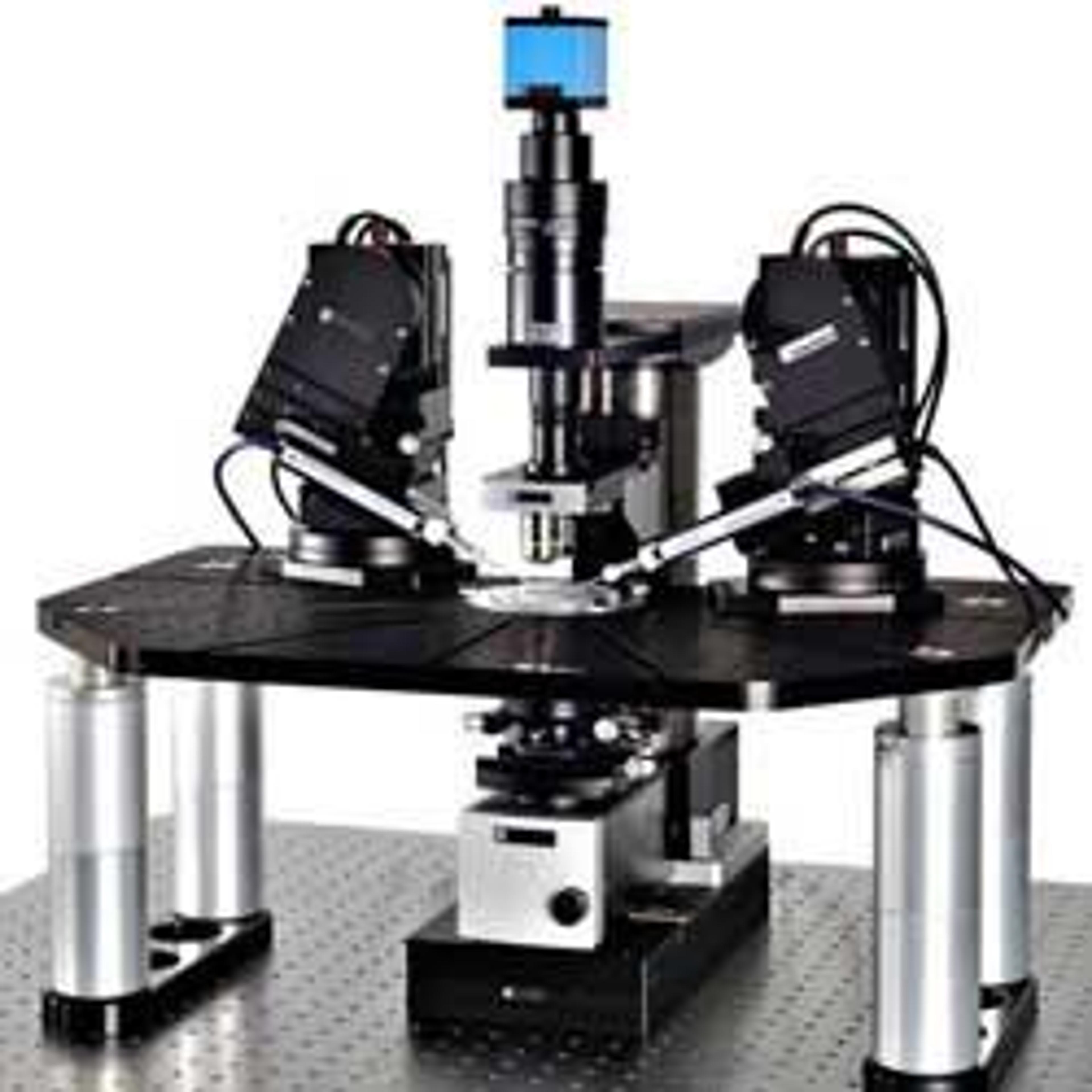
SliceScope Pro Microscopy Systems
Scientifica LtdSliceScope Pro systems are a range of fully integrated and motorised patch clamping solutions. They allow the user to control manipulators, microscope platforms, microscope condenser and objective focus all from simple user consoles. The SliceScope a fixed stage, focussing nosepiece microscope. This defines a new bench mark in imaging and electrophysiology research. Developed in collaboration with the world leading researchers, the SliceScope provides a very stable, compact platform with outstanding optics for efficient automated microscopy. The SliceScope’s smart design is ready to accept a wide range of accessories and light sources to best suit your application. The SliceScope frame is interchangable between in-vivo and in-vitro configurations, making the SliceScope Pro systems very versatile, ideal for saving space and money. Features • Motorised focus control: provides fine focus control • Motorised condenser control: for optimal Kohler illumination • Unique “follow software”: to keep pipettes within field of view as you move around your sample • Narrow, ultra stable profile: extra room for equipment around your experiment • Choice of contrast allows you to use your preferred contrast method such as Oblique, IR DIC and Dodt Contrast • Ultra-low noise electronics: Suitable for demanding single channel electrophysiology recordings • Modular design: easily configured into two-photon, confocal and in-vivo designs by the user • Easy pipette exchange: increased productivity and reproducible positioning