G:BOX Chemi XT4 for Whole Plant Imaging Application
11 Jun 2012Product news
Syngene, a manufacturer of image analysis solutions, has announced that its advanced G:BOX Chemi XT4 imaging system can be used to safely visualize and analyze fluorescently labeled proteins in whole plants, allowing scientists to accurately detect how much of their marker proteins are being expressed, and where.
Using a G:BOX Chemi XT4 system fitted with safe, blue LED lighting and filter (Syngene UV, or short pass filters), fluorescent proteins such as GFP fusions of microtubule binding proteins are easily visualized in whole plants. Since the light tight cabinet which comes with the G:BOX Chemi XT4 system is spacious, plants, including Arabidopsis, will easily fit into the cabinet.
To make plant imaging quick and simple, the G:BOX Chemi XT4 system is controlled by Syngene’s intuitive GeneSys software, which automatically sets up the optimum imaging conditions for every fluorescently labeled protein. This means plant biologists can capture a high-quality whole plant image in seconds, with minimum training. To save time with quantifying proteins, the G:BOX Chemi XT4 system also includes GeneTools image analysis software, enabling researchers to instantly determine how much of their marker proteins are being expressed.
Laura Sullivan, Syngene’s Divisional Manager explained: “Many researchers want to trace their plant proteins and record the results easily but until now there hasn’t been an imaging system that could do this quantitatively. We’re delighted that our technical team has worked on this problem to determine not only the best, but also the safest imaging conditions for visualising fluorescently labeled plant proteins. This new application means the Syngene G:BOX Chemi XT4 is the best system for plant biologists wanting a simple method of pinpointing where and how much of their fluorescent proteins are being expressed in whole plants.”
Related products
Request Quote for All Products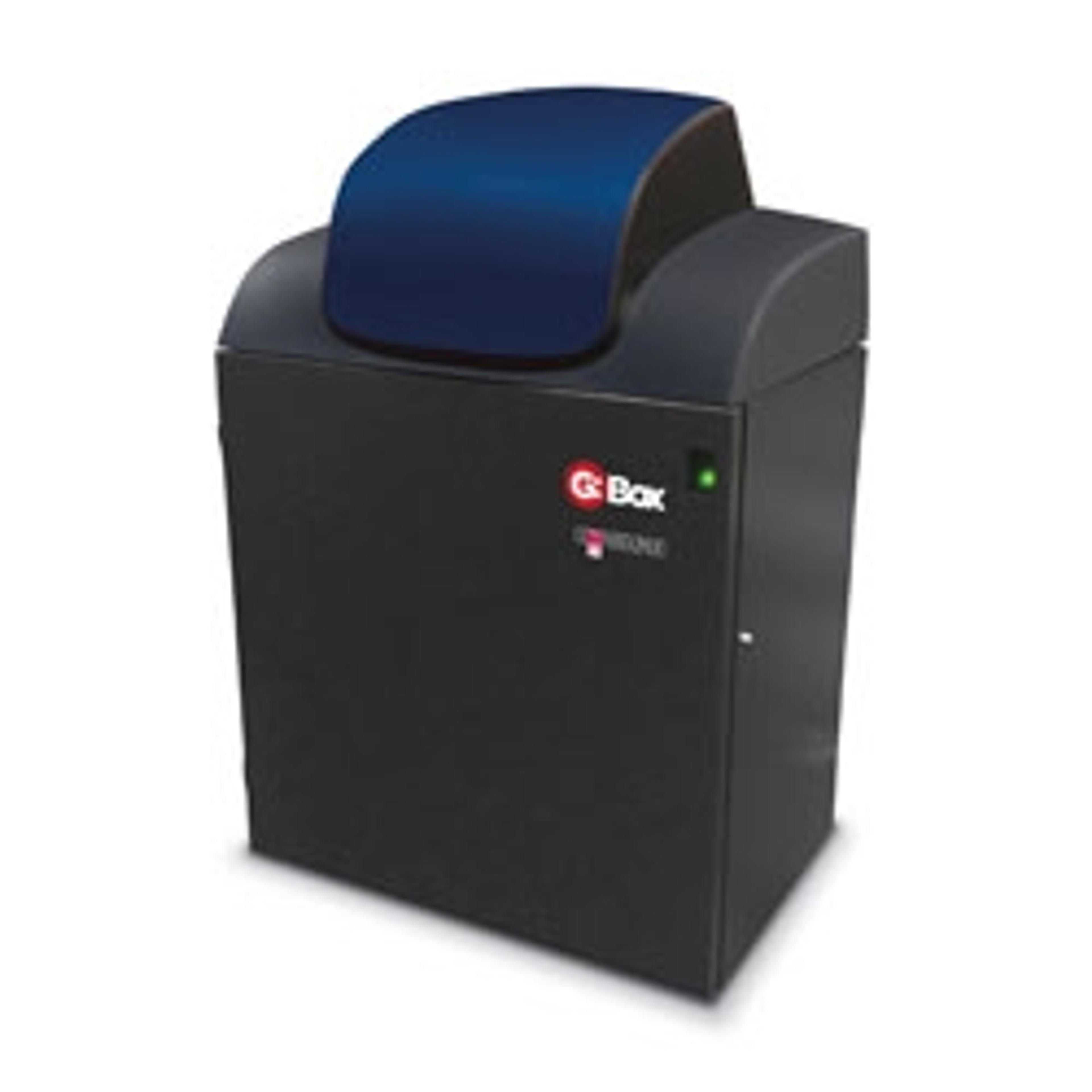
G:BOX Chemi XRQ
SyngeneG:BOX Chemi XRQ is a cost-efficient chemiluminescence imaging and gel documentation system. For a laboratory that needs hassle-free chemiluminescent detection, as well as routine gel documentation, using the G:BOX Chemi XRQ’s powerful GeneSys software to switch between applications is simplicity itself.
G:BOX Chemi XX6
SyngeneG:BOX Chemi XX6 gel imaging system has a high resolution camera for imaging multiple sample types and sizes, from fluorescence 1D to 2D gels to chemiluminescent blots. Your lab’s imaging system shouldn’t control how you detect proteins on Western blots. Chemiluminescence is great if you want sensitive detection of picogram or femtogram amounts, while fluorescence lets you quantify and detect multiple different proteins on one blot.