FluoView FVMPE-RS Multiphoton System - Two New Microscopes for Live Cell and In-Vivo Imaging
20 Feb 2015Product news
Olympus has announced the release of two new configurations of the FluoView FVMPE-RS multiphoton laser scanning microscope series - a Gantry microscope frame and an inverted microscope frame.
The flagship offering in the range of Olympus biological microscopes, the FluoView FVMPE-RS series is widely used in life science research. Its high-speed scanner allows observation of ultra-rapid biological responses, and the system can obtain vivid images from as deep as 8 mm below the tissue surface. Two new configurations now expand the FluoView FVMPE-RS series beyond the original upright frame version introduced in 2013 to offer even greater scope and flexibility for research. The new configurations also open up a wide range of observation possibilities suited to imaging a greater variety of biological specimens.
The Gantry microscope frame features an ultra-stable arch-like structure that allows considerable space beneath the objective to accommodate experiments of varied sizes. A volume of 640 mm wide, 355 mm high and 520 mm deep is available if the stage is removed, providing sufficient space for the researcher’s own experimental apparatus and providing flexibility to suit different observation purposes.
The inverted microscope frame is ideal for observation of cells in three-dimensional (3D) cultures, where multiple layers of cells are cultured in a petri dish or similar vessel. Because inverted microscopes allow the researcher to observe the sample from below, this configuration is optimized for 3D-culture imaging and allows cells that have adhered to the base of the petri dish to be observed without culture fluid touching the objective.
Multiphoton imaging is a microscopy technique used in the life sciences to elucidate the role and function of proteins or nerves, and to seek out important data related to drug discovery. For example, in the case of cerebral nerves, scientists need to observe biological responses at sites far below the tissue surface, both to study the nerve mechanism and to develop therapeutic drugs or other treatments for diseases such as cancer or Alzheimer’s. Relying on Olympus’s renowned optical and digital technologies, the FluoView FVMPE-RS multiphoton laser scanning microscope systems meet the needs of researchers by helping them capture images and movies of rapid biological responses deep within samples.
As a direct result of researchers requesting support for a wider range of applications and specimens, Olympus has now added the Gantry microscope and inverted microscope frames to complement the original upright system. Delegates of Focus on Microscopy at Göttingen, Germany, March 29 – April 1 2015, will have a first chance to see mock-ups of the new configurations at booth 36.
Related products
Request Quote for All Products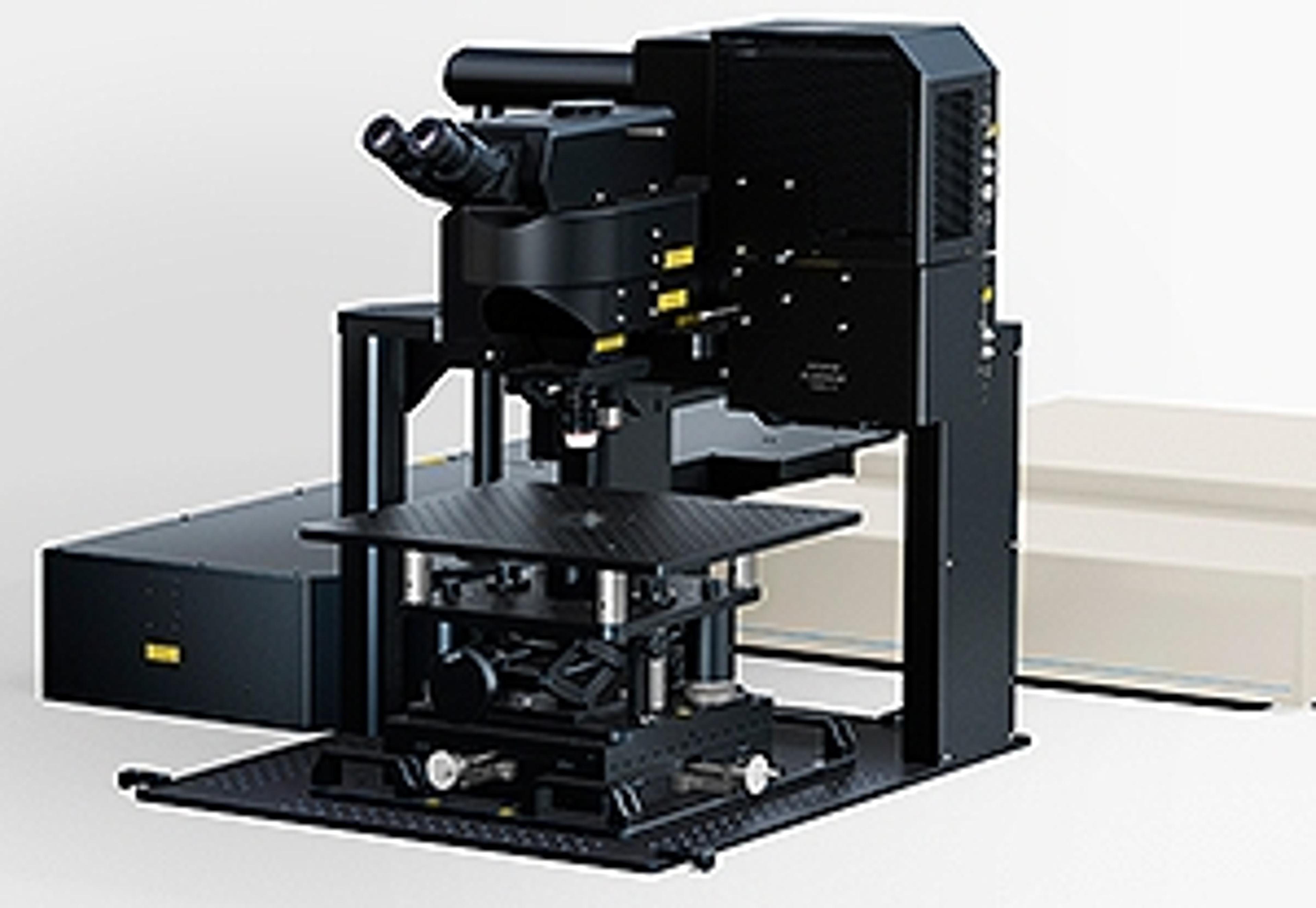
FVMPE-RS Multiphoton System - Two New Frames for Live Cell and In-Vivo Imaging
EVIDENTExpanding the possibilities of multiphoton microscopy, Olympus presents two new configurations of its FluoView FVMPE-RS multiphoton laser scanning microscope series: the Gantry microscope frame to accommodate large samples and setups, and the inverted microscope frame for 3D cell culture. The Olympus FluoView FVMPE-RS multiphoton laser scanning microscope series is optimised for life science research. Its high-speed scanner allows observation of ultra-rapid biological responses, and the system can obtain vivid images from as deep as 8 mm below the tissue surface. As a direct result of researchers requesting support for a wider range of applications and specimens, Olympus has now expanded the range beyond the original upright frame version. The new Gantry microscope and inverted microscope frames offer even greater scope and flexibility for research, opening up a wide range of observation possibilities suited to imaging a greater variety of biological specimens. The Gantry microscope frame features an ultra-stable arch-like structure that allows considerable space beneath the objective to accommodate experiments of varied sizes. A volume of 640 mm wide, 355 mm high and 520 mm deep is available if the stage is removed, providing sufficient space for the researcher’s own experimental apparatus and providing flexibility to suit different observation purposes. The inverted microscope frame is ideal for observation of cells in 3D cultures, where multiple layers of cells are cultured in a petri dish or similar vessel. Because inverted microscopes allow the researcher to observe the sample from below, this configuration is optimised for 3D cell culture imaging and allows cells that have adhered to the base of the petri dish to be observed without culture fluid touching the objective. Such capabilities meet the needs of researchers, enabling them to capture rapid biological responses deep within samples. Features: Gantry frame accommodates large samples and setups Three-dimensional Adjustment - A removable manual XYZ stage enables height adjustments. Changing between thin sample and whole animal imaging can be easily accomplished. Large Workspace Applications: Multiphoton confocal laser scanning microscopy Optogenetics Electrophysiology In vivo imaging with large samples Live cell imaging including 3D cell culture