ResourceSpectroscopy
Oridonin Perfusion Causes Cytotoxicity in U-2 OS Cells
11 Jul 2016This application note describes using the ONIX2 System in conjunction with the Lionheart™ FX Imager to rapidly image and analyze perfused tissue culture cells in multiple fluorescent colors and brightfield.
Related products
Request Quote for All Products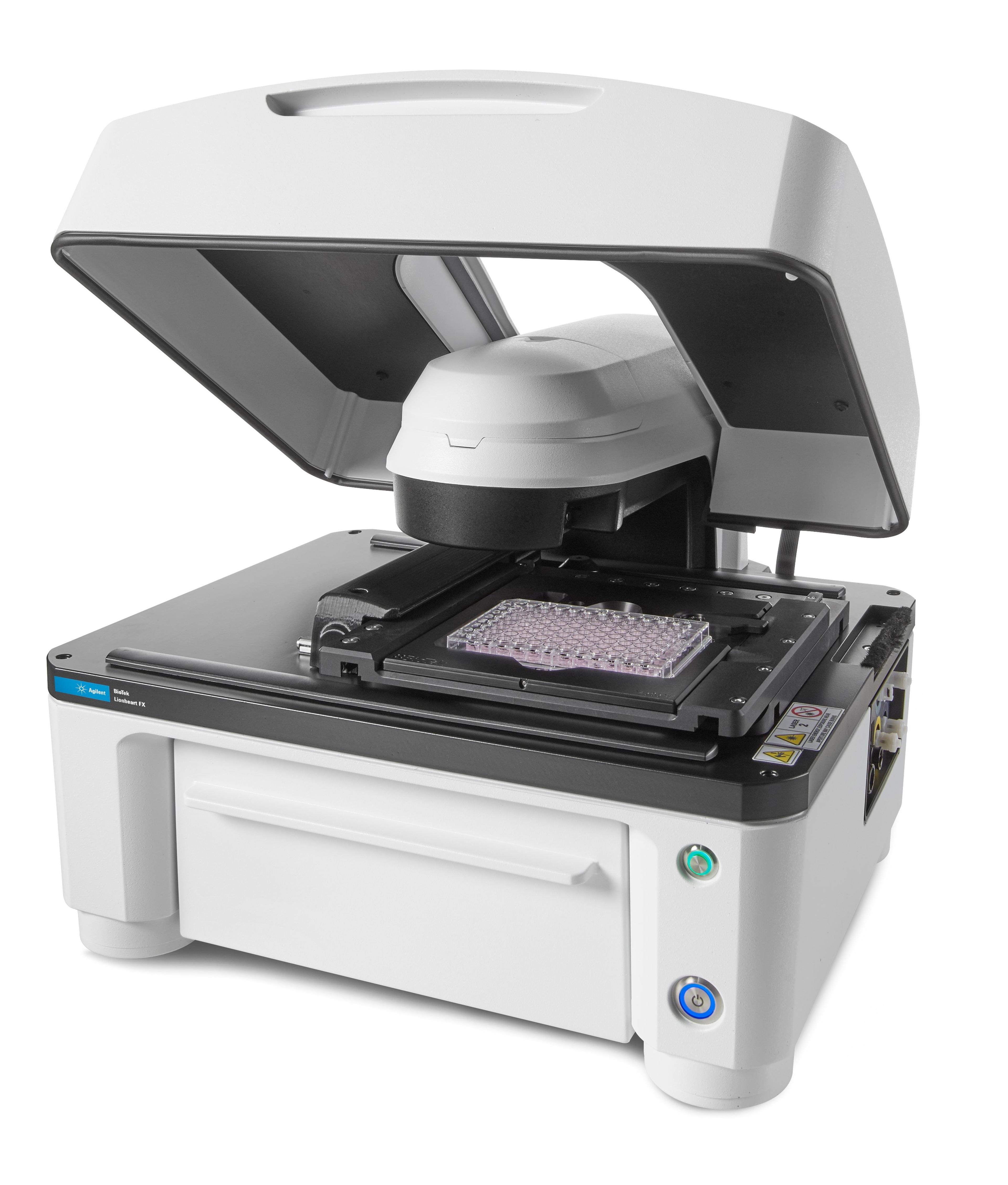
Agilent BioTek Lionheart FX Automated Microscope
Agilent TechnologiesAgilent BioTek Lionheart FX Automated Microscope is an inclusive microscopy system for fixed and live-cell applications.
Links
Tags
Sample PreparationSample preparation can improve the quality and speed of separation techniques. Products to assist sample preparation include filtration equipment, evaporators, membranes and sieves.Fluorescence SpectroscopyFluorometers and spectrofluorometers (also called fluorescence spectrometers) are used to measure the intensity and wavelength of fluorescent light emitted from a sample after excitation by illumination. Spectrofluorometers utilize monochromators to select the desired wavelengths, whereas filter fluorometers employ a set of filters. Spectrofluorometers for measuring steady-state fluorescence and lifetime fluorescence (or time-resolved fluorescence) are available, as well as fluorescence microscopes and microplate readers. Find the best fluorescence spectroscopy products in our peer-reviewed product directory: compare products, check customer reviews and receive pricing direct from manufacturers.Cell / Tissue CultureCell culture or tissue culture is used to study the biology of cells or tissues and to isolate cellular products in an environment which can be manipulated and well defined. Accurately control your culture environment with bioreactors or culture incubators, bind your cells to a surface or together with an extracellular matrix. Distinguish cell types with differential media or proliferate cells with certain characteristics using selective media. Enrich your media with supplements such as growth factors, sera and vitamins. Find the best cell and tissue culture products, kits and equipment in our peer-reviewed product directory: compare products, check customer reviews and receive pricing direct from manufacturers.Gel Doc / Image AnalysisGel documentation (gel doc) or gel imaging systems are used for the analysis of proteins, antibodies and nucleic acid immobilized in polyacrylamide or agarose gels, membranes or microarrays. Explore a range of a gel imaging systems, densitometers, scanners, transilluminators or UV lamp + CCD cameras for your image analysis solutions. Colorimetric, fluorescent and/or radioisotopic samples can be visualized and documented for further analysis. See gel doc / Image analysis software for quantitative 1D and 2D analysis of your samples. Find the best gel doc / image analysis products in our peer-reviewed product directory: compare products, check customer reviews and receive pricing direct from manufacturers.Flow Cytometry / Cell CountingFlow cytometers are used to count, sort and examine multiple characteristics of cells. Other cell analysis equipment includes image cytometers, cell counters, fluorescence-activated cell sorters (FACS), magnetic-activated cell sorters (MACS), and a range of flow cytometry assay kits. Flow cytometers can reveal information on cell viability, cell proliferation, apoptosis and cell cycle progression, as well as identify cell populations and intracellular or cell-surface molecules. Additionally, some flow cytometers, known as FACS, have an additional sorting function after analysis. Cell counters and image cytometers count live and dead cell populations and can also conduct cell proliferation assays. Find the best flow cytometers, cell counters and cell sorters in our peer-reviewed product directory: compare products, check customer reviews and receive pricing direct from manufacturers.Cell-Based AssaysCell-based assays are used to monitor the presence, quantity and activities of a desired cellular analyte including drug molecules or biomarkers. This can reveal information on cell health (apoptosis, cytotoxicity, viability and proliferation assays), cell metabolism, cell migration and cell signaling mechanisms. Find the best cell-based assay products, kits and equipment with our peer reviewed product directory: compare products, check customer reviews and receiving pricing direct from manufacturers.In Vivo Imaging Systems<i>In vivo</i> imaging systems, including pre-clinical imaging systems and medical imaging systems are used to non-invasively visualize and capture images of live animals and plants. Monitor the natural processes or diseases of your subjects using small-animal pre-clinical imaging systems, including single photon positron emission tomography (SPECT), positron emission tomography (PET), computed tomography (micro-CT), magnetic resonance imaging (MRI), X-ray radiography, ultrasound, fluorescence and bioluminescence imagers. Multimodal systems and software solutions are also available for correlative analysis of organ, tissue, cell, or molecular-level processes. Find the best in vivo imaging products in our peer-reviewed product directory: compare products, check customer reviews and receive pricing direct from manufacturers.Toxicology / Drugs of Abuse TestingToxicology and Drugs of Abuse Testing is the investigation into toxic and illegal substances found at the scene of a crime or from a suspect and / or victim associated with a scene of a crime. Analyzers, immunoassays and drugs of abuse test kits and presumptive test kits are an essential aspect of toxicology investigation.Light MicroscopyLight microscopes or optical microscopes are used to visualize microscale objects under magnification, including cells, clinical specimens and materials. Lab equipment for light microscopy includes confocal microscopes, fluorescence microscopes, zoom and stereo microscopes. Microscope slides and imaging reagents are available for visualizing samples, as well as various microscope stages and incubators for large or temperature-sensitive samples. Find the best light microscopes in our peer-reviewed product directory: compare products, check customer reviews and receive pricing direct from manufacturers.CytotoxicityCytotoxicity assays measure the toxic effects of substances on cells, often used in drug testing and environmental studies. These tests are crucial in determining the safety of chemicals and pharmaceutical compounds. Explore cytotoxicity testing tools in our peer-reviewed product directory; compare products, check reviews, and get pricing directly from manufacturers.FluorescenceThe emission of fluorescence occurs when a photon of energy is supplied to a fluorescent chemical compound by an external source, causing it to become excited. Fluorescence can be detected and measured for different purposes using microplate readers, fluorescence microscopes, fluorescence scanners, and flow cytometers.Cell AnalysisThe analysis of cells allows researchers to understand the factors which contribute to cell health and function. These influencing processes can then be predicted and altered, leading to the development of medication and disease treatments.Live Cell ImagingLive cell imaging is the study of living cells using microscopes and high-content imaging systems. This technique provides in-depth insight into fast and complex biological processes, by allowing dynamic imaging of living cells instead of acquiring an individual image at a single point in time.MicroscopyMicroscopy is a technique used to observe small objects in detail, from cells to materials, using light or electron microscopes. It enables researchers to examine structures with high resolution, aiding in fields such as biology, medicine, and materials science. With advanced microscopy techniques, scientists can gain insights into cellular processes, tissue structures, and material properties. Explore the best microscopy solutions in our peer-reviewed product directory, compare products, read customer reviews, and get pricing directly from manufacturers.