Pushing the Boundaries of 3D Microtissue Analysis using High Content Imaging
Pushing the Boundaries of 3D Microtissue Analysis using High Content Imaging
22 Oct 20153D cell culture methods are widely accepted as being more physiologically relevant than conventional 2D cell culture methods, and are believed to improve the prediction of drug candidates at an early stage in the drug development process. The visualization of 3D structures is challenging, for example when imaging microtissues there is light scattering and absorption which prevents imaging deep into the center of the microtissue. This application note demonstrates the imaging of microtissue using the Opera® High Content Screening System (equipped with a water objective lens) in combination with a microtissue pretreatment.
Related products
Request Quote for All Products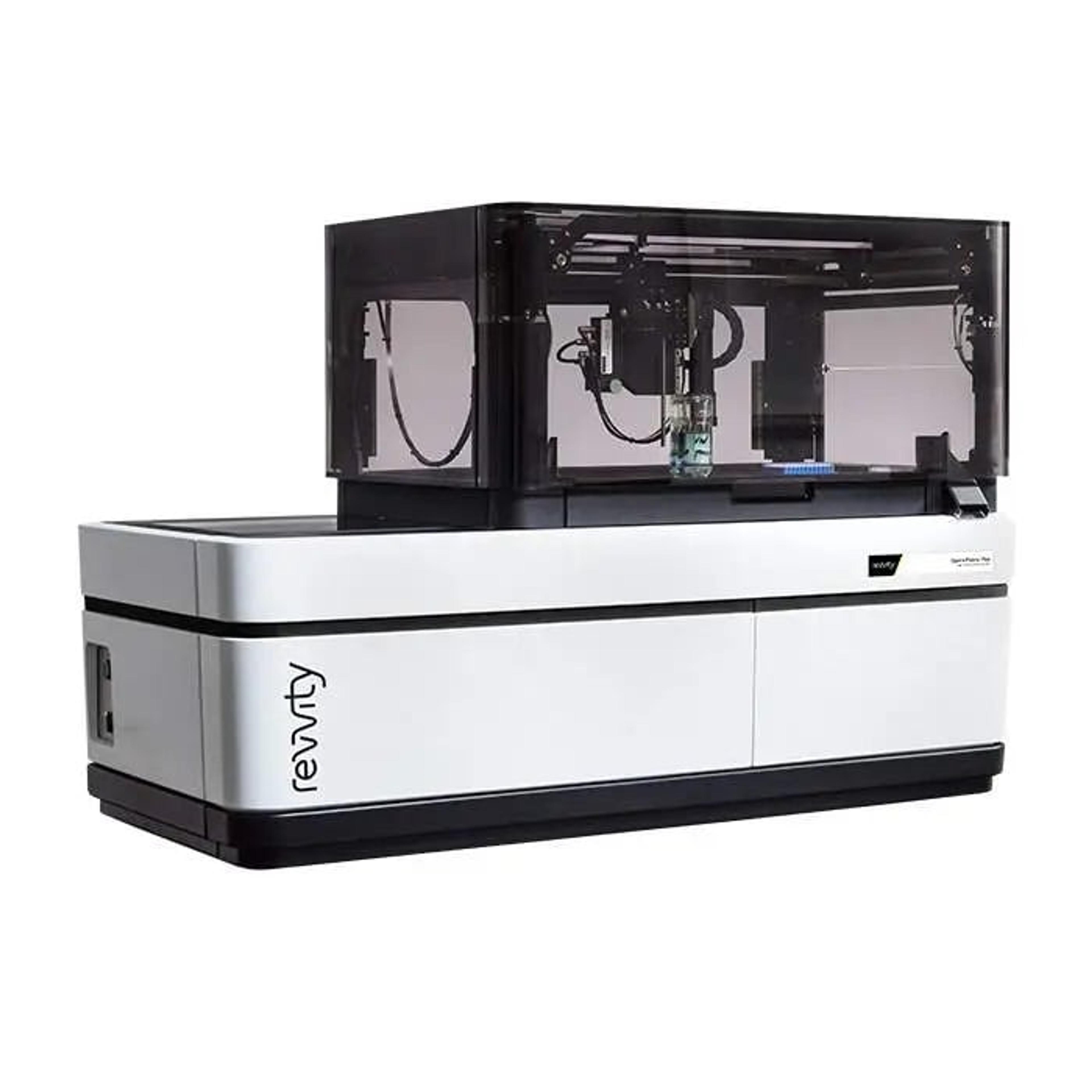
Opera Phenix High Content Screening System
RevvityThe Opera Phenix ® Plus High-Content Screening System is the premier confocal solution for today' most demanding high content applications. Drawing on over a decade of experience with the industry-leading Opera ® and Opera ® Phenix system, the Opera Phenix Plus is designed for high-throughput high-content assays, phenotypic screening, assays using complex disease models, such as live cells, primary cells and microtissues, and fast-response assays, such as Ca2+ flux.