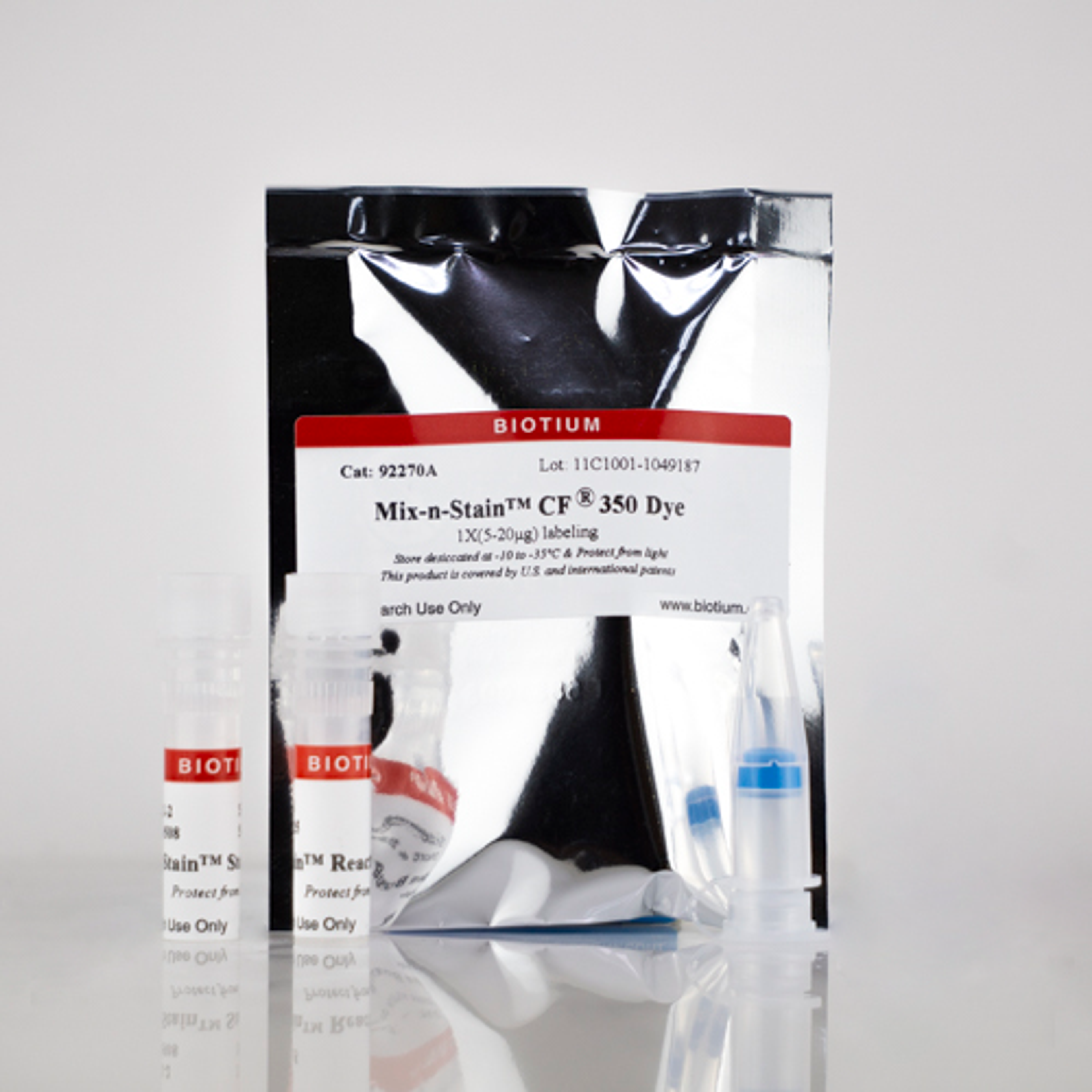
Mix-n-Stain™ CF® Dye Antibody Labeling Kits
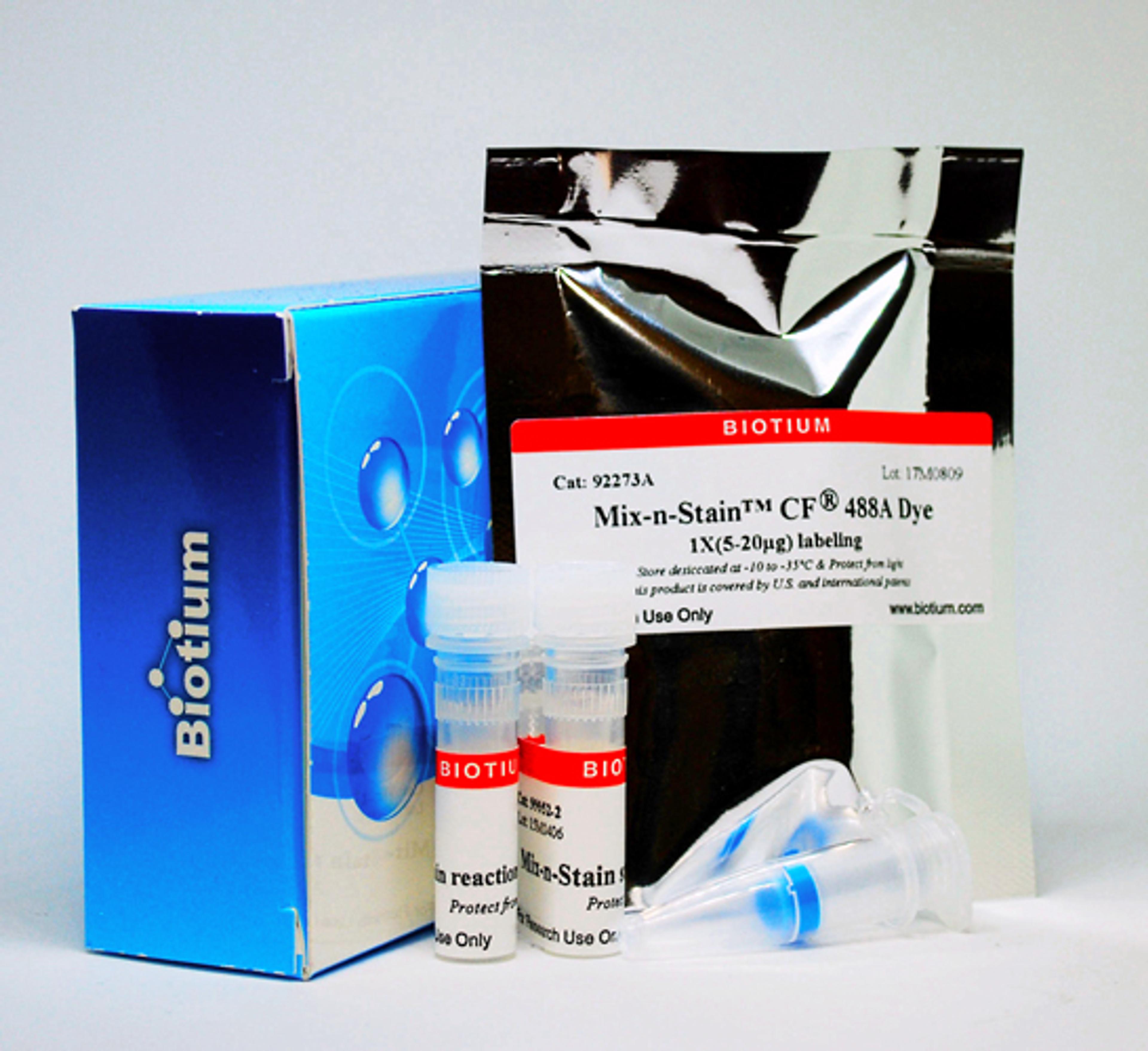
BiotiumLabel your IgG antibody with one of our superior CF® Dyes in as little as 15 minutes without a purification step. Labeling tolerates many common buffer components including BSA and ascites.
Label your IgG antibody with one of our superior CF® Dyes in as little as 15 minutes without a purification step. Labeling tolerates many common buffer components including BSA and ascites.
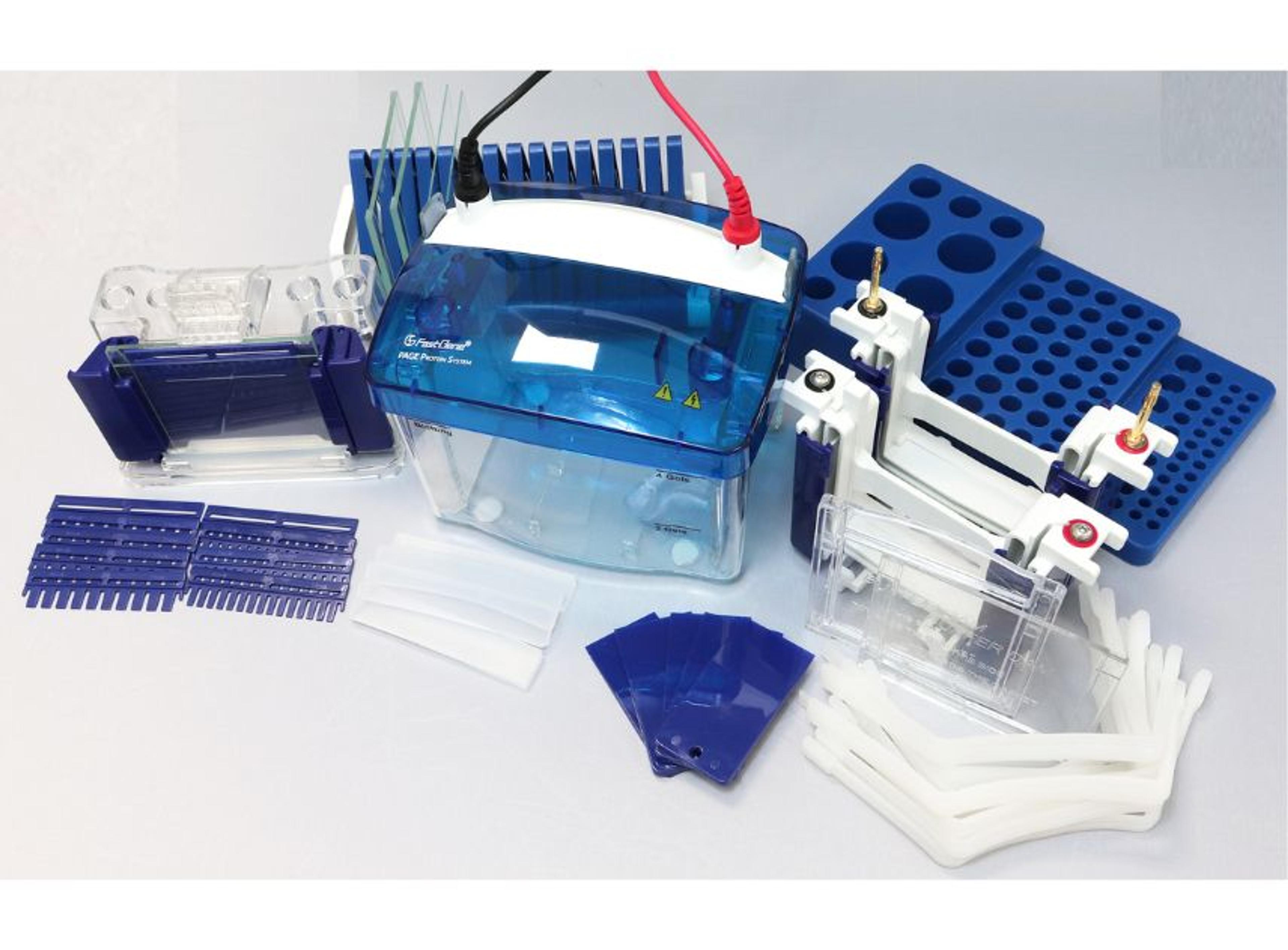
Complete PAGE protein analysis system
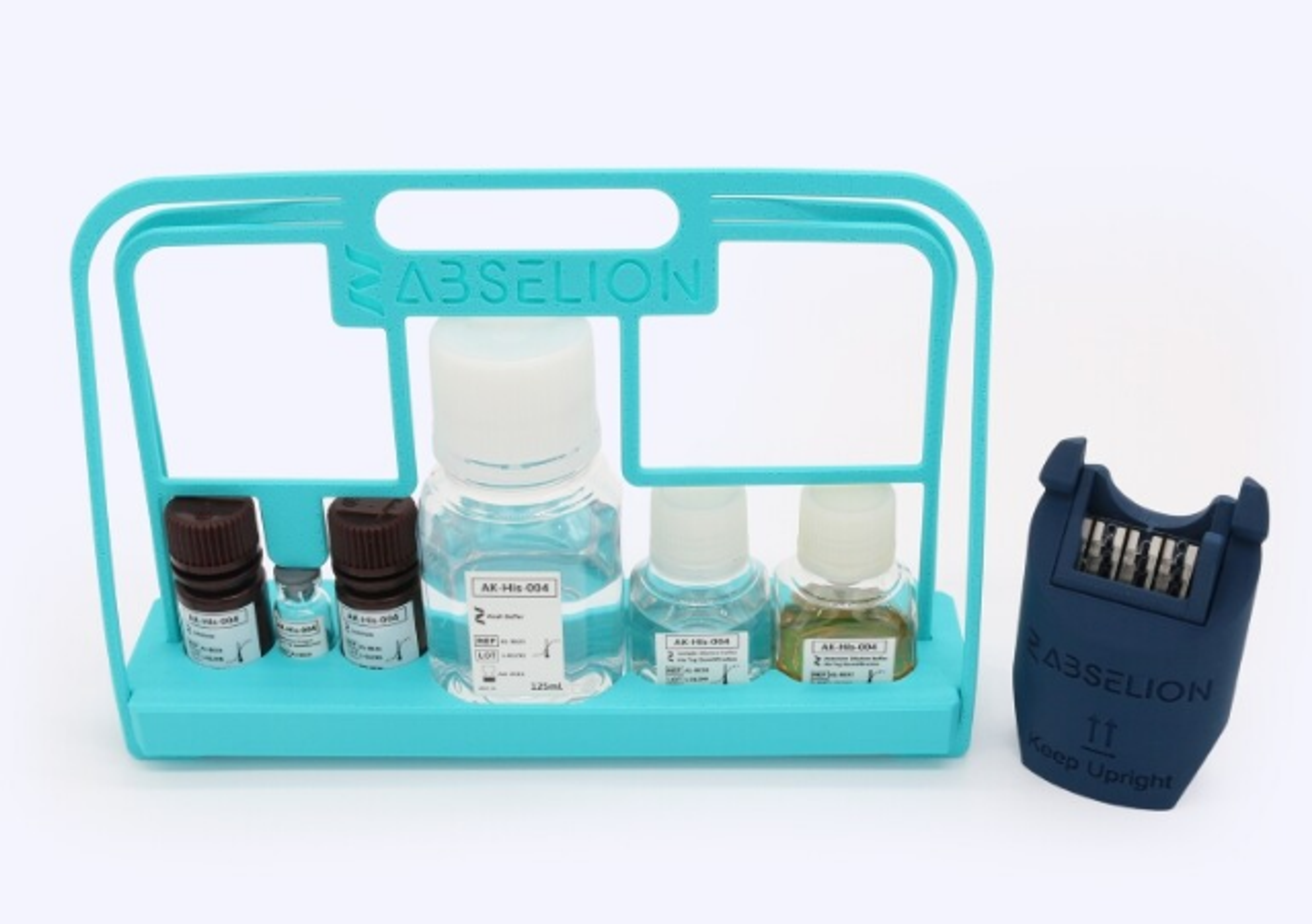
The His-Tag Protein Quantification Kit extends the Amperia™ Protein Quantification System for direct, tag-specific measurement of His-tagged recombinant proteins. Using a premix competition format with GenScript’s THE™ His Tag Monoclonal Antibody, the kit delivers accurate, reproducible results across both crude lysates and purified samples, with up to 80 tests per kit.
Label your IgM antibody with one of our superior CF® Dyes in 15-30 minutes without a purification step.
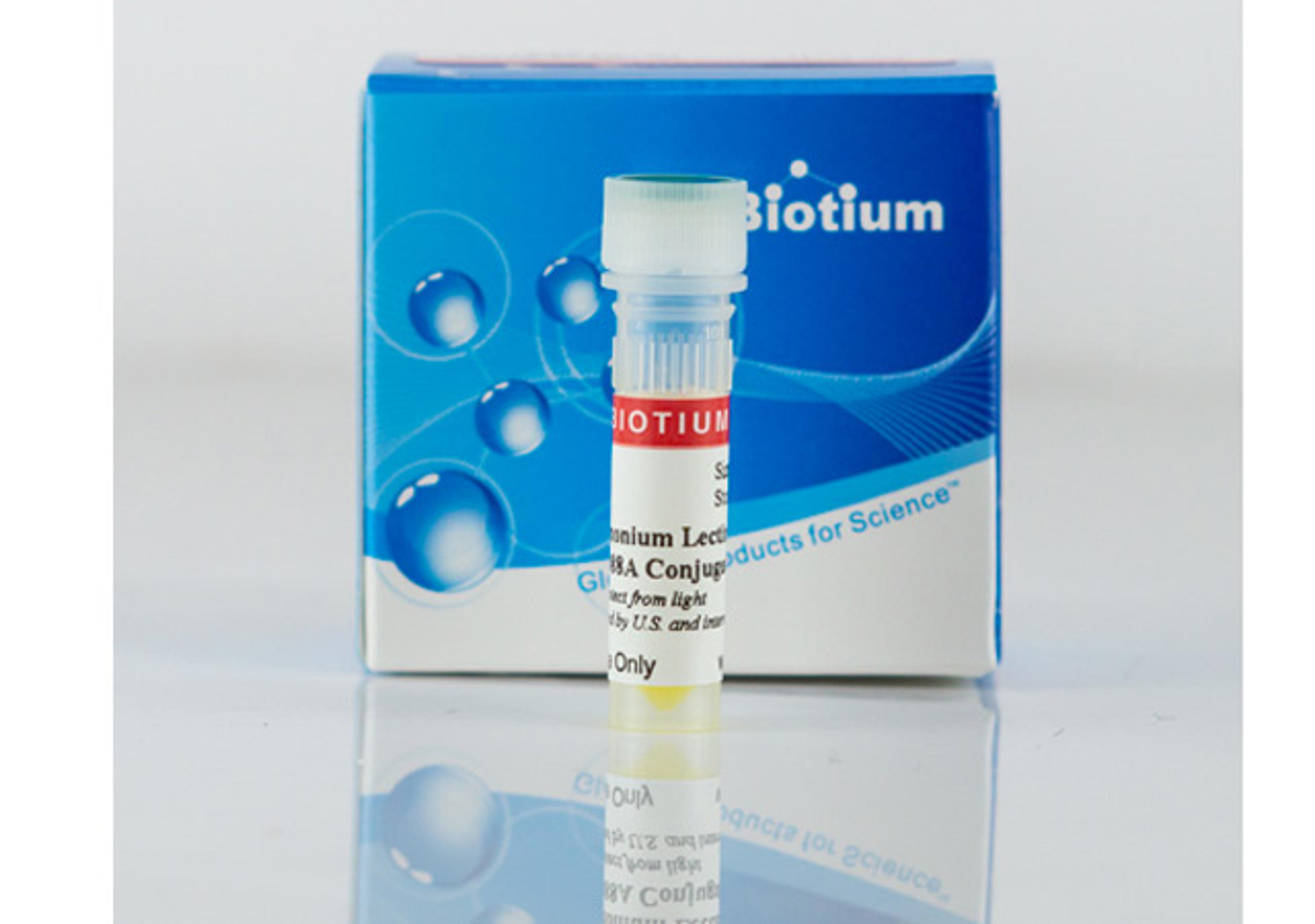
DSL binds to (beta-1,4) linked N-acetylglucosamine oligomers, preferring chitobiose or chitotriose over single N-acetylglucosamine residues. Biotium offers DSL conjugated to biotin and a selection of 6 bright and photostable CF® Dyes.
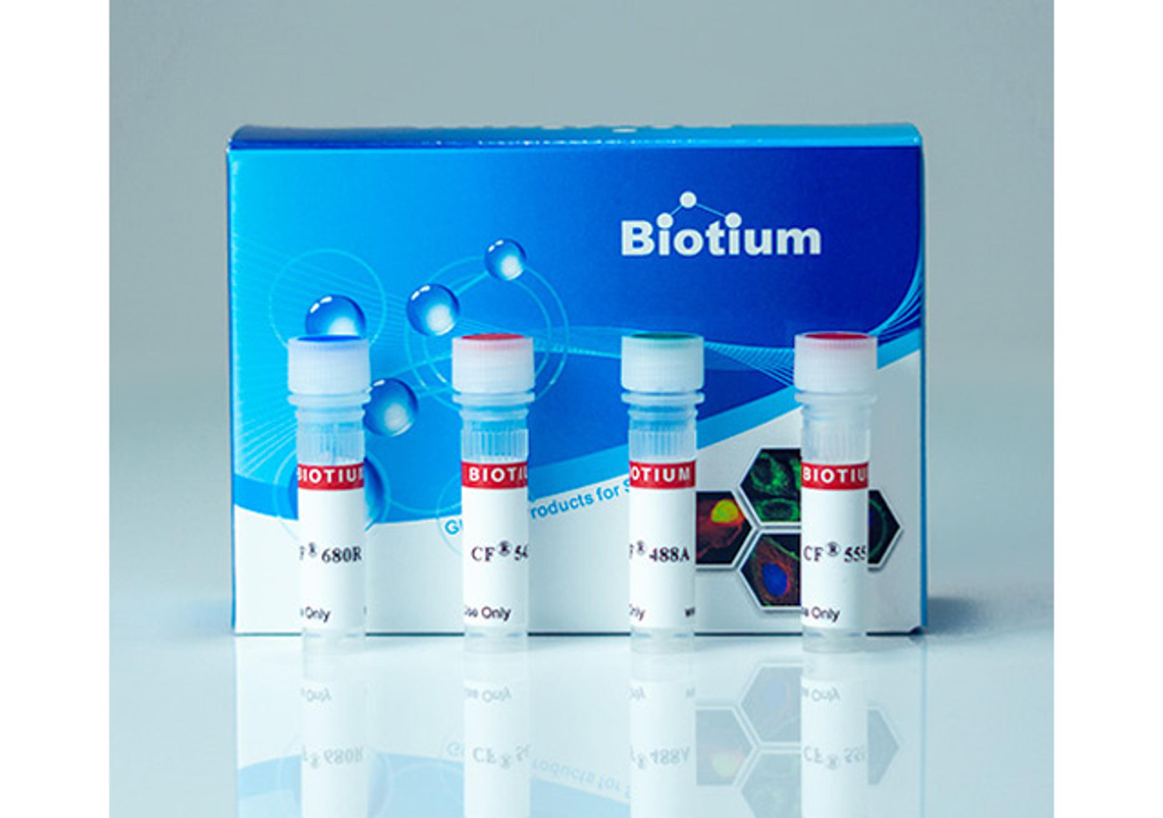
A lectin labeled with our superior fluorescent CF® dyes is an effective and widely used cell surface stain for yeast, fungi, and mammalian cells.
These kits allow optimal labeling of single-chain nanobodies with CF® dyes or biotin in 30 minutes without a purification step.
Label your antibody with one of our CF® Dyes specialized for STORM super-resolution microscopy. Labeling takes just 30 minutes without a purification step, and yields an antibody conjugate with a low degree of labeling optimal for STORM.
The FRETWorks™ S•Tag™ Assay is a FRET-based method that enables extremely sensitive detection of S•Tag fusion proteins in minutes with a homogeneous format.
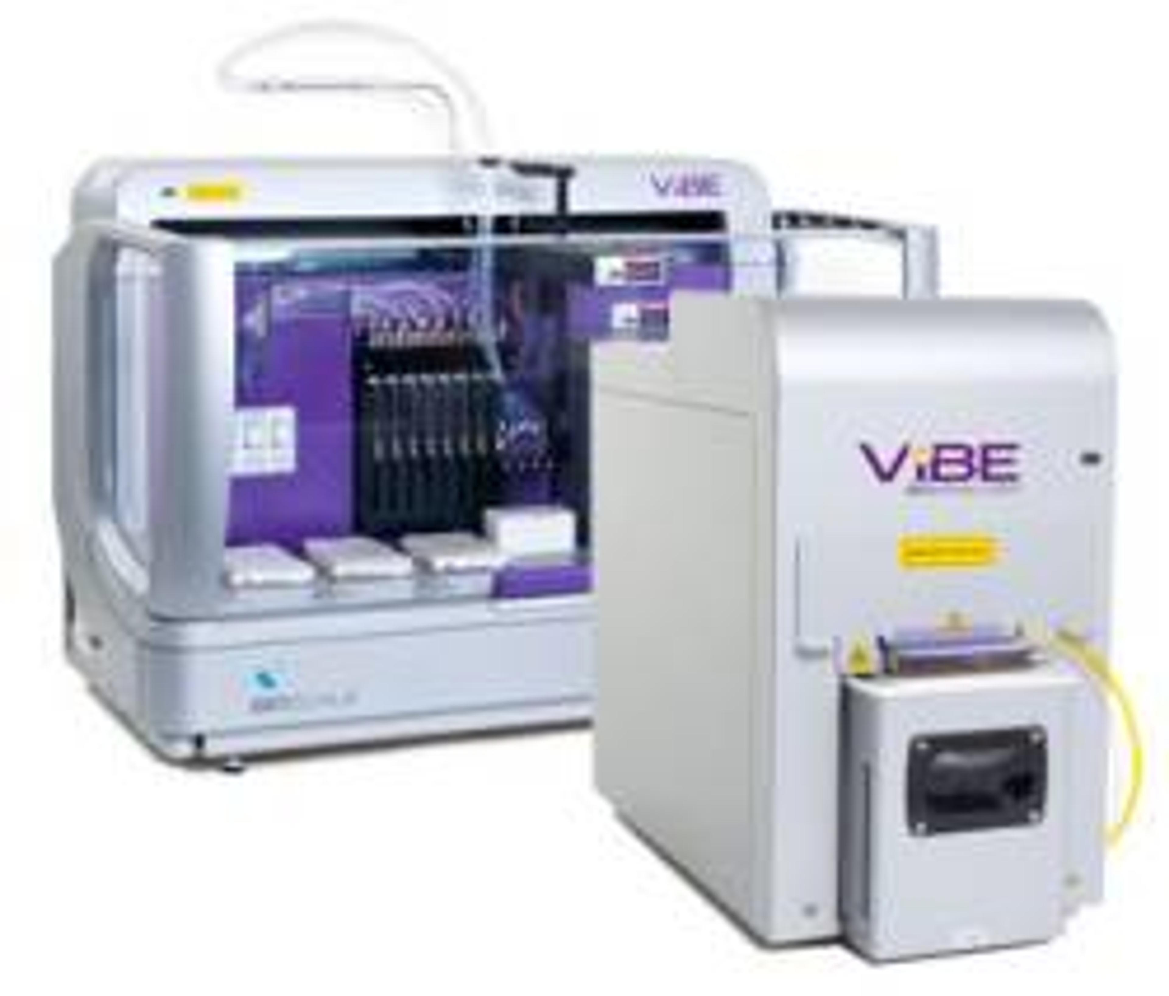
ViBE Protein Analysis PlatformPowered by AMMP Technology, the ViBE platform allows rapid design and configuration of protein assays to achieve highly-sensitive detection and quantitation of proteins in complex biological samples with real-time results. The ViBE platform … • Open assay design allows use of your own antibodies and the ability to define sensitivity, dynamic range, and speed requirements for efficient process…
The H2A.X Phosphorylation Assay Kit (Flow cytometry) is a cell based assay formatted for flow cytometric detection of levels of phosphorylated Histone H2A.X. Cells are cultured in microplates, treated with agents that induce DNA damage or apoptosis, which stimulates H2A.X phosphorylation. Cells are then fixed and permeabilized in preparation for staining and detection. Histone H2A.X phosphorylated at serine 139 is detected by…
The TRAPeze® Telomerase Detection Kit is a highly sensitive in vitro assay system for detecting telomerase activity. The assay is a one-buffer, two-enzyme, system utilizing PCR to enhance the sensitivity of telomerase detection in small samples.
The TRAPEZE® XL Kit is a highly sensitive in vitro assay for the fluorometric detection of telomerase activity. As in the original TRAPEZE® Kit, primer sequence modifications that reduce amplification artifacts and an internal PCR control are included. In addition, the TRAPEZE® XL Kit uses fluorescence energy transfer (ET) primers to generate fluorescently labeled TRAP products which permit nonisotopic, quantitative analysis o…
GST fusion-protein, corresponding to the p21-binding domain (PBD, residues 67-150) of human PAK-1 provided bound to glutathione agarose. Specifically binds to and precipitates Rac-GTP and cdc42-GTP from cell lysates.
GST-tagged fusion protein, corresponding to residues 7-89 of mouse Rhotekin Rho Binding Domain, expressed in E.coli and bound to glutathione-agarose. Specifically binds to and precipitates GTP-Rho, but not GDP-Rho from cell lysates.
A GST-tagged fusion protein, corresponding to residues 7-89 of mouse Rhotekin Rho Binding Domain, expressed in E. coli. Provided bound to glutathione-agarose. Purity 90%.
This kit contains nonradioactive components for radiolabeling and purifying histone H4 peptide with [3H]-acetyl CoA. The radiolabeled peptide is a suitable substrate for assay of HDAC activity. HeLa nuclear extract, which is rich in histone deacetylase activity, is included as a positive control. The HDAC inhibitor sodium butyrate is provided to demonstrate specificity of the deacetylase activity.
Accessory reagent kit for the analysis of histone methyltransferase activity.
This assay is a simple two-step procedure performed in a microtiter plate. In the first step samples are incubated with the HDAC assay substrate, allowing deacetylation of the fluorometric substrate. Next, Activator Solution releases a fluorophore from the deacetylated substrate or standard.
This kit is designed to measure the phosphotransferase activity in a kinase cascade reaction initiated by activated Raf-1. Recombinant unactive MEK1 is the substrate for Raf-1. Once activated by Raf-1, MEK1 can phosphorylate and thereby activate another kit component, recombinant unactive MAP Kinase 2/Erk2. Activation of MAP Kinase 2/Erk2 is determined by phosphorylation of myelin basic protein (MBP), in vitro. Phosphorylation…
The assay kit is designed to measure B-Raf dependent phosphotransferase activity in a kinase cascade reaction using recombinant MEK1, unactive as a B-Raf substrate. Recombinant MAPK2/Erk2, unactive is phosphorylated and activated by the activated MEK1 leading to phosphorylation of a MAP Kinase substrate, myelin basic protein (MBP). [γ-32P]ATP is the final phosphate donor.
Intended for the assay of recombinant B-Raf (supplied) kinase activity with experimental variables as determined by the user. For assay of B-Raf activity from a cell lysate, we recommend an immunoprecipitation-kinase assay, using anti-B-Raf antibody (07-453).
Intended for the assay of recombinant Raf-1 (supplied) kinase activity, with experimental variables as determined by the user. For assay of Raf-1 activity from a cell lysate, we recommend an immunoprecipitation-kinase assay, using anti-Raf-1 antibody (07-396).
The 384 well format kit is a newer version of the tried and true non-radiographic HDAC assay. As in the previous kit, samples are first incubated with the HDAC Assay substrate, allowing deacetylation of the fluorometric substrate. After incubation, the activator solution is added, and releases a fluorophore from the deacetylated substrate or standard. The sample is then put into a microplate reader and fluorescence (i.e free/c…