SPATIAL
ariadne.aiAn intuitive browser-based tool for spatial omics data analysis. Simple to use with datasets created by any microscope, SPATIAL features best-in-class image alignment, cell and tissue segmentation.
Imaging Systems
An intuitive browser-based tool for spatial omics data analysis. Simple to use with datasets created by any microscope, SPATIAL features best-in-class image alignment, cell and tissue segmentation.
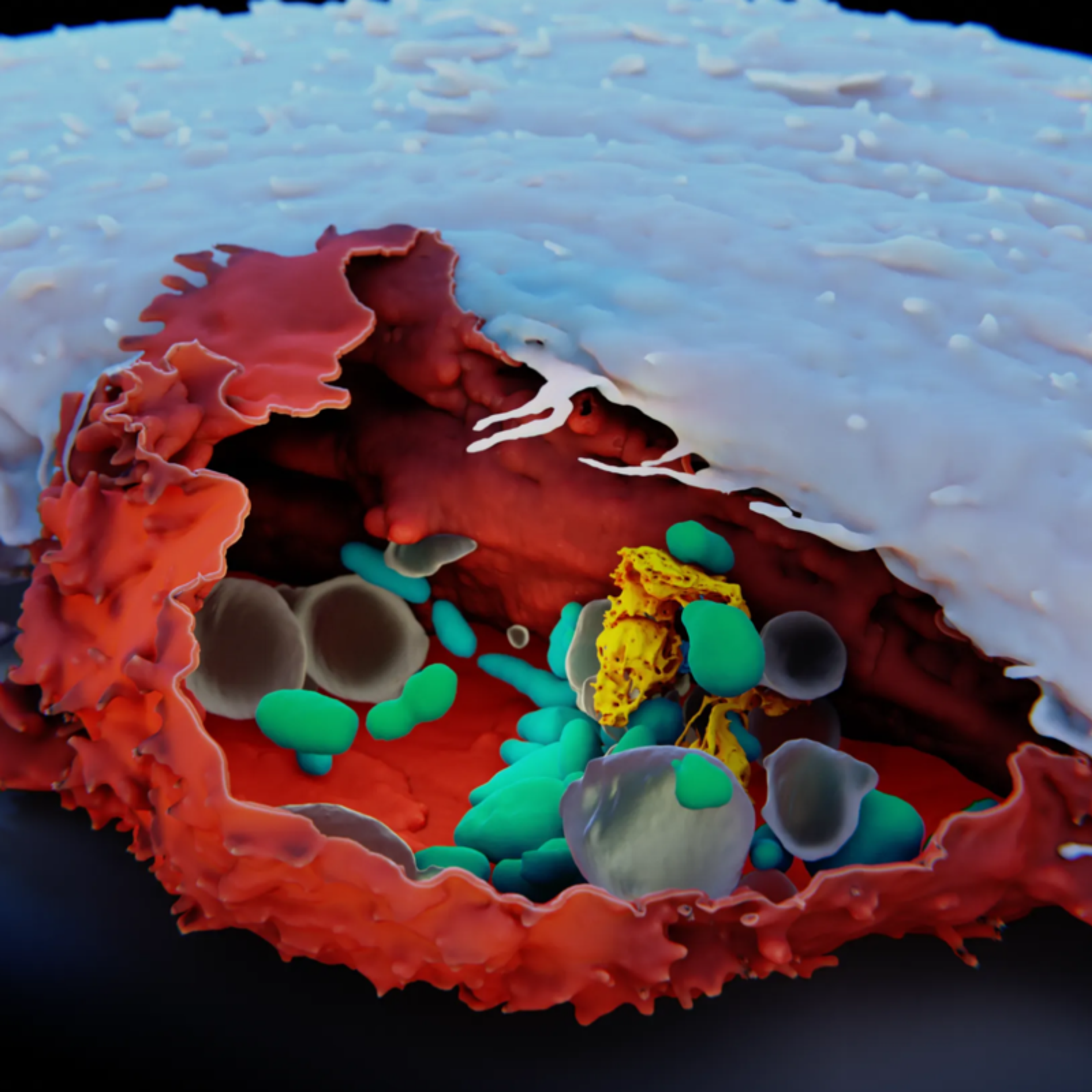
Segmentation of subcellular structures from 3-dimensional electron microscopy (EM) data
TauTec’s TriMScope, a state-of-the-art multifocal multiphoton microscope, is based on a patented beam divider that splits up an incoming laser beam into up to 64 beamlets which are scanned simultaneously in the object plane. This results in either 64 times brighter images or 64 times higher image rates compared to standard single beam multiphoton scanning microscopes. The foci in the object plane are aligned in a single line a…
Modular microscope-based high content screening solution

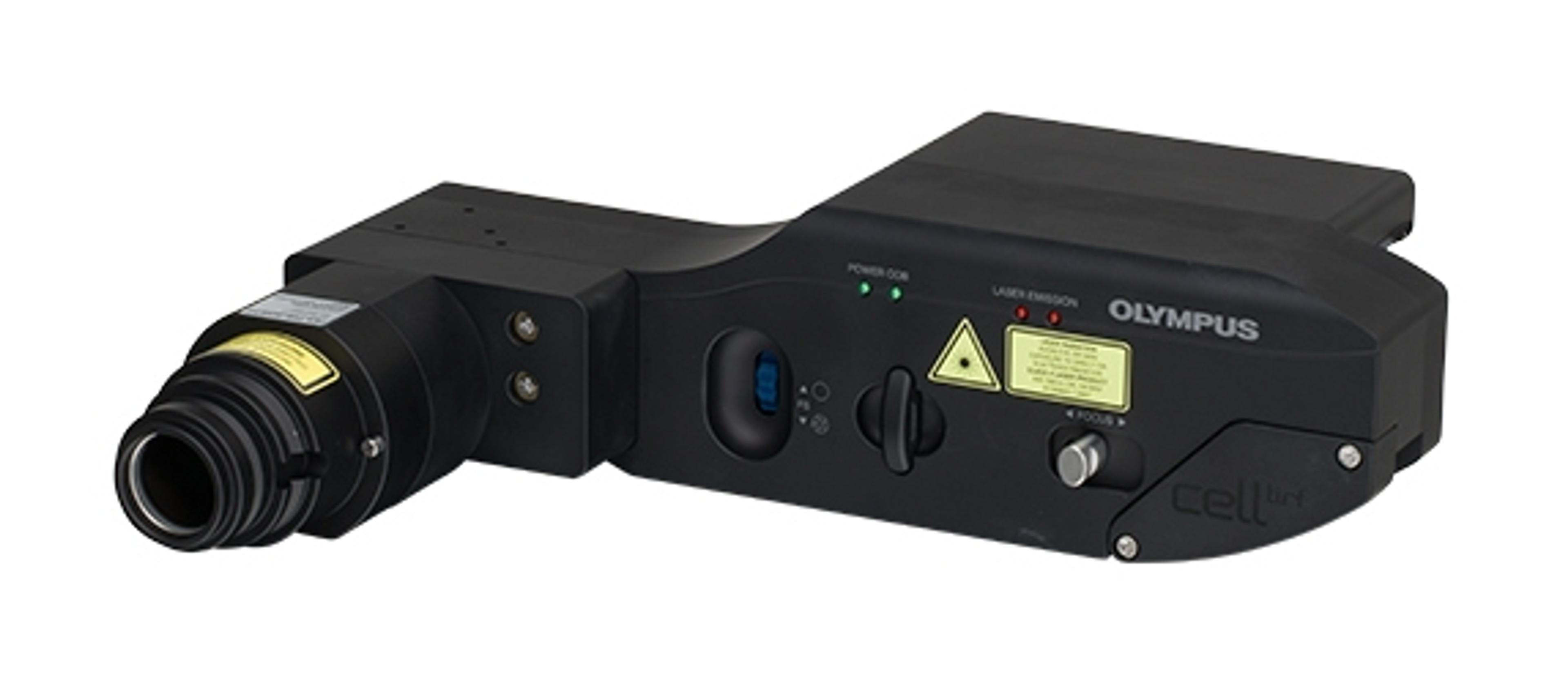
Total internal reflection fluorescence microscopy (TIRFM) has firmly established itself over the last few years as a key technique in the investigation of molecular interactions at or near the cell surface.Olympus is experienced in providing advanced TIRFM solutions and cell^tirf takes this technology to the next level with a series of peerless features such as highly advanced optics, unique independent laser control and excep…
The VisiTIRF Fluorescence Imaging System from Visitron Systems is the ideal solution for the analysis of finest structures or surfaces. The flexibility of the VisiTIRF system and the use of scientific digital CCD camera systems with optimal resolution, speed and sensitivity allows for the adaptation of customized application and configuration. For fluorescence illumination Visitron use solid state lasers or laser diodes . The…
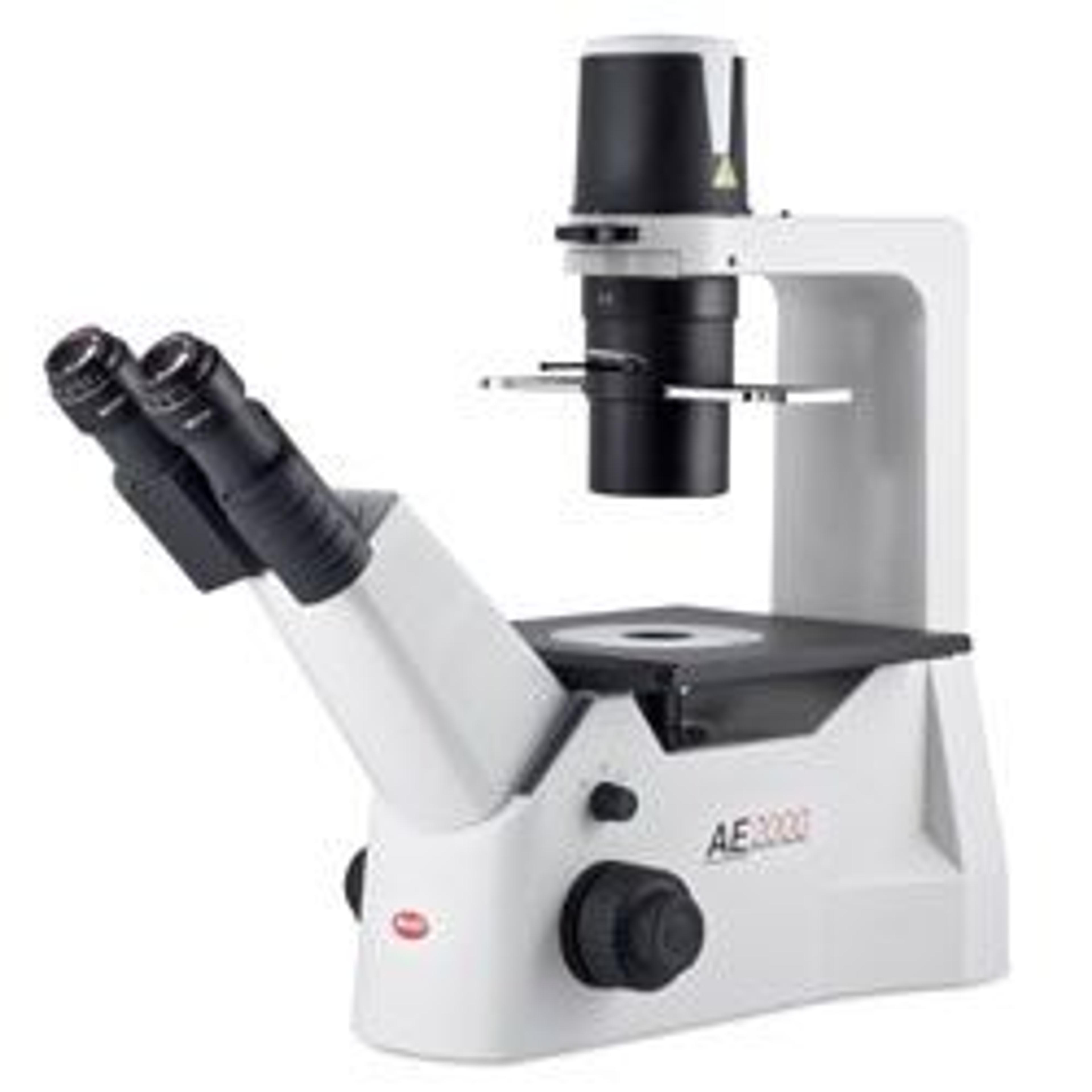
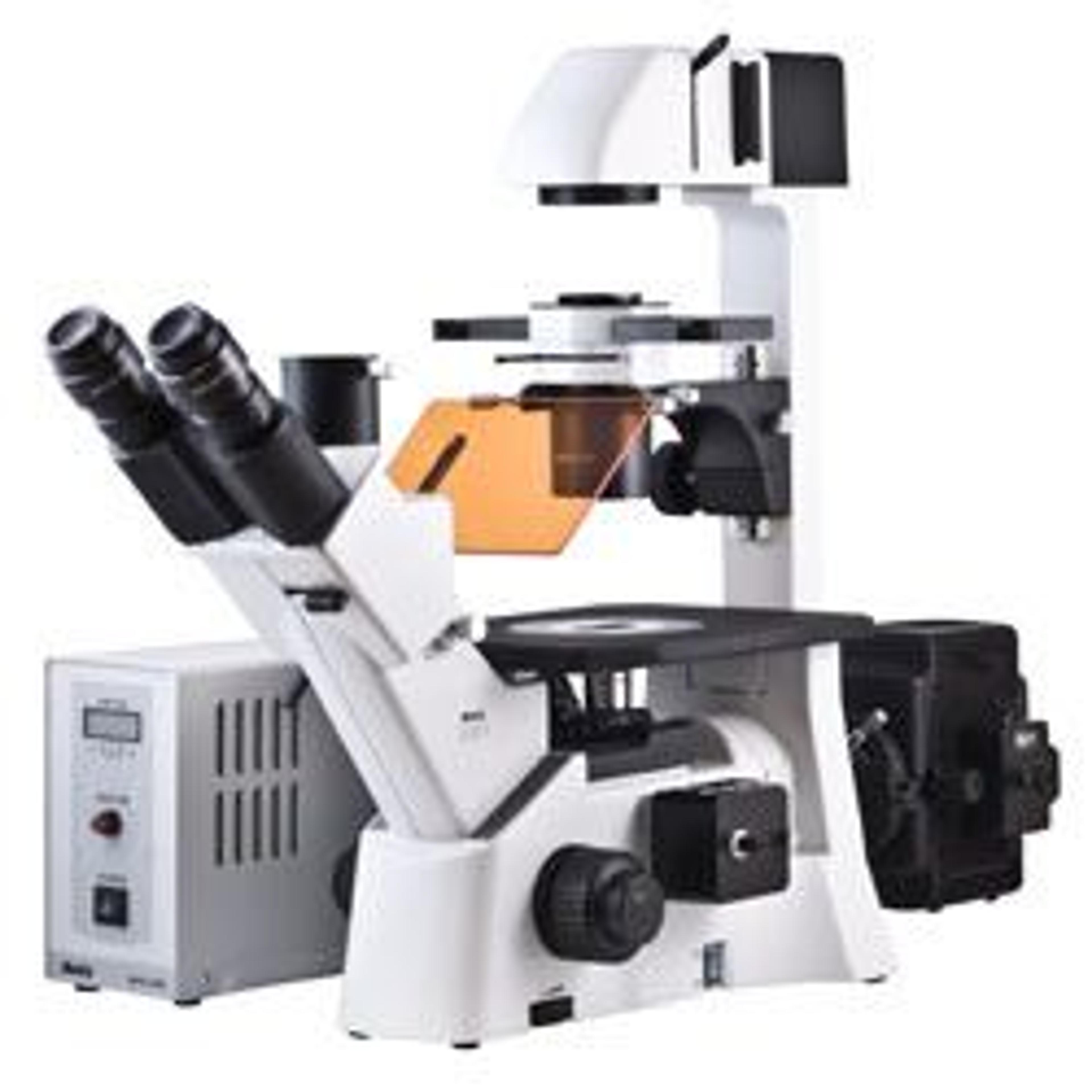
Motic´s new AE2000 Series Inverted Microscope is the ideal instrument for routine live cell inspection in both educational and high grade professional applications.The uniquely integrated feature Auto cut-off mode (“sleep mode”) is focused on enhancing its performance during the rigors of daily use.Designed for routine-lab or clinical work, as well as research requirements in Pharmaceutical Laboratories or Universities, the AE…
With the introduction of its premium AE30 Series of Inverted Microscopes, Motic joined the group of manufacturers capable of providing High-End optics, ergonomic design and craftsmanship as well as durable product quality. Motic doing so, however, all at an affordable price point. The AE30 Series quickly became the perfect solution for all kind of routine microbiological work in clinical and pharmaceutical laboratories as well…
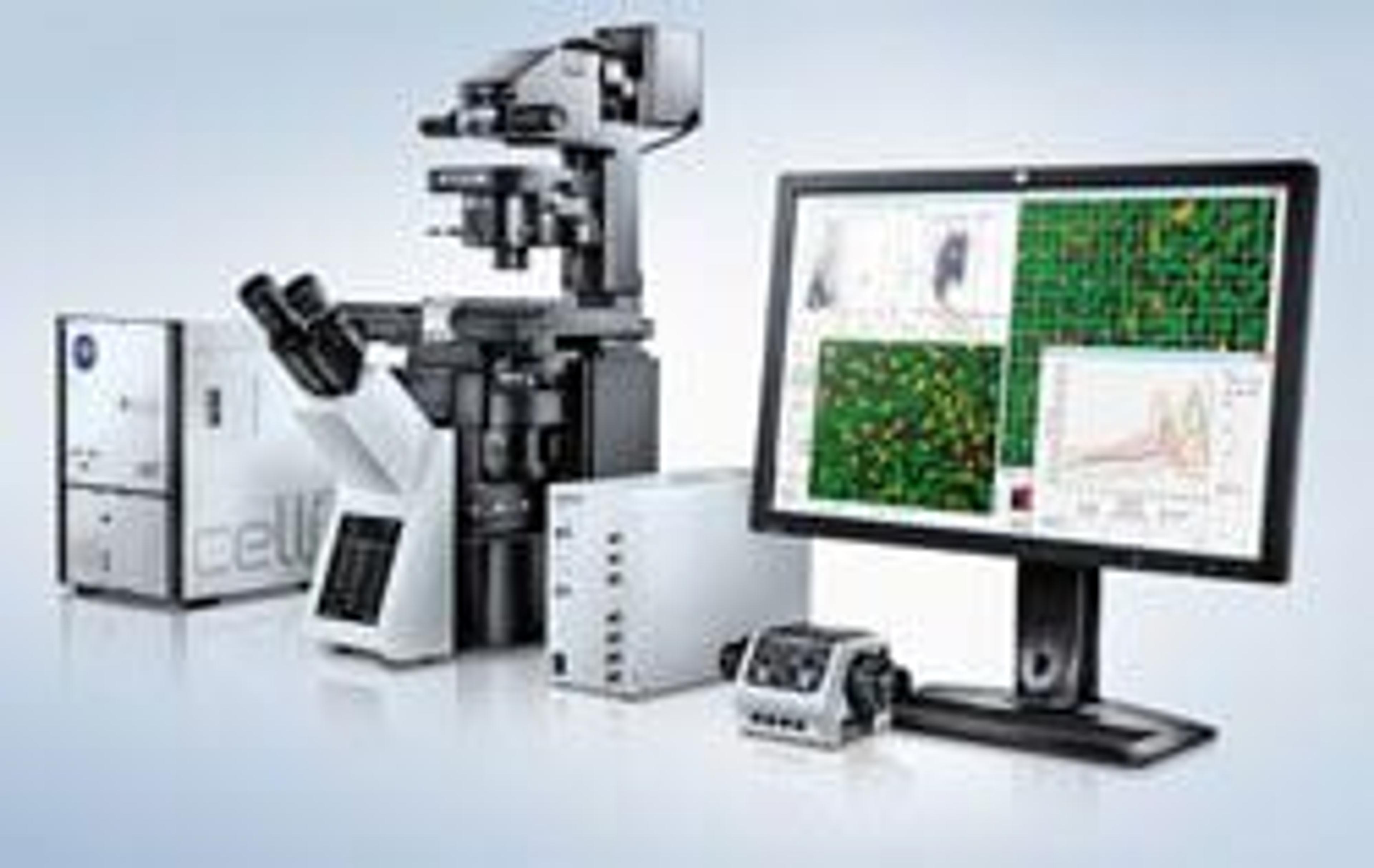
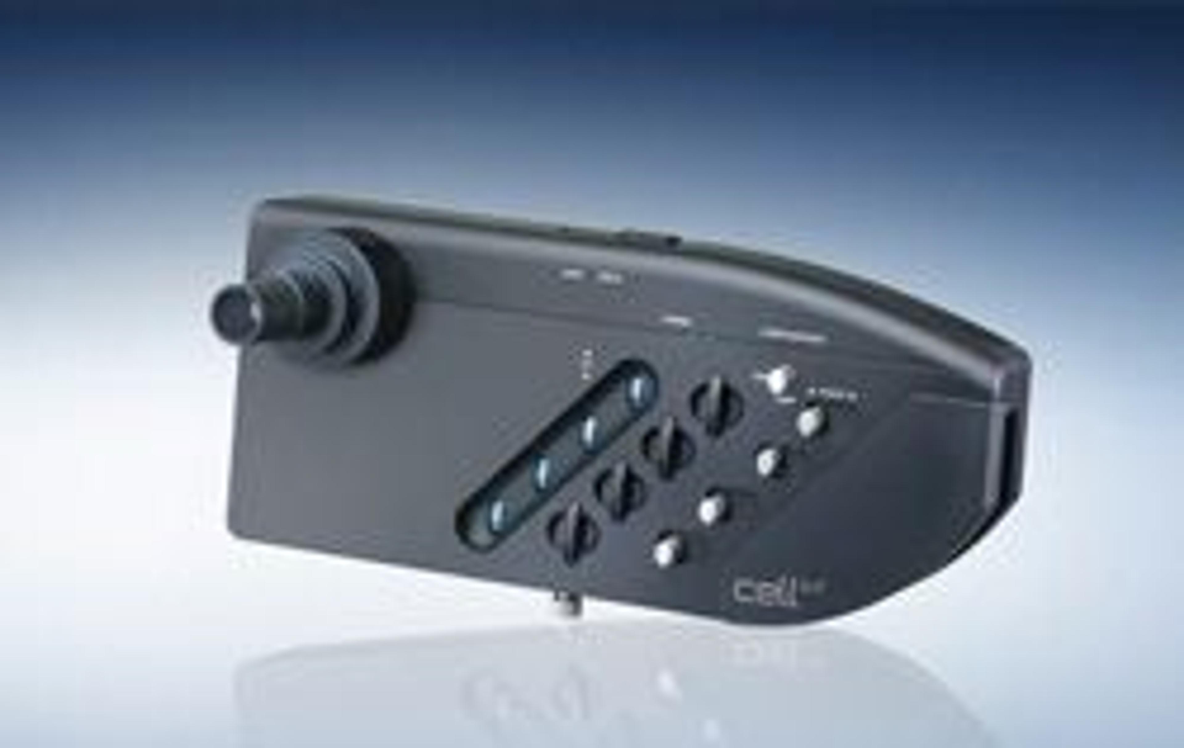
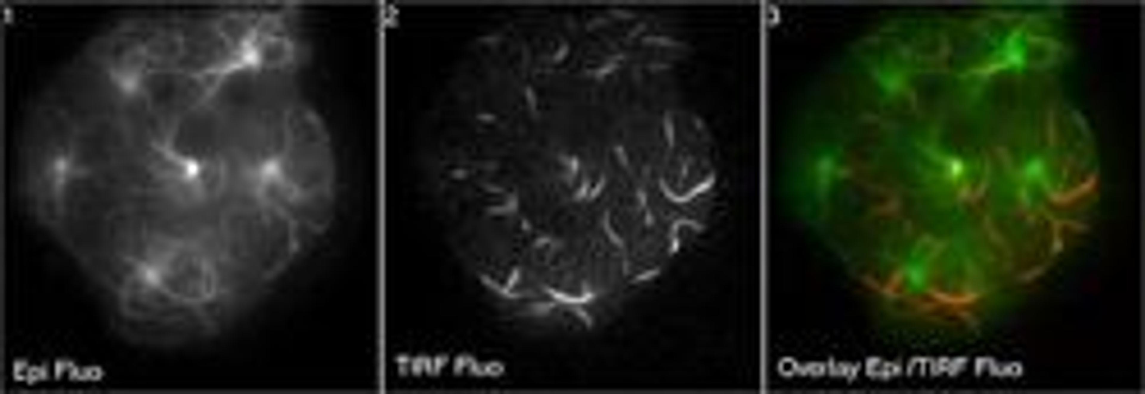
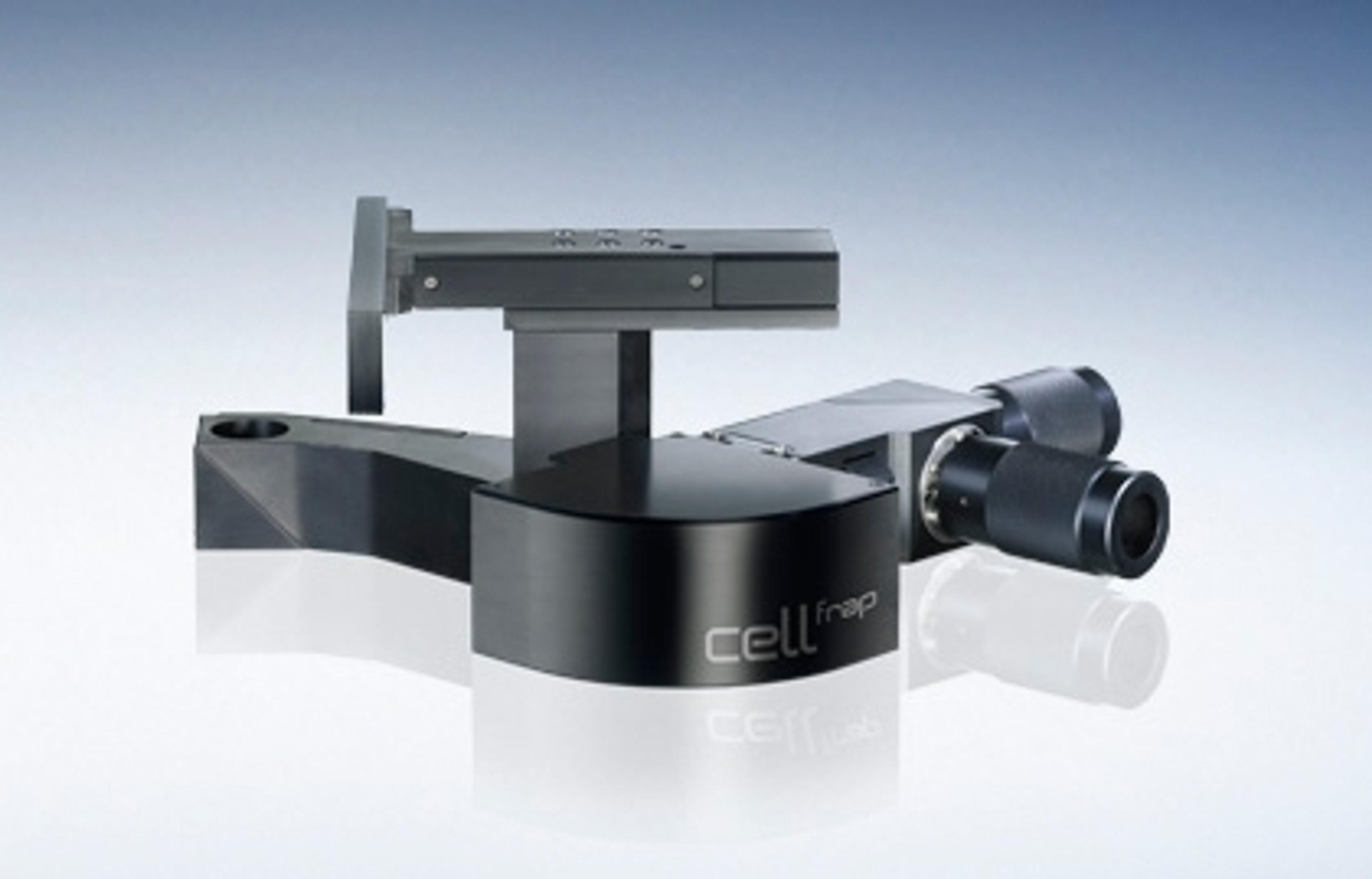
The cellFRAP system provides a versatile platform for a wide range of experimental needs, from basic bleaching to advanced protocols including FRAP, iFRAP, FLIP and FLAP. The system is also ideal for processes such as photo−conversion, photo−activation, pattern bleaching, laser cutting and trapping. An independent light path enables simultaneous imaging and bleaching and the module integrates seamlessly with other imaging m…
Designed to remove all of the inherent complexities of capturing and processing three color fluorescent cell images
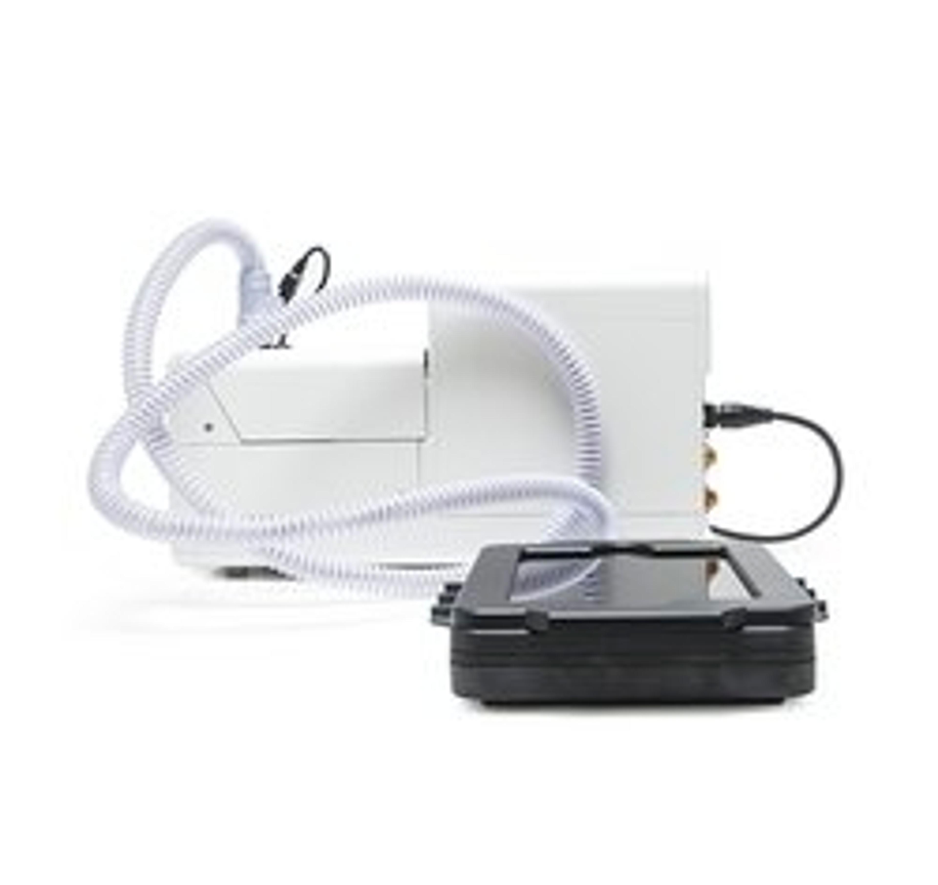
Enables precise control of temperature, humidity, and three gases for time-lapse imaging of live cells
Real-time fluorescence imaging of 3D tissue sections typically used in neuroscience and histology research
Fast and easy 3D exploration of whole organisms for developmental or molecular biology research
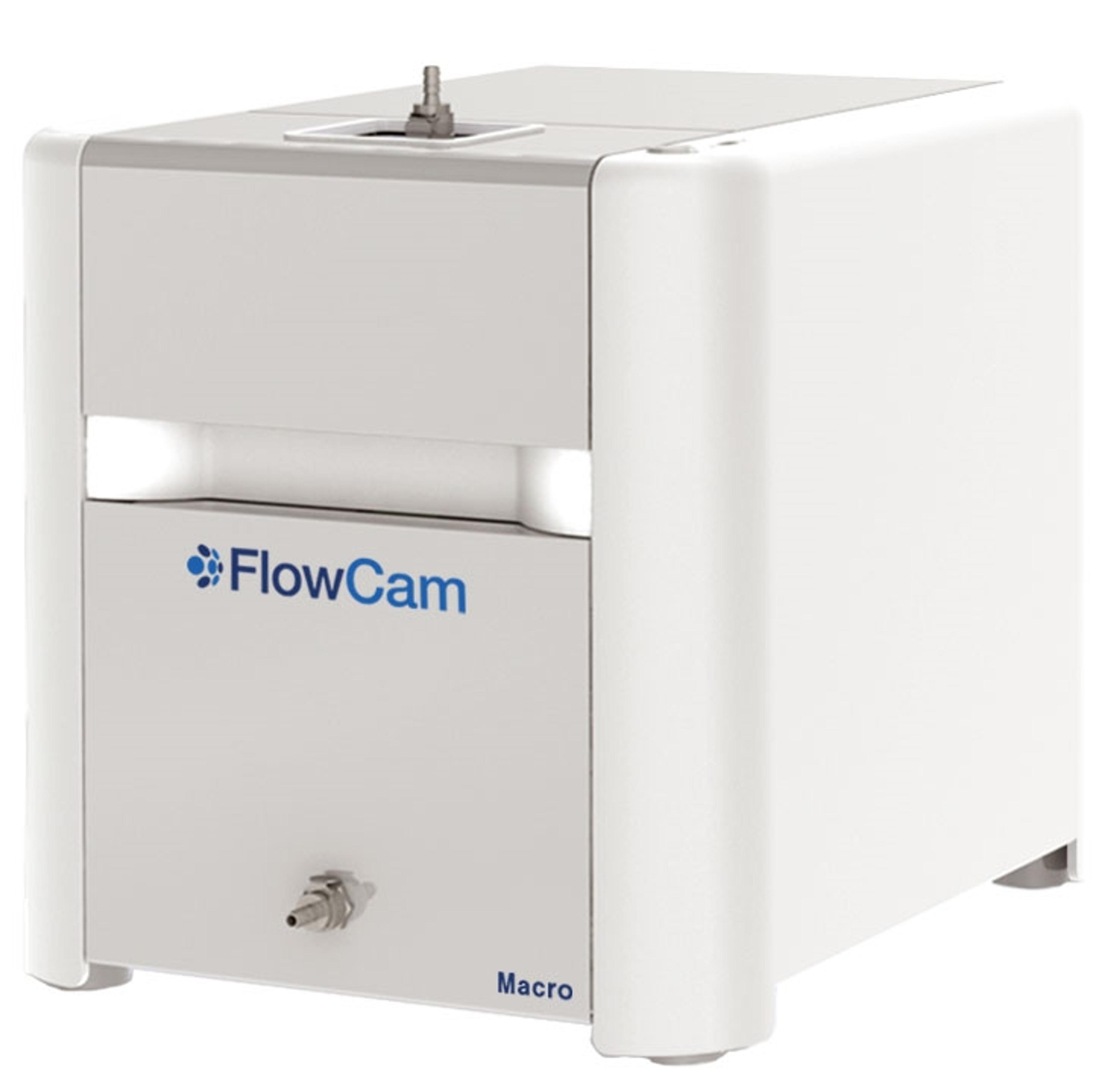
Extend your particle imaging capabilities from 300 μm to 5 mm with FlowCam® Macro for aquatic and environmental research and materials characterization. Obtain detailed morphological data along with accurate counting and sizing measurements to enable differentiation of diverse zooplankton species and particle types.
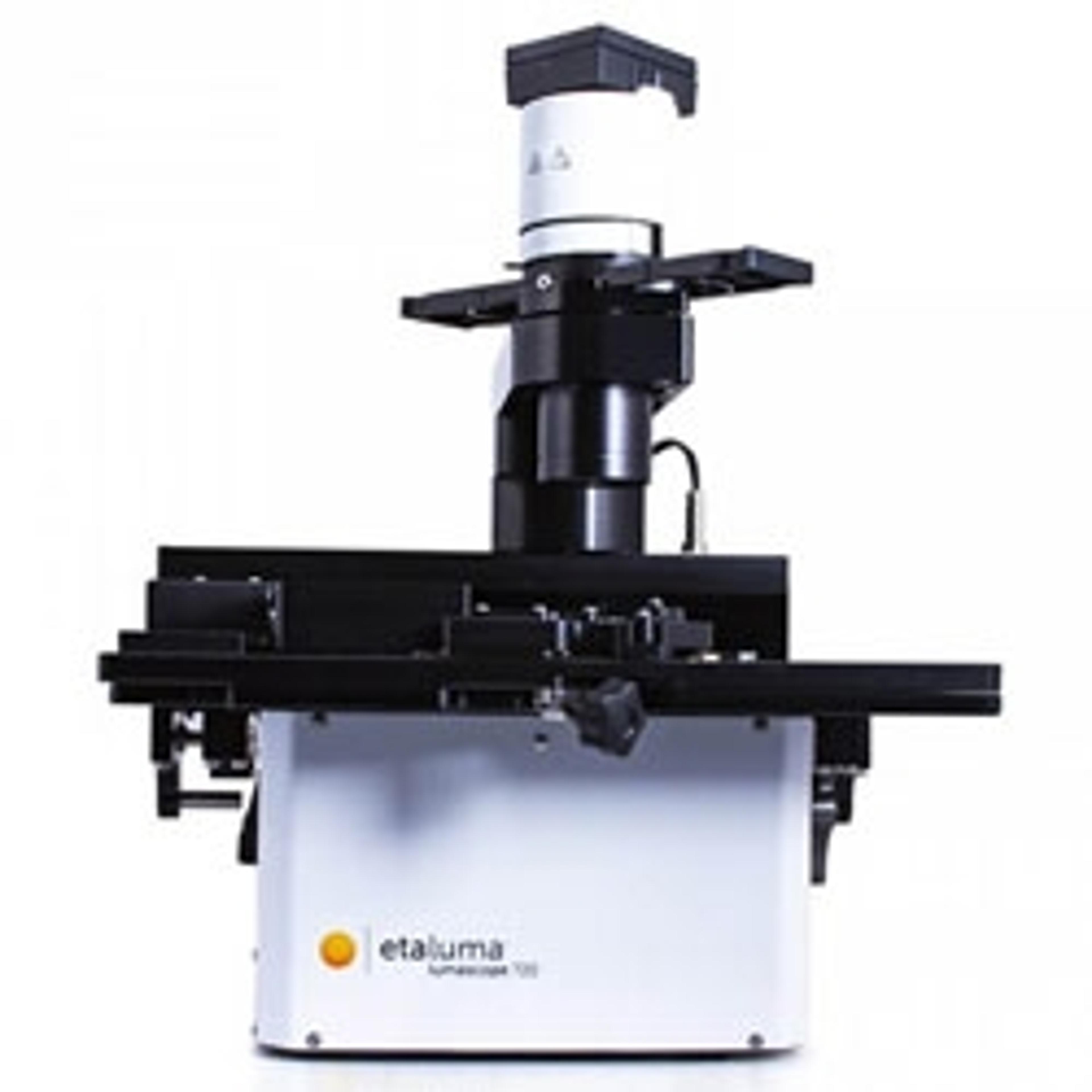
The Etaluma Lumascope range provides a unique approach to live cell imaging. These compact and durable systems offer high quality brightfield, phase-contrast and fluorescence imaging. Lumascopes are engineered to withstand the most demanding environments including incubators, hoods and workstations.
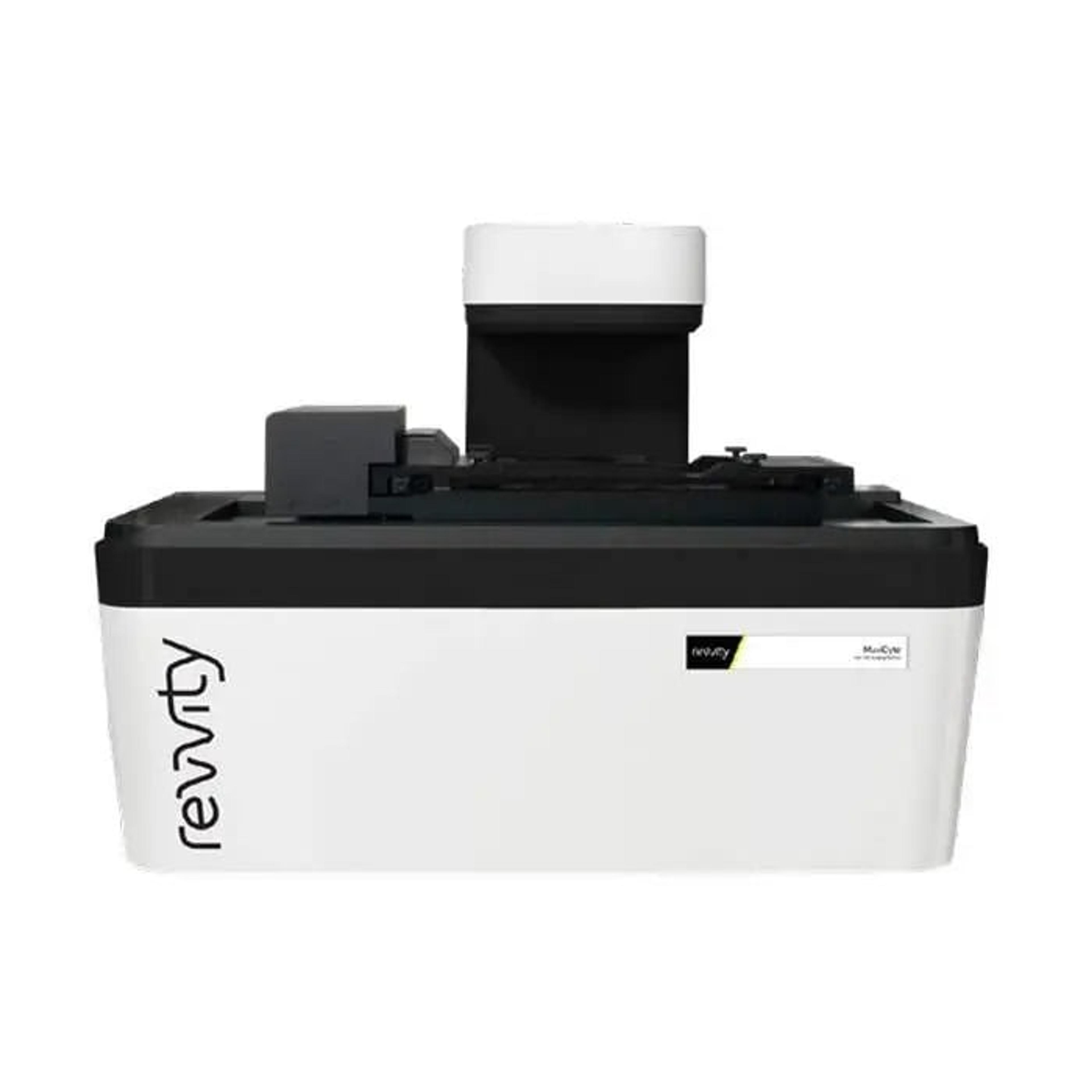
The MuviCyte™ live-cell imaging system is designed to operate inside your cell-culture incubator, enabling you to maintain your cells under optimal conditions and perform a wide range of assays in a variety of culture vessels.
Premium digital microscope for live imaging, scanning and real-time diagnostics of multiple slides
Intuitive interfaces, intelligent design and powerful image analysis tools leading to meaningful dynamic measurements at the cell-, well- and field-level.
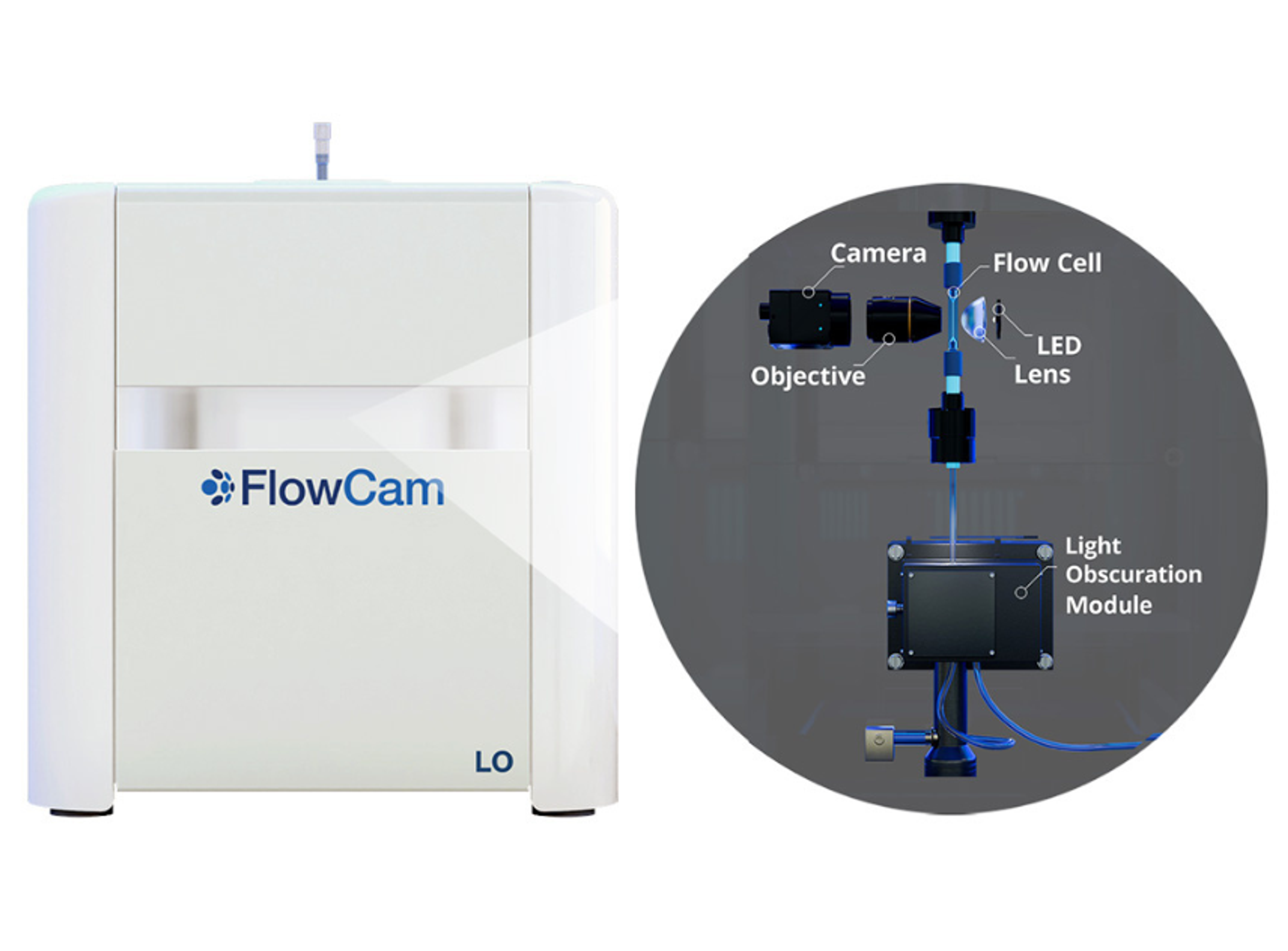
Innovative particle characterization with FlowCam® LO combines flow imaging microscopy (FIM) and light obscuration (LO) into a single analytical solution. Beyond the compendial light obscuration method to fulfill USP <787> and <788> requirements, flow imaging microscopy provides an orthogonal method for quality control of subvisible particulate matter.
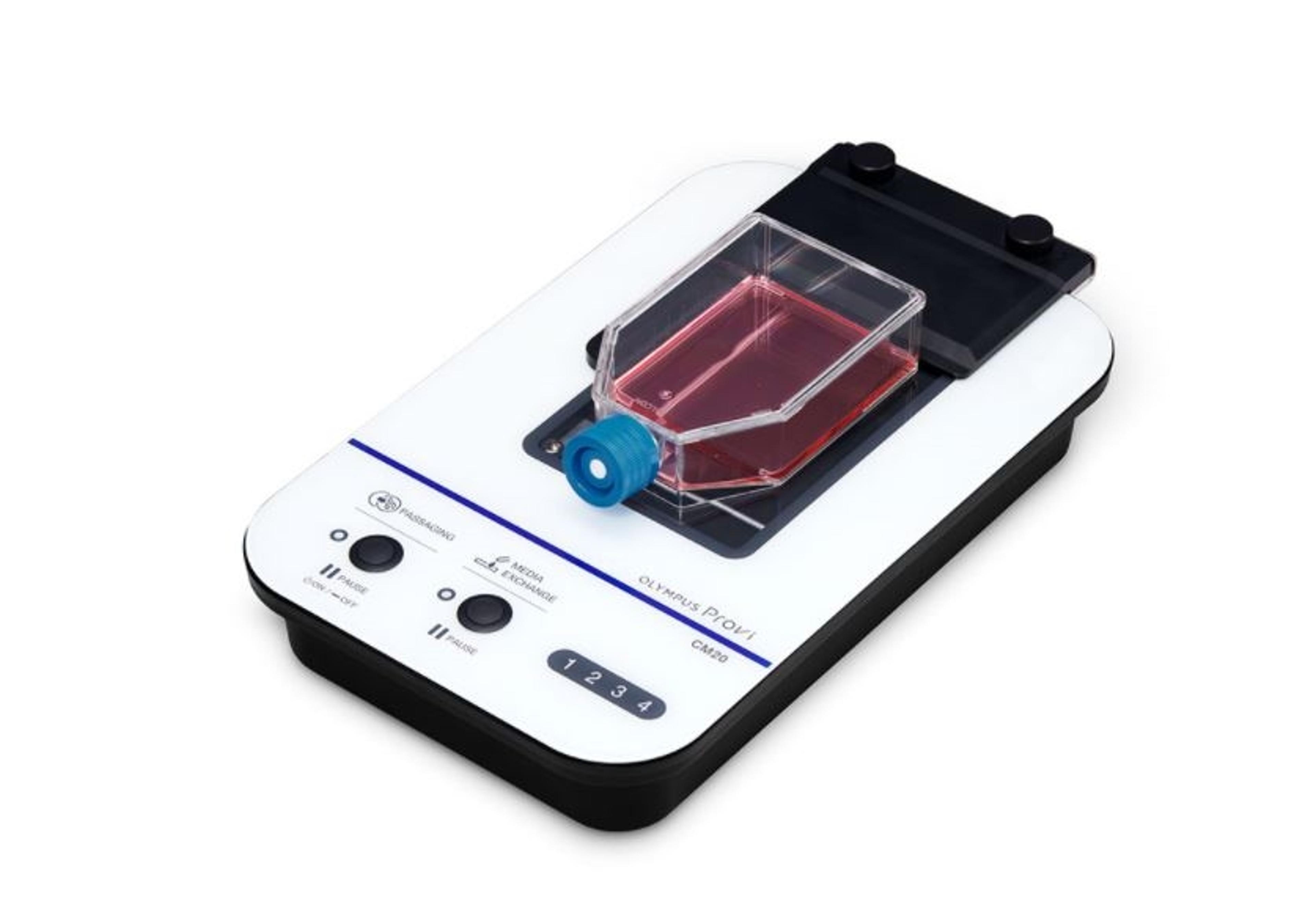
The CM20 system provides quantitative data remotely - place the head and your cell cultures in the incubator, and the system will periodically scan it, count the number of cells, and determine confluency. The data are wirelessly communicated to a PC or a tablet through an optional router, so you can monitor your cultures' progress without entering a clean room.
STELLARIS 8 FALCON (FAst Lifetime CONtrast) is the future of functional imaging. Harness the power of fluorescence lifetime to investigate cellular physiology and explore dynamics in living cells.