Microscope Adapters
EppendorfFor all workstations, additional microscopes, adapters, video equipment and application-specific accessories may be required.
For all workstations, additional microscopes, adapters, video equipment and application-specific accessories may be required.
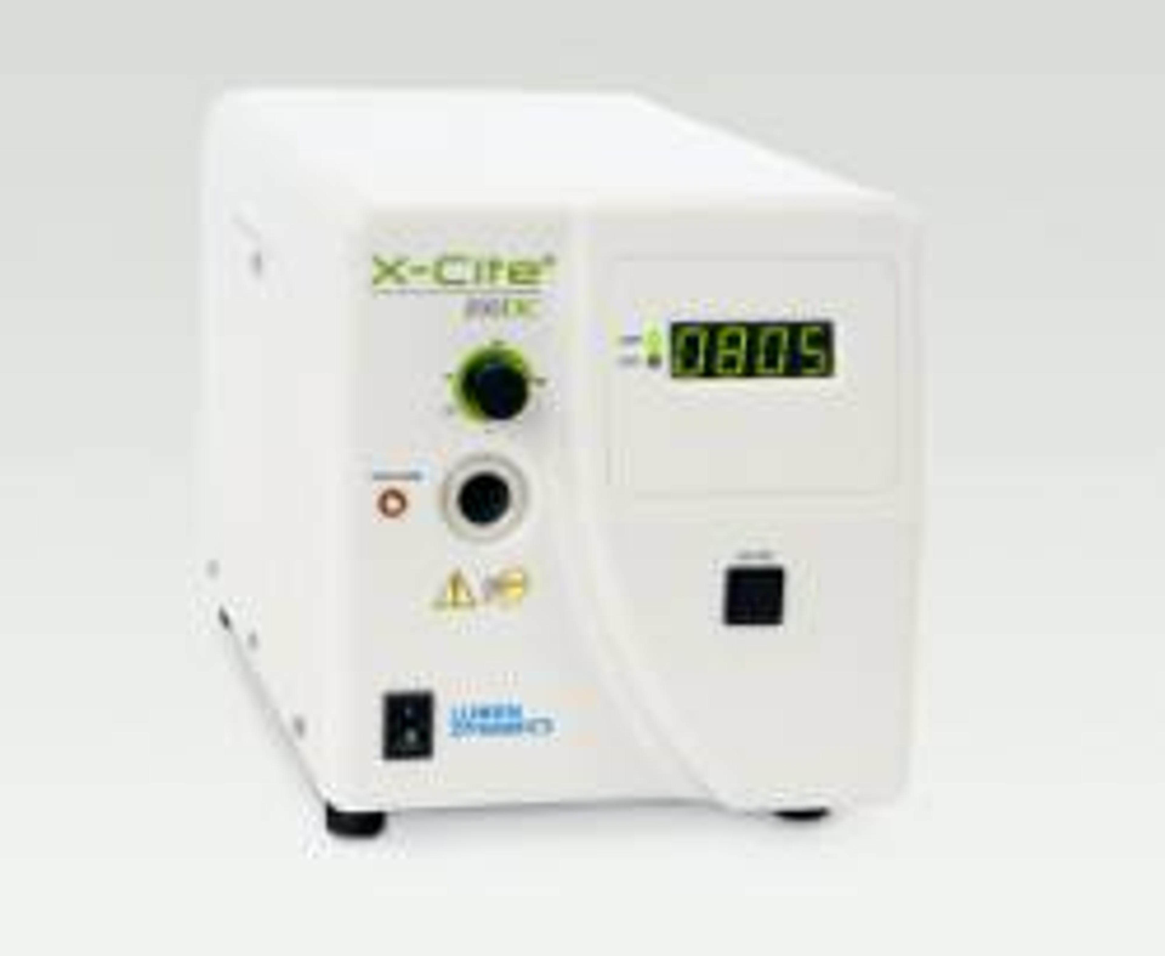
The X-Cite® 200DC offers the ultimate combination of optical performance, short term stability and a built-in fast shutter. With the convenience and superior illumination uniformity found in all X-Cite® systems, the X-Cite® 200DC's intuitively easy-to-use design also includes light guide auto-detection to ensure optimized light coupling - every time. Novice and experienced microscopists will enjoy the flexibility provided by t…
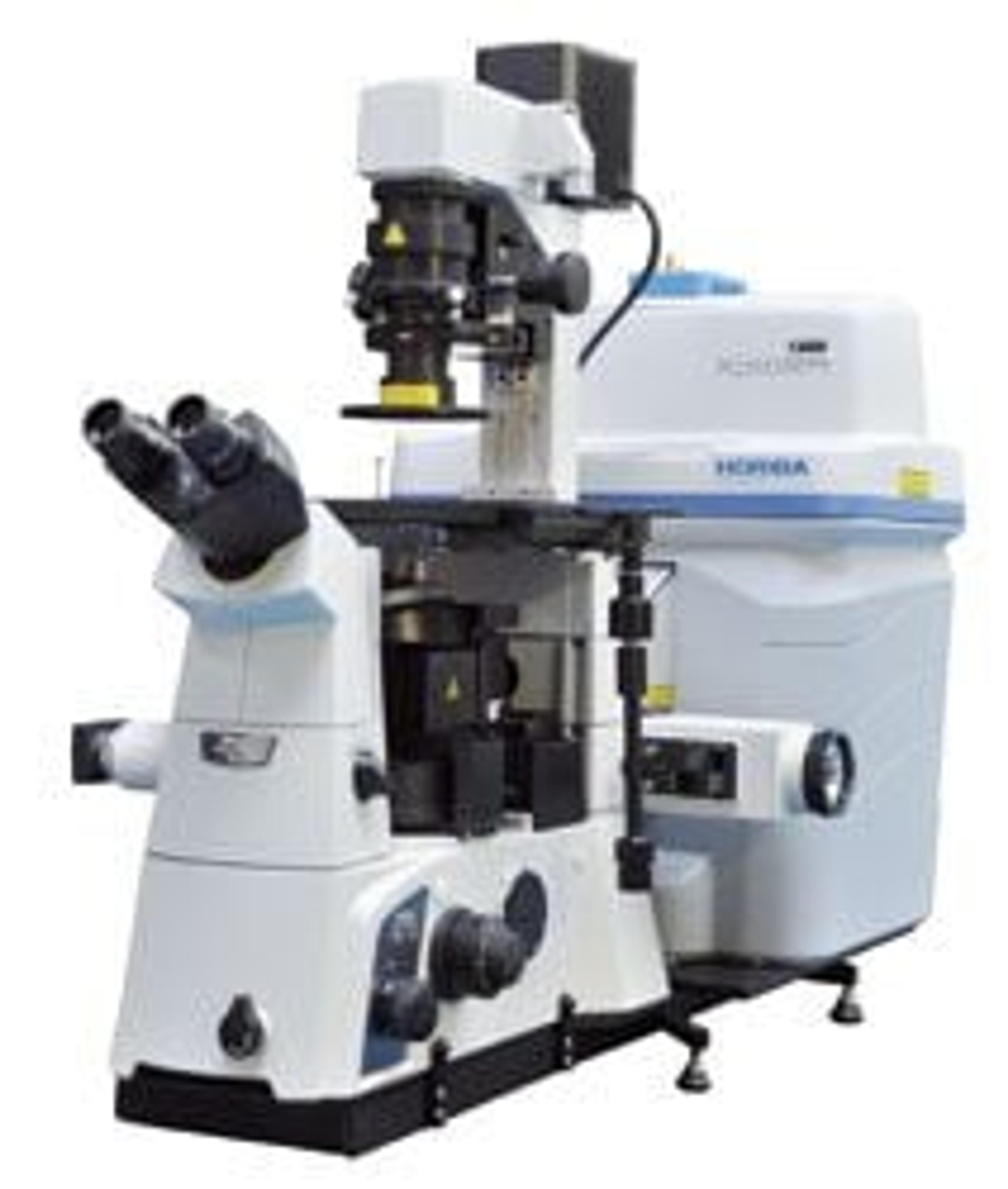
Optimized Raman Solution for Life Sciences from HORIBA Scientific The XpIoRA INV is an inverted Raman microscope designed specifically for use with biological samples requiring analysis from the bottom and/or open access to the sample from the top. Like all other XpIoRA series of Raman microscopes, the INV features a small footprint, complete automation (autofocus, autoexposure, autovalidation, autocalibration), and ease of us…
TauTec’s TriMScope, a state-of-the-art multifocal multiphoton microscope, is based on a patented beam divider that splits up an incoming laser beam into up to 64 beamlets which are scanned simultaneously in the object plane. This results in either 64 times brighter images or 64 times higher image rates compared to standard single beam multiphoton scanning microscopes. The foci in the object plane are aligned in a single line a…
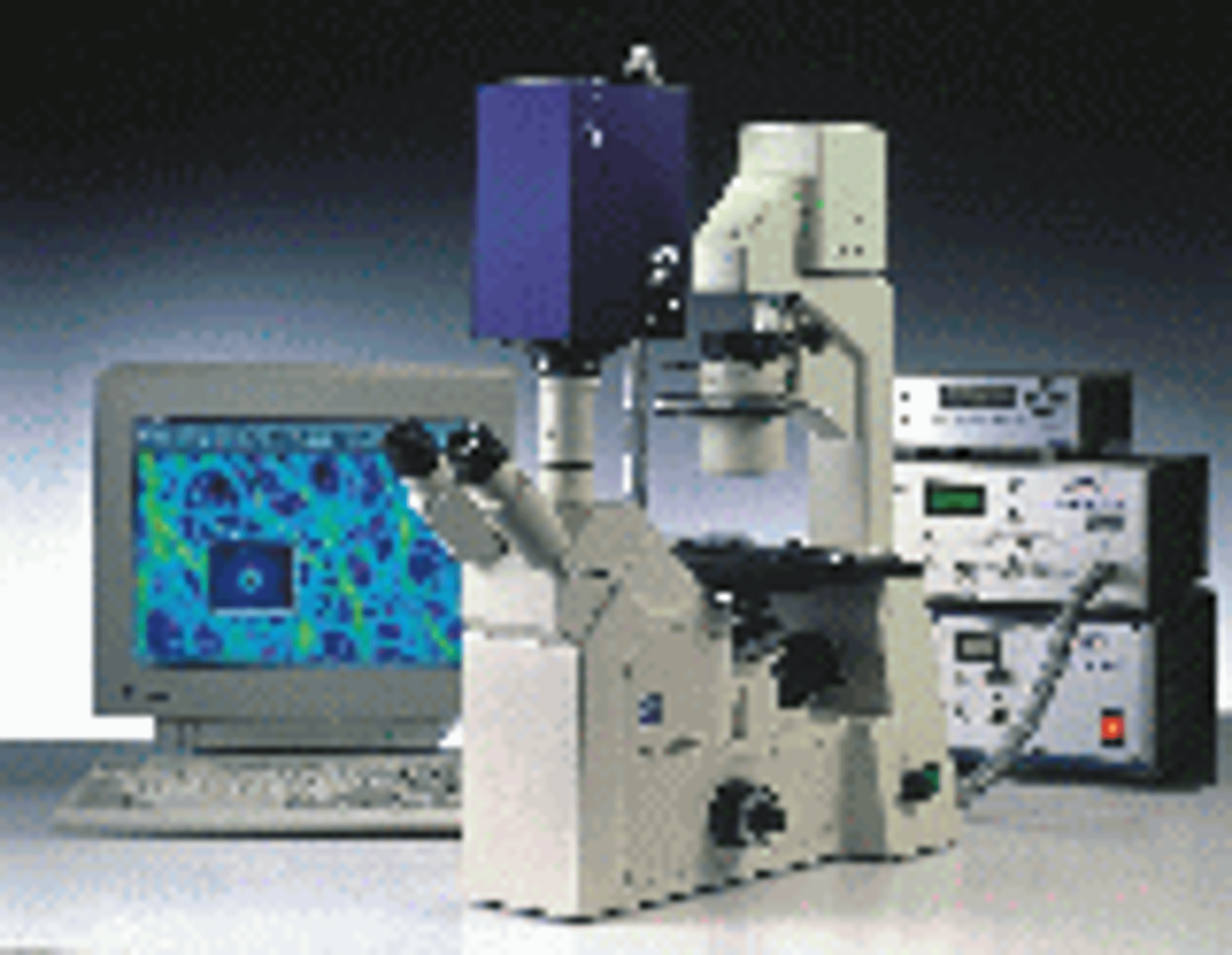
TauTec is now offering customized, turn-key time-resolved imaging systems incroporating state-of-the-art intensified, picosecond gated ICCD camera, choice of excitation laser source (mode-locked lasers up to 100 MHz), optical microscope (inverted or upright), imaging spectrograph and the required accessory electronics. The modular system is suitable for various applications such as FLIM (Fluorescence Lifetime Imaging Microscop…
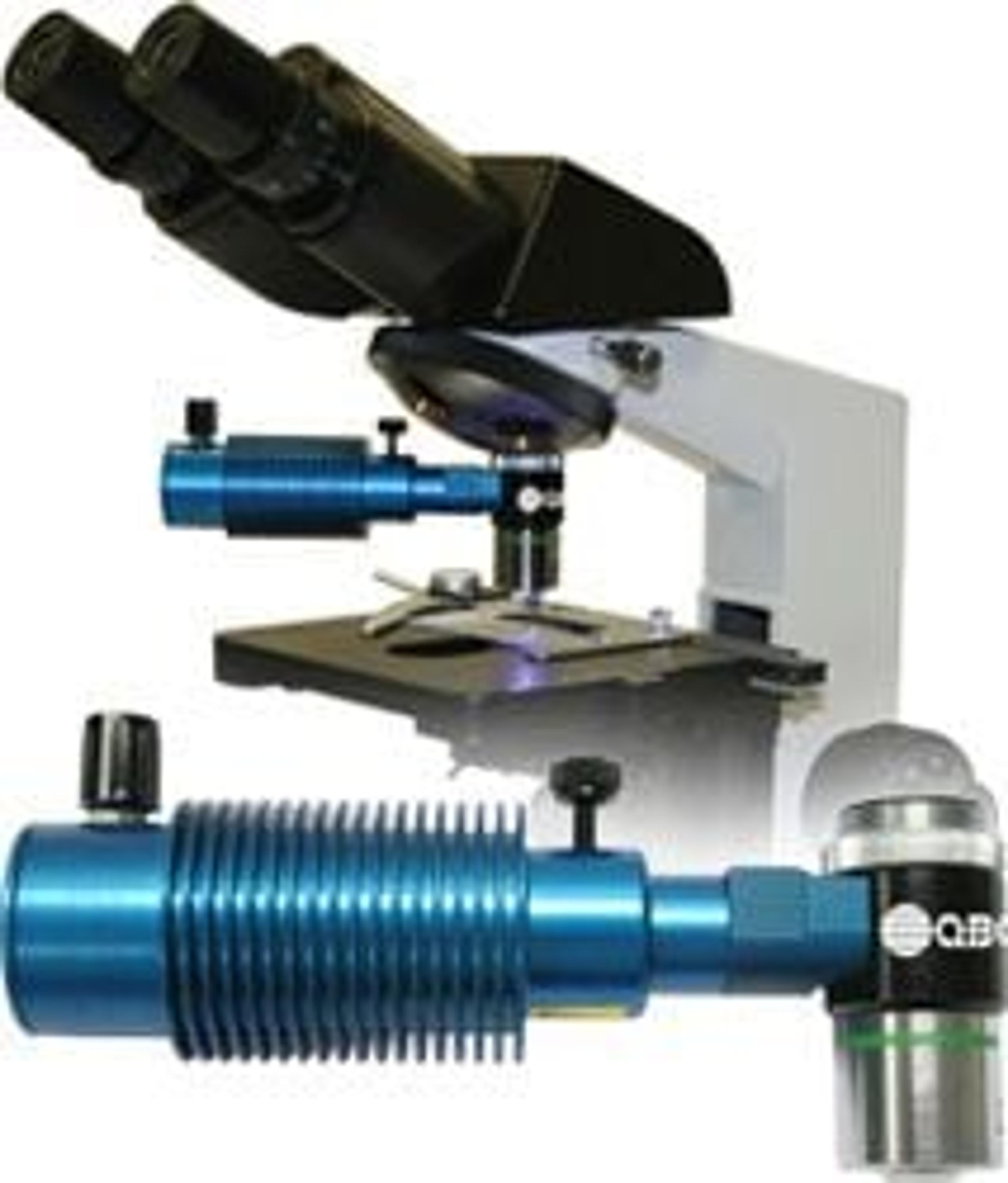
The QBC ParaLens Advance microscope attachment is a unique solution to fluorescence microscopy in that it combines the critical components of an epi-fluorescence microscope, namely the light source and filter set, with a microscope objective in a compact, durable format that can transform virtually any compound light microscope into an epi-fluorescence microscope. Utilising LED technology, the ParaLens light source lamp has a…
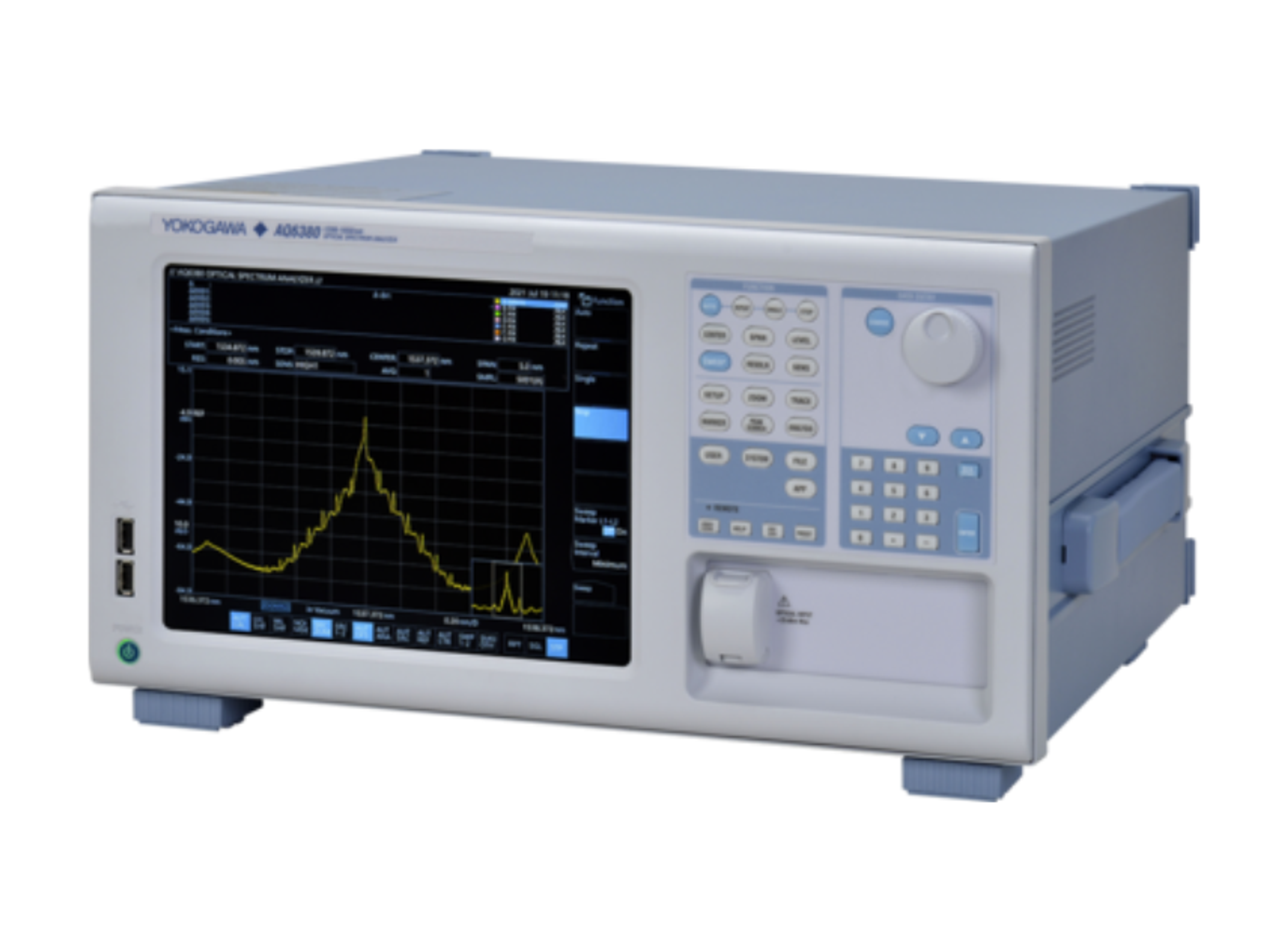
Yokogawa's Optical Spectrum Analyzers deliver high quality, cutting edge technology with dependability, performance and flexibility. They are designed to meet the performance requirements of research, development, verification or production.
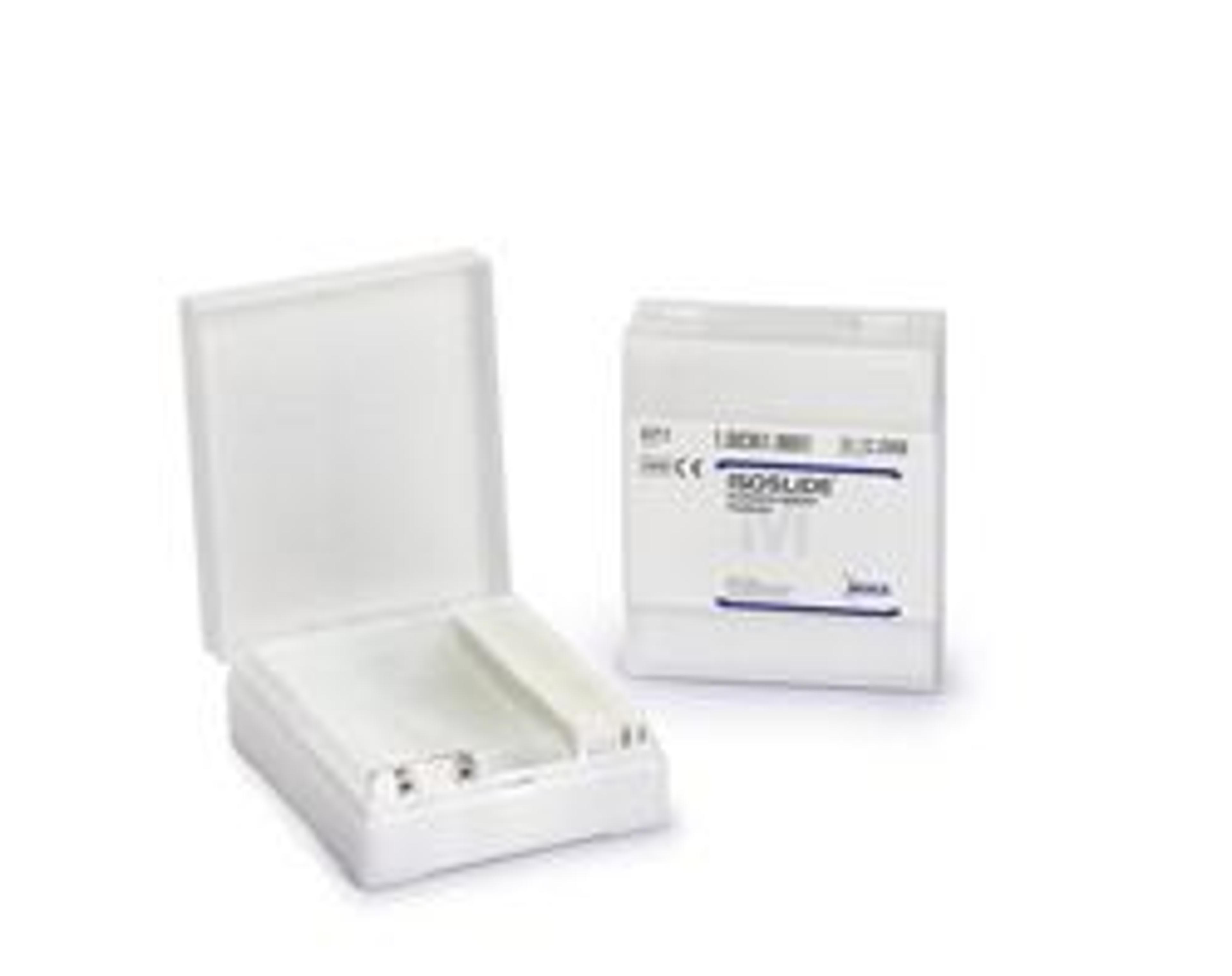
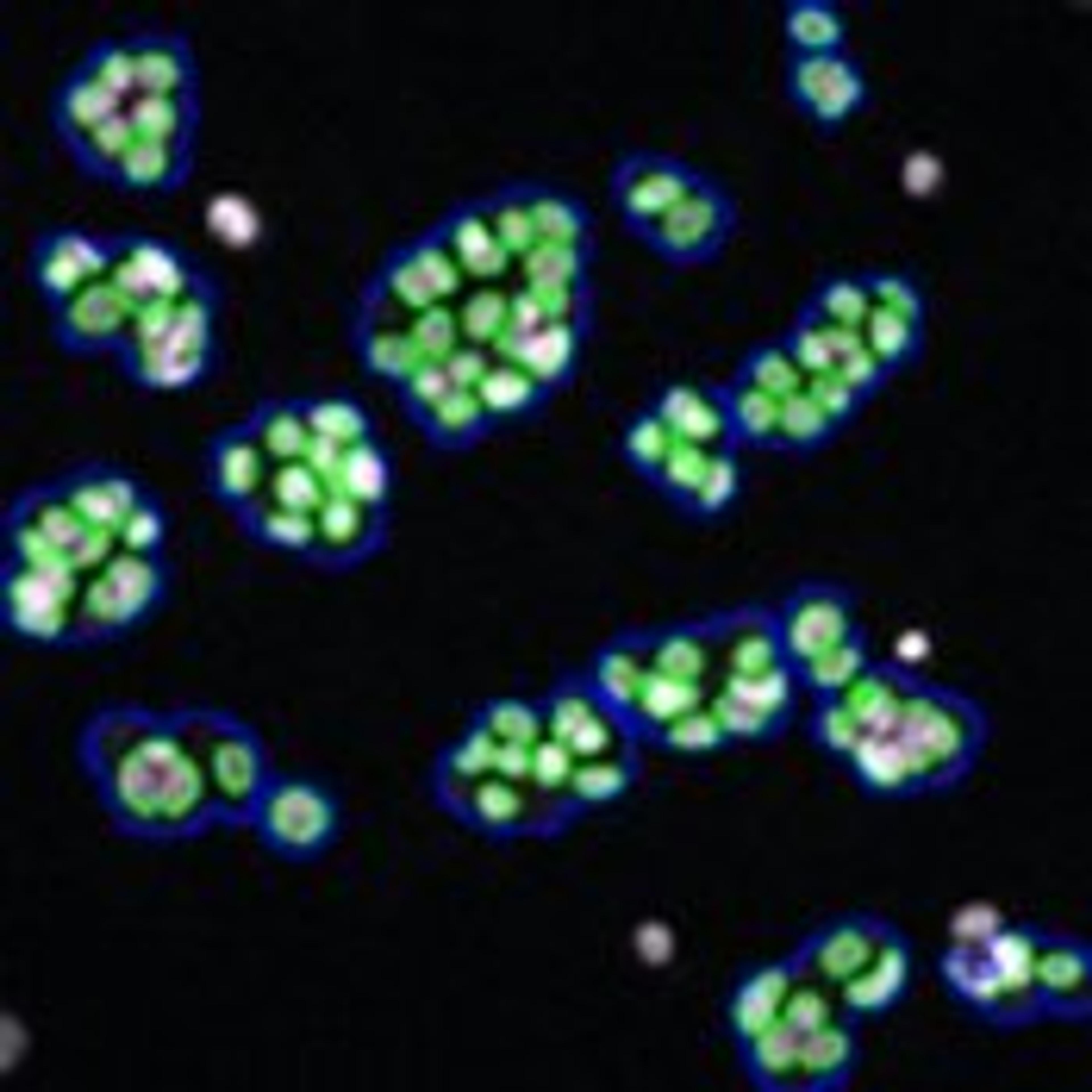
To ensure that the test preparations contain the material relevant for diagnostics and the target structures can be visualized by staining with good differentiation, it is recommended to use a control preparation. ISOSLIDE® control slides enable a direct comparison of typical control material with laboratory-internal specimens.Reticular fibers consist of thin bundles of fine fibrils of type III collagen. These collagen fibrils…
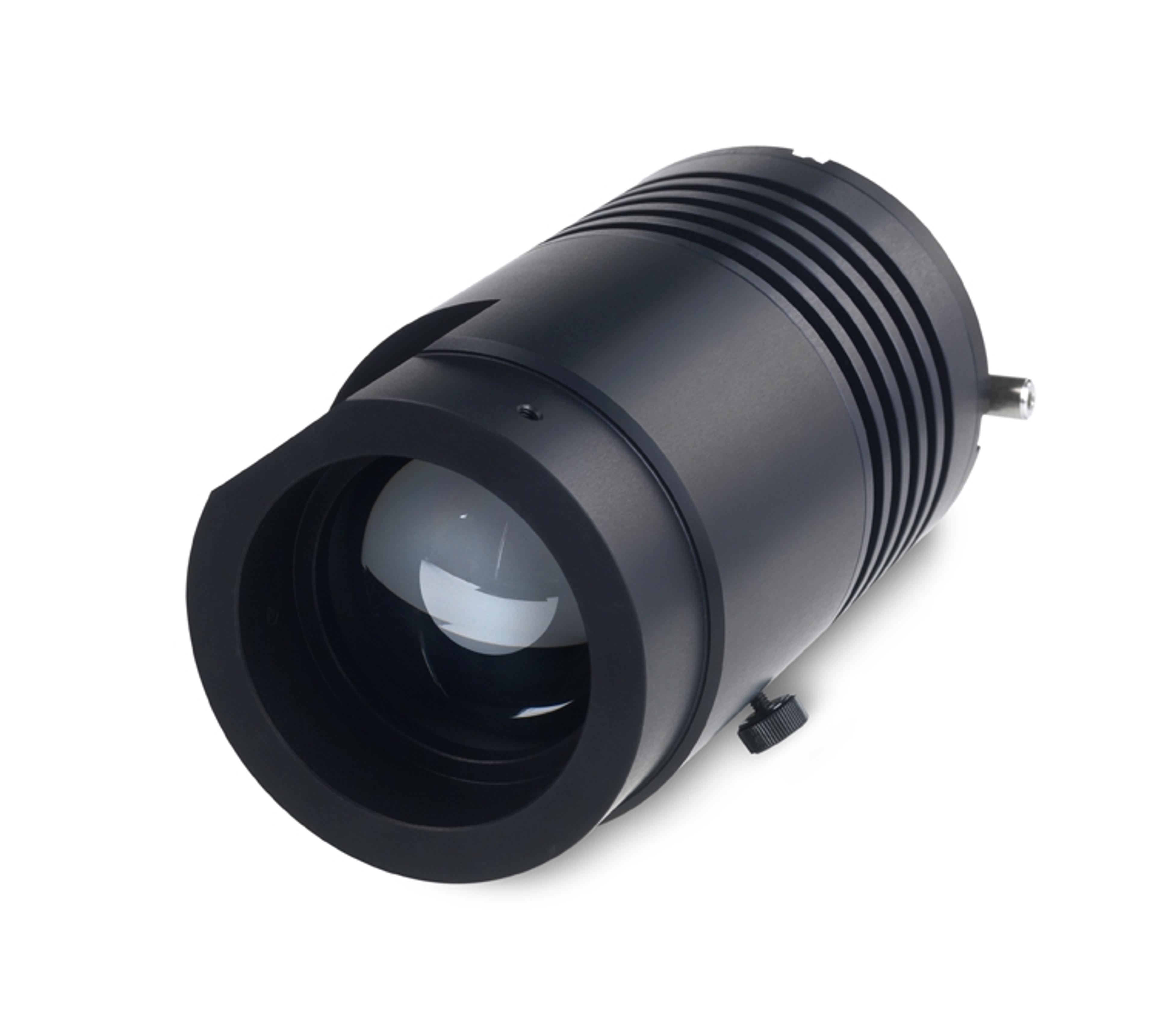
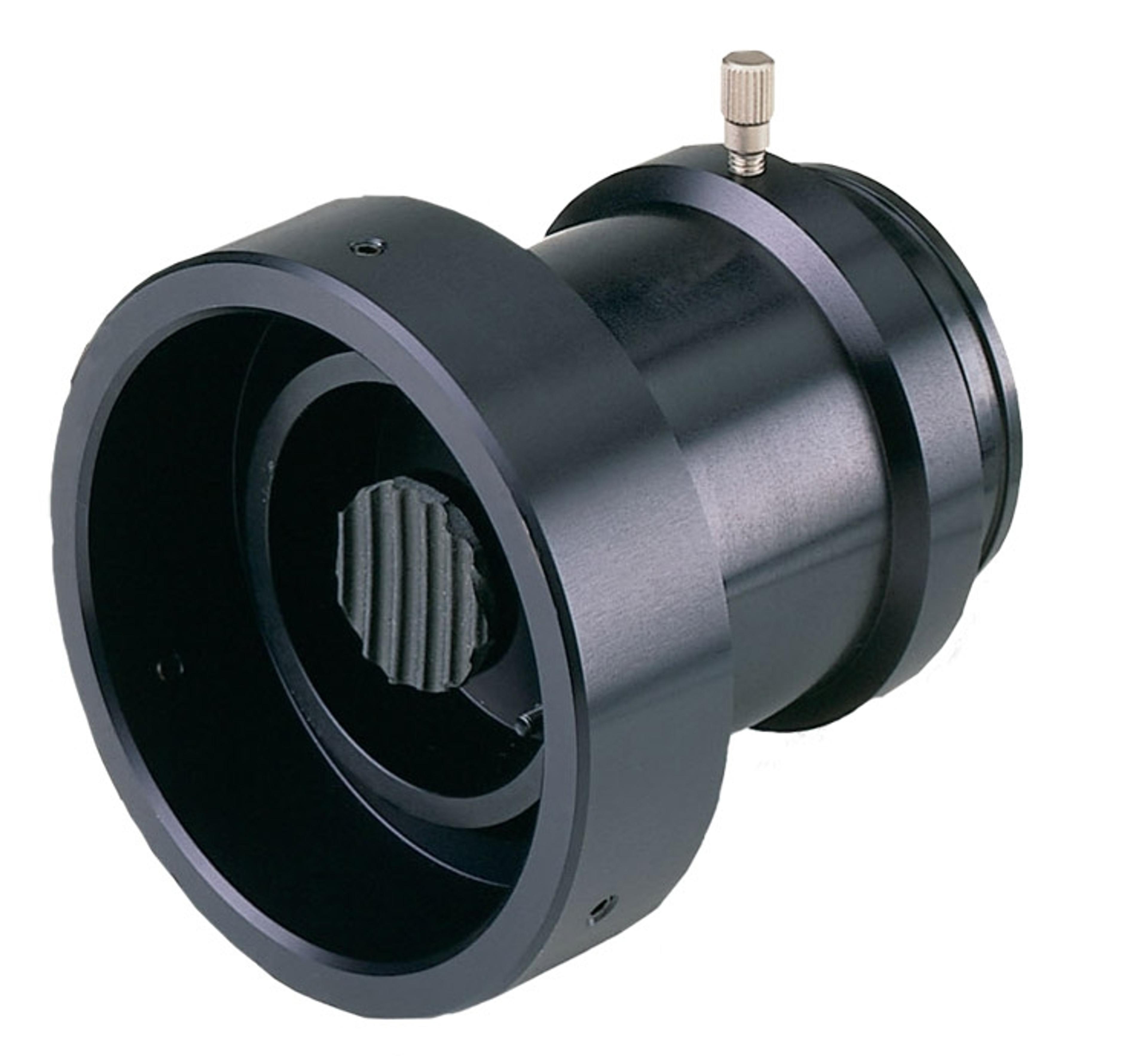
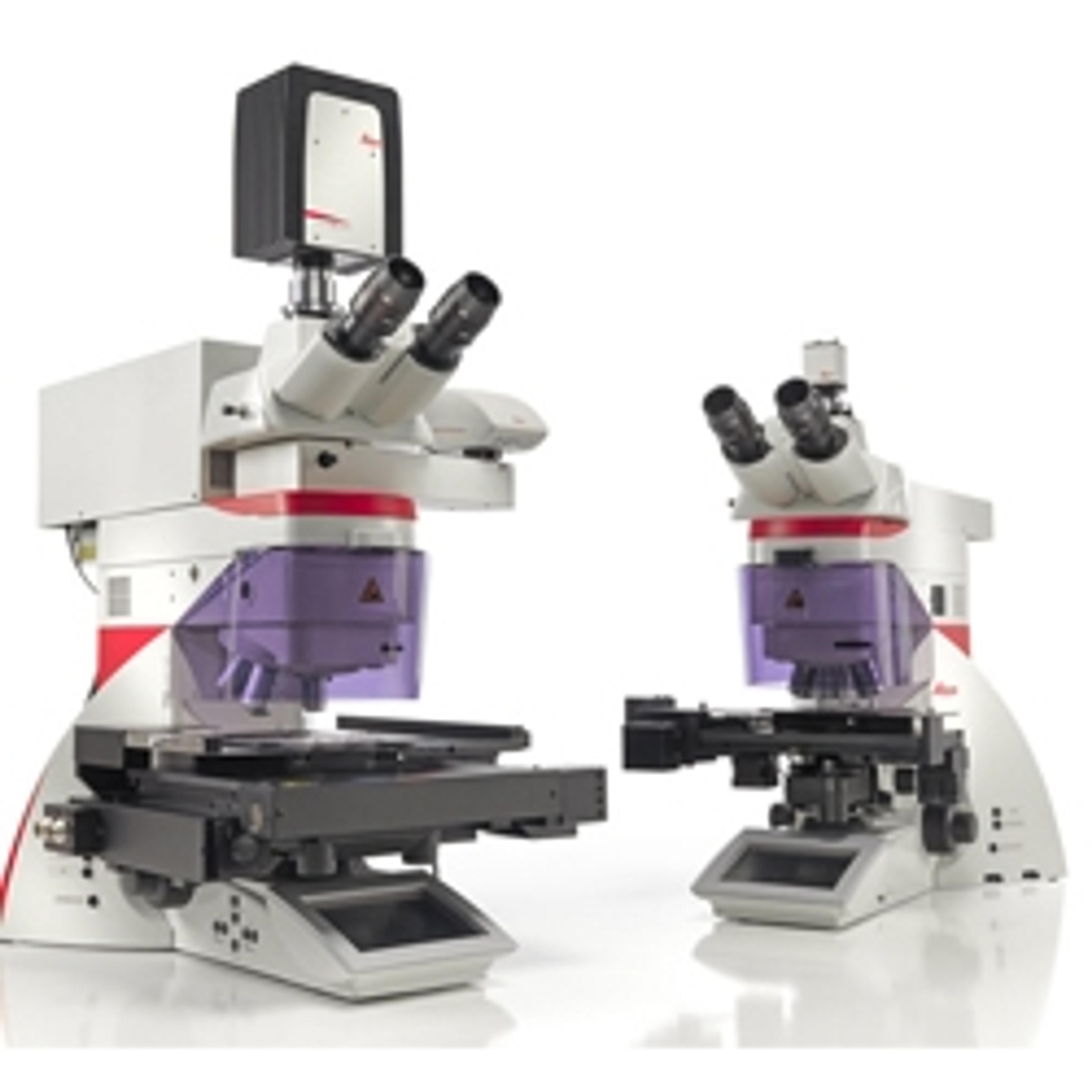
Multiphoton objectives for looking deep within specimens
With high-performance optics, ergonomic design, and 5W LED illumination, the Leica DM IL LED is ideal for cell culture, micromanipulation, documentation of immunostained specimens, and routine live cell examinations.
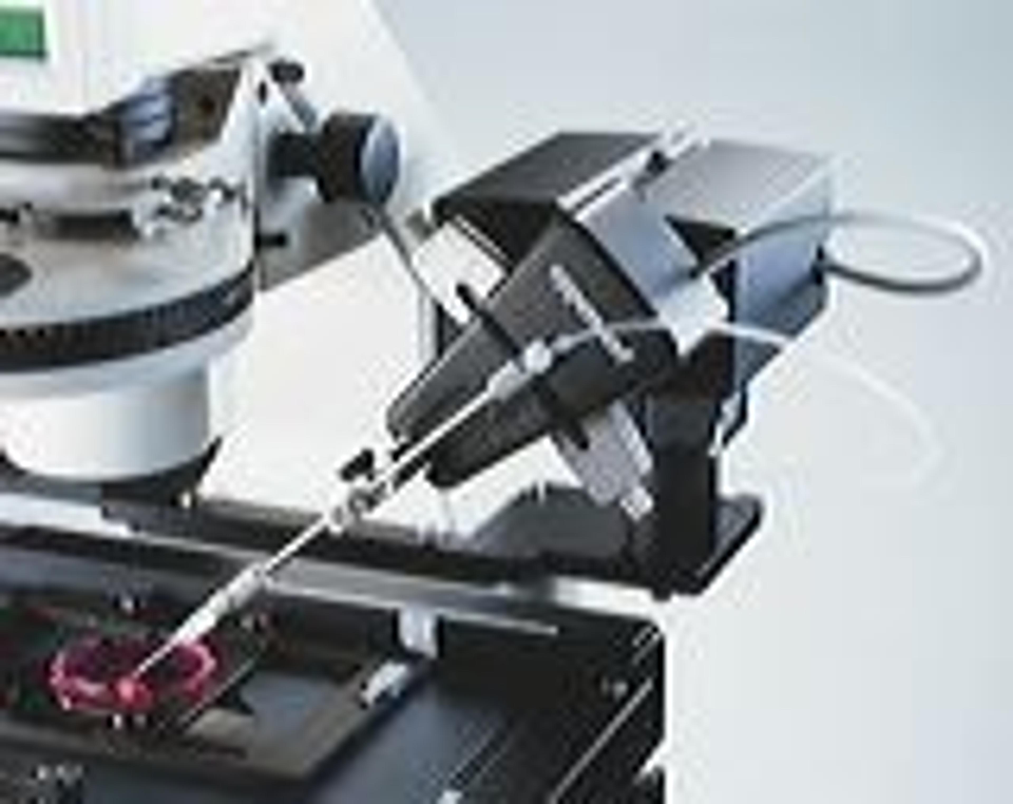
Powered by a unique laser design and dynamic software, Leica LMD systems allow users to easily isolate Regions of Interest (ROI) from entire areas of tissue down to single cells or even subcellular structures such as chromosomes. Intuitive software combined with adjustable lasers and optics allow more precise cutting and collection of a diverse array of sample types.
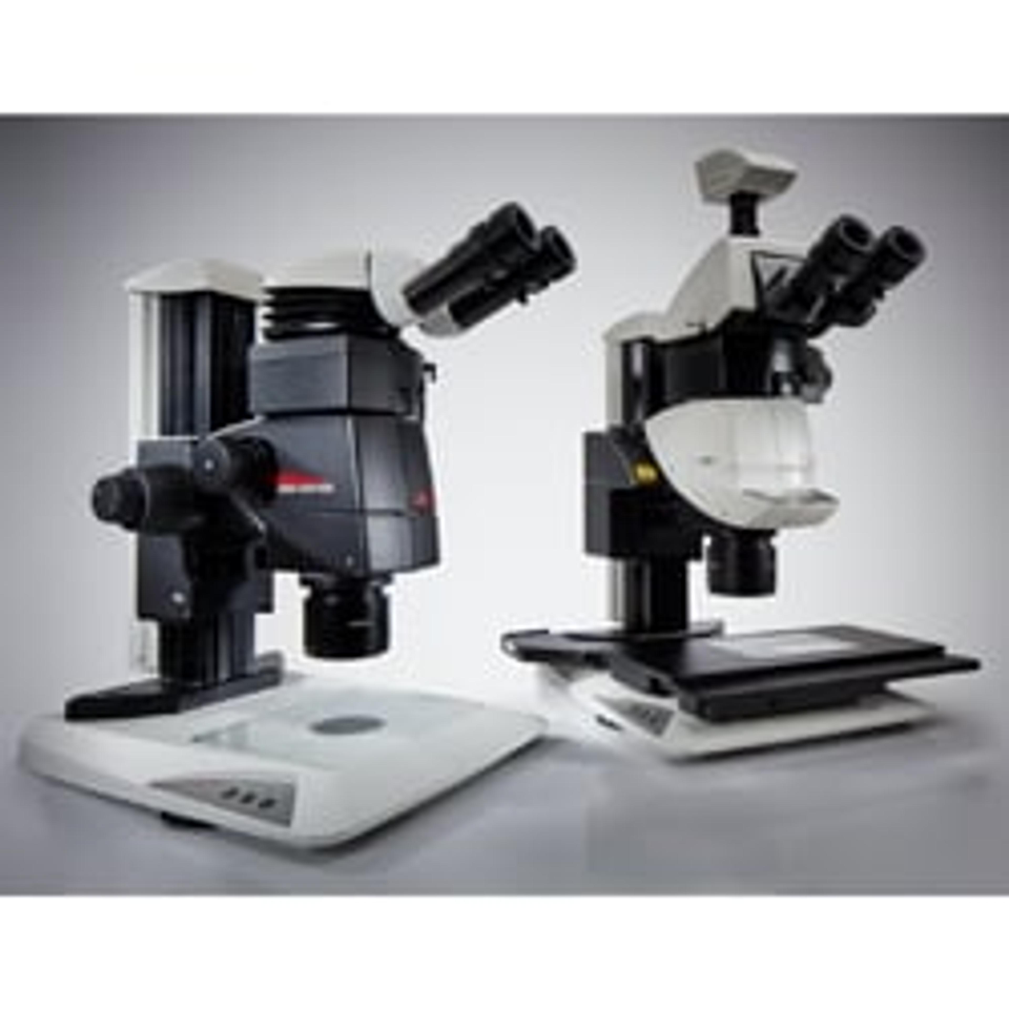
Leica Microsystems has developed the M205 FA and M205 FCA fluorescence stereo microscopes to enable you to detect transgenic expression like GFP and mCherry in early stages, allowing you to select the right sample to successfully base your studies on.
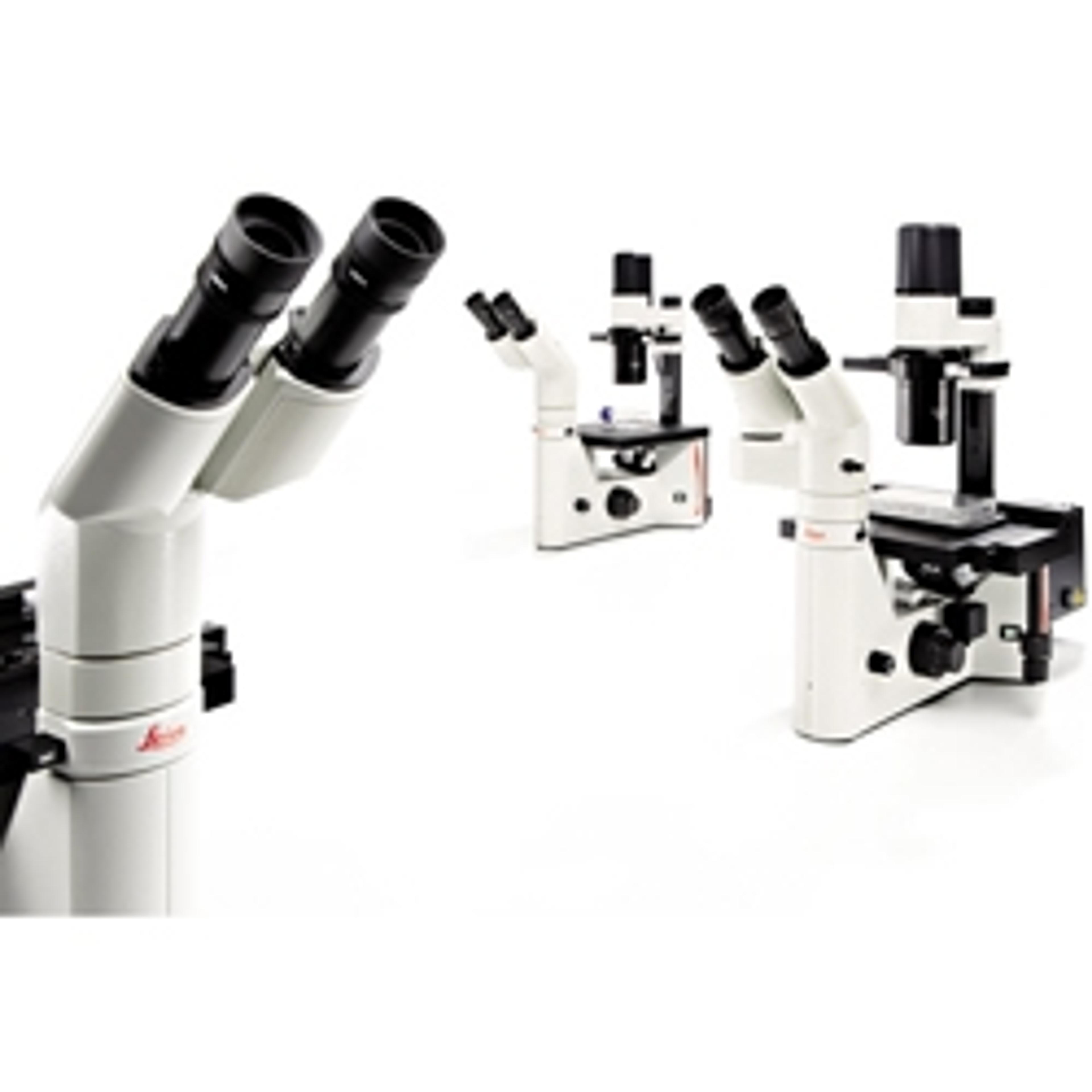
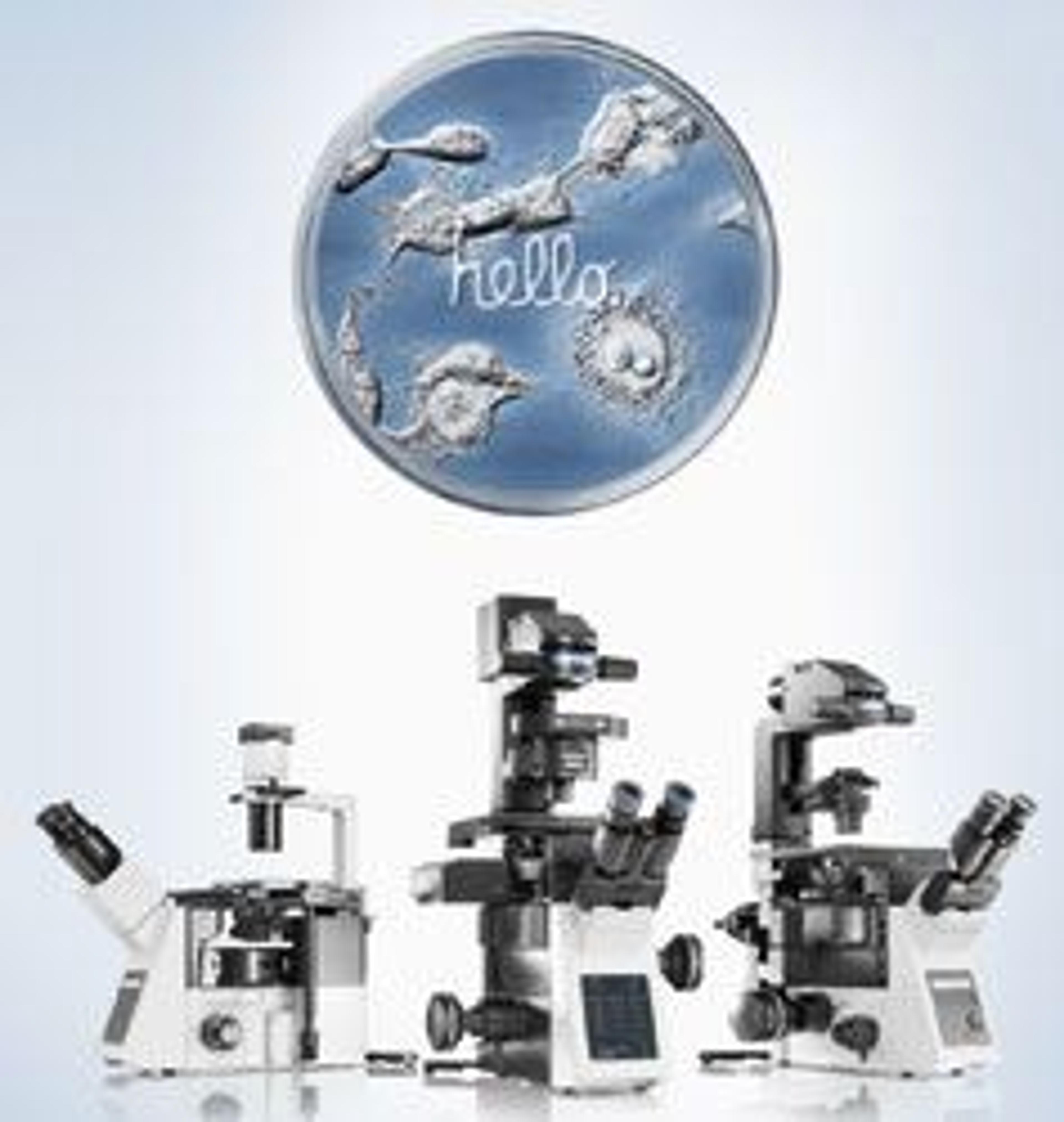
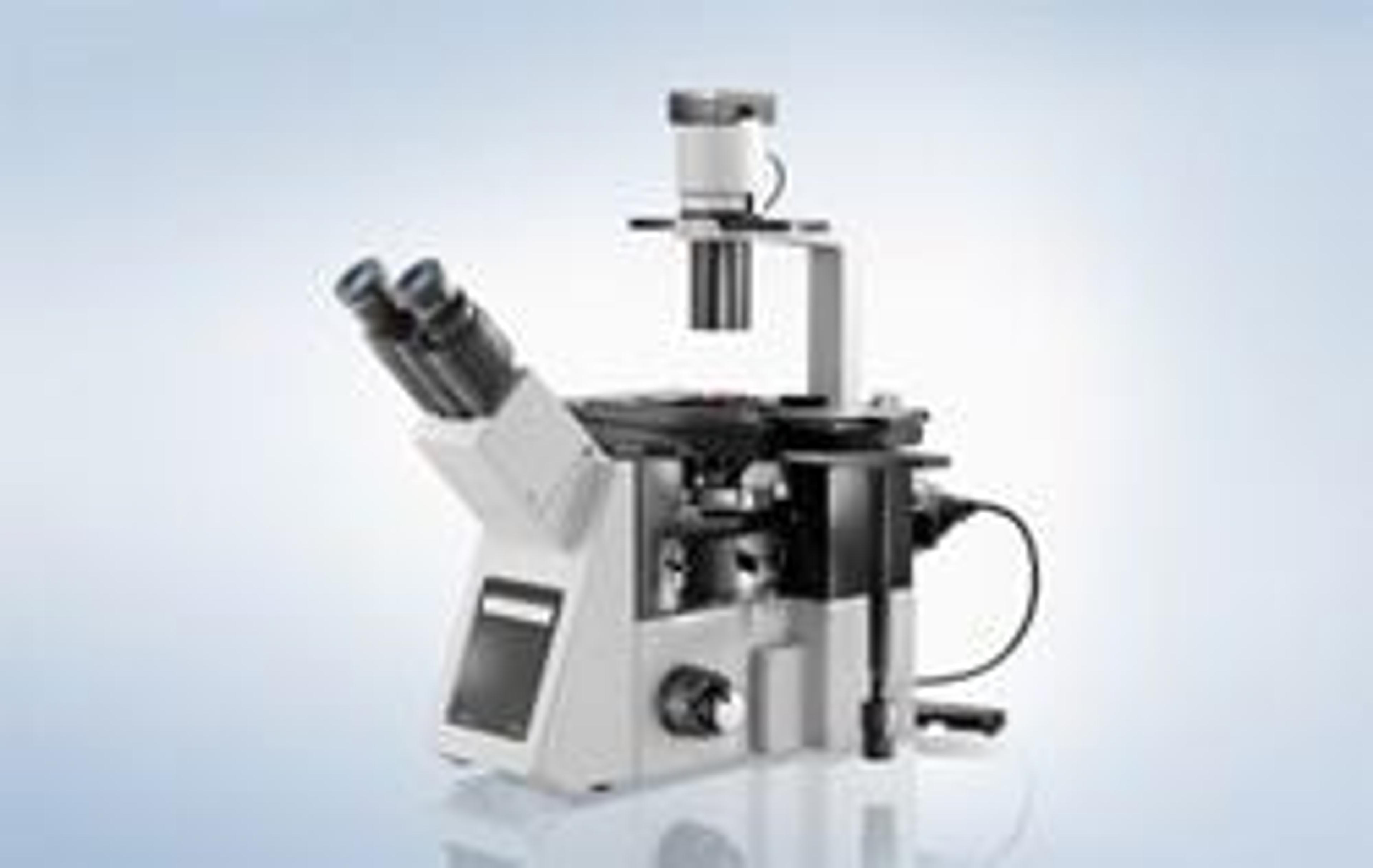
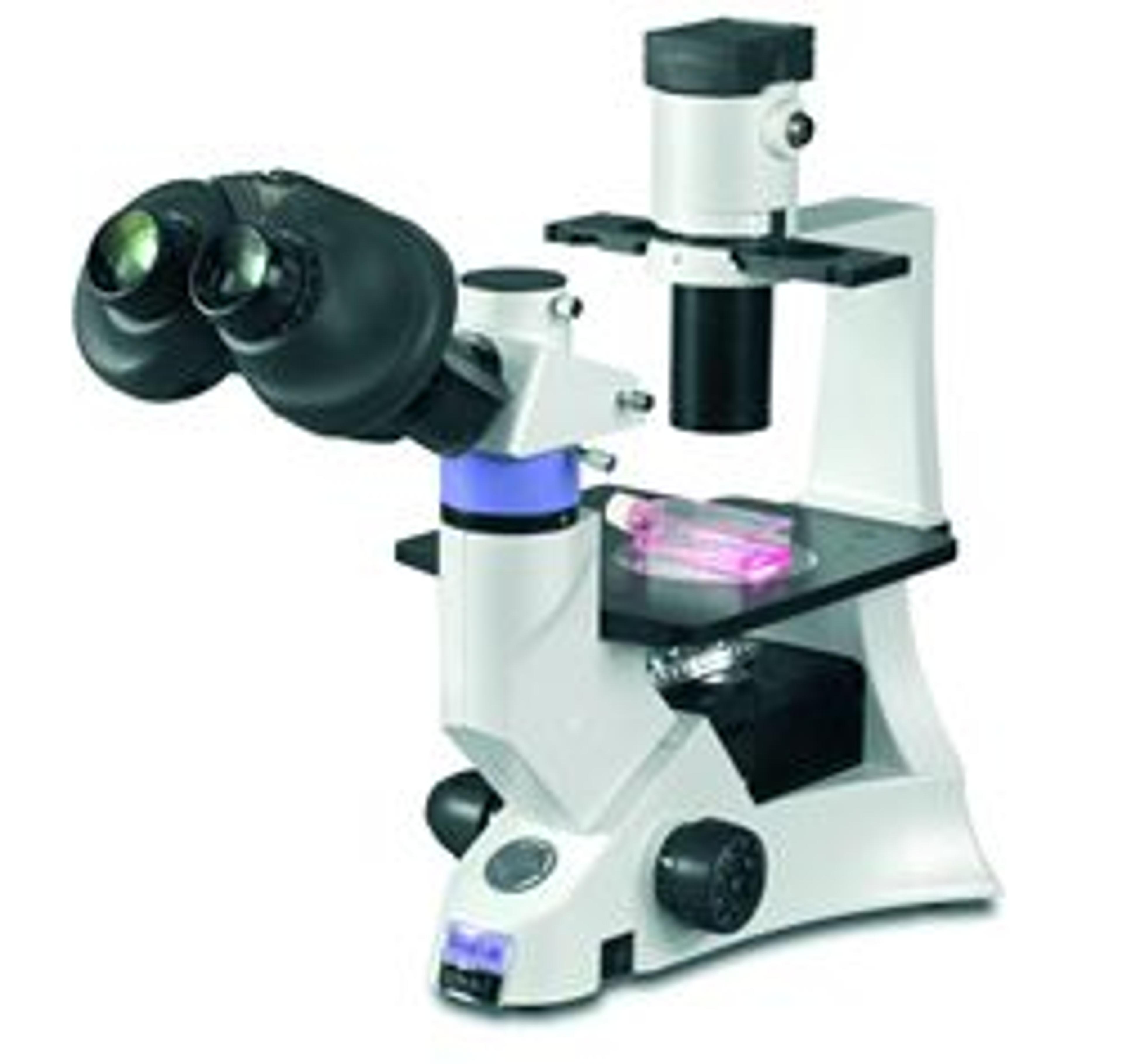
The series of flexible inverted microscope frames for live cell imaging
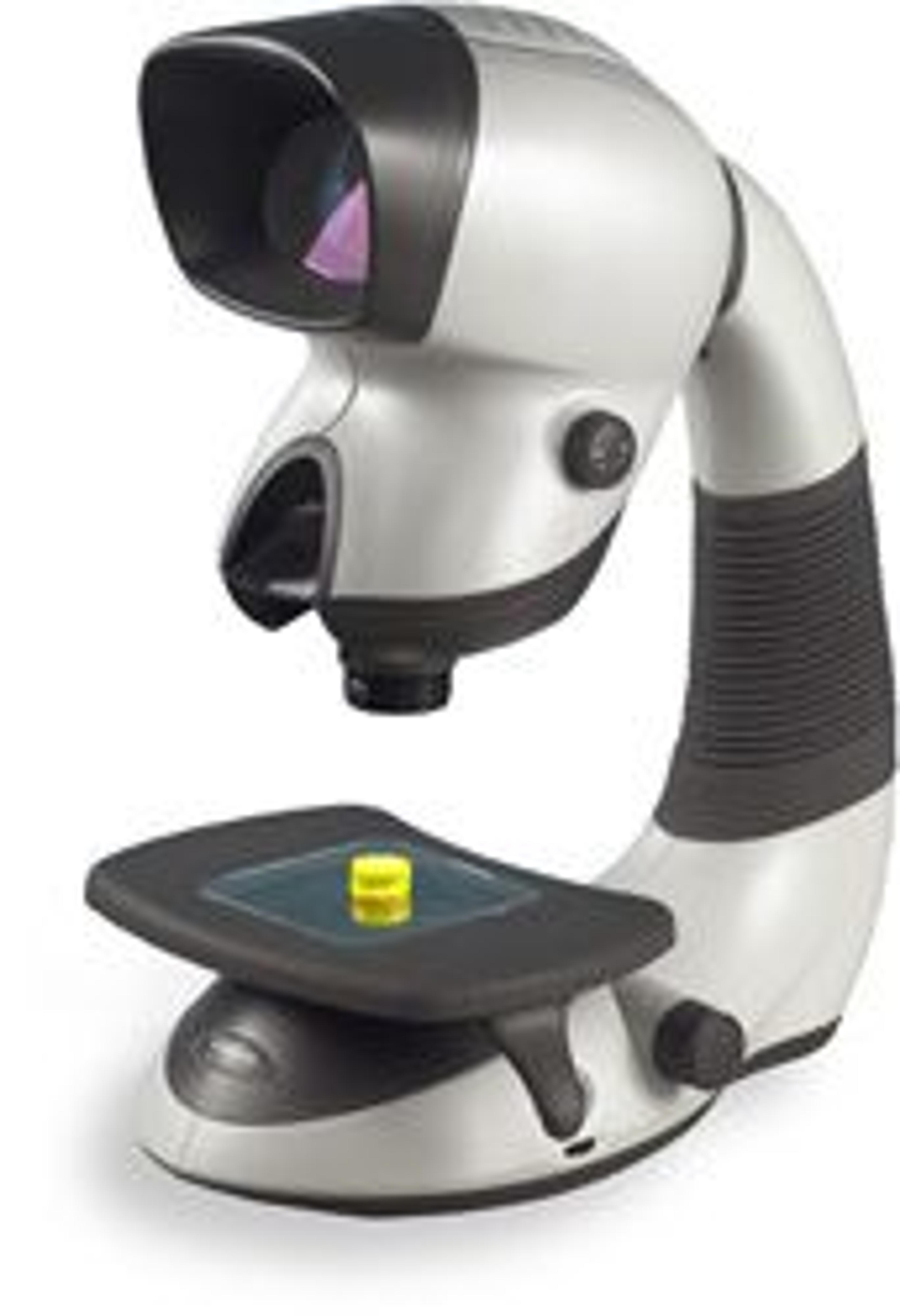
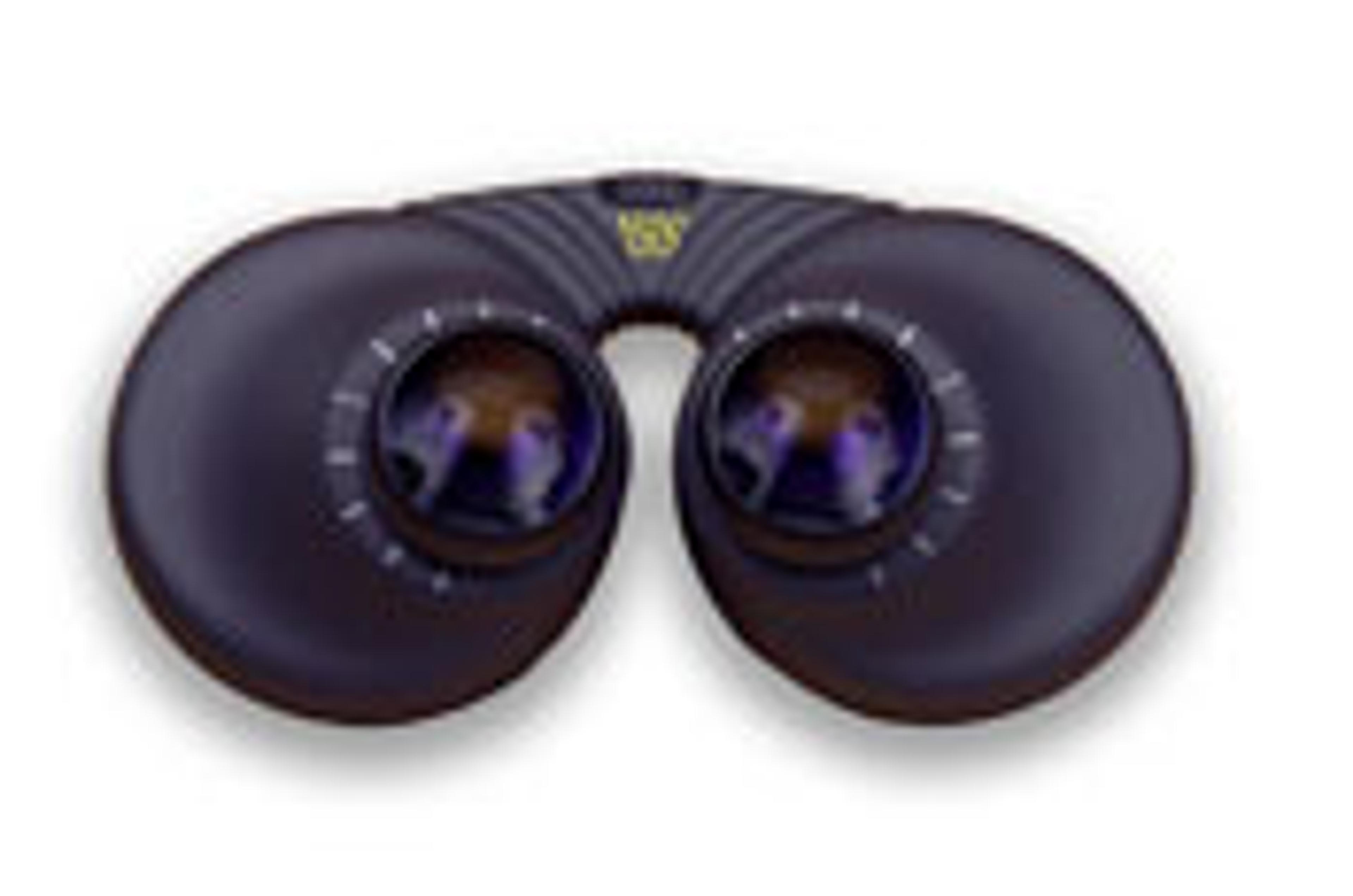
With in excess of 150,000 installed units worldwide, Mantis has become the low magnification stereo viewer of choice in a vast range of laboratory and industrial applications. All of these applications require bright, well contrasted images that are easy to view, especially for prolonged periods. The patented spatial imaging technology used by Mantis provides true optical 3D images, together with large working distances an…
The DX41 offers excellent optical performance with a stable compact design and reduced footprint providing ample bench space for accessories. Vision Biomed’s infinity corrected optics provide outstanding contrast and resolution. As a system microscope, Vision Biomed’s DX41 is particularly well suited to clinical and research microscopy applications. A modular design adds to its versatility with optimised ergonomics making f…
The DX61 offers life science professionals convenience, performance and flexibility combining infinity corrected optics with innovative mechanical design and is particularly suited to most tissue culture applications. - Outstanding optical performance - Infinity optical system- Ergonomically optimised and compact design- Optional ISIS Expanded-Pupil eyepieces increase eye-reliefVision Biomed have developed the DX61 with the…
The DX21 raises the bar in educational and clinical examination. The infinity optical system provides clear sharp images and Vision Biomed ’s ergonomic design ensures comfortable viewing over extended periods of use.- Superb ergonomics for optimum operator comfort- Outstanding optical performance - Modular design allows for easy addition of accessories - Compact stable design The DX21 is a robust, simple to use, purpose built…
Isis expanded pupil is newly patented multi-lenticular technology to a new device designed to enhance the ergonomics of all conventional binocular microscopes. Vision’s Isis accessory fits directly into the microscope’s eyepiece tubes to deliver Expanded Pupil technology, offering an exit ray bundle twelve times larger than a traditional eyepiece.Expanded Pupil technology allows a much greater freedom of operator head movement…
Morphology Explorer V2 is a powerful general-purpose image analysis application for measuring size, shape, and distribution of cells, colonies, and subcellular components. It performs sophisticated calculations in a turn-key, automated manner allowing higher throughput of complex cell-based assays. This application has been biologically validated, enhancing confidence in your image-based data. Available for the Cellomics…