iLite® ADCC Bioassays
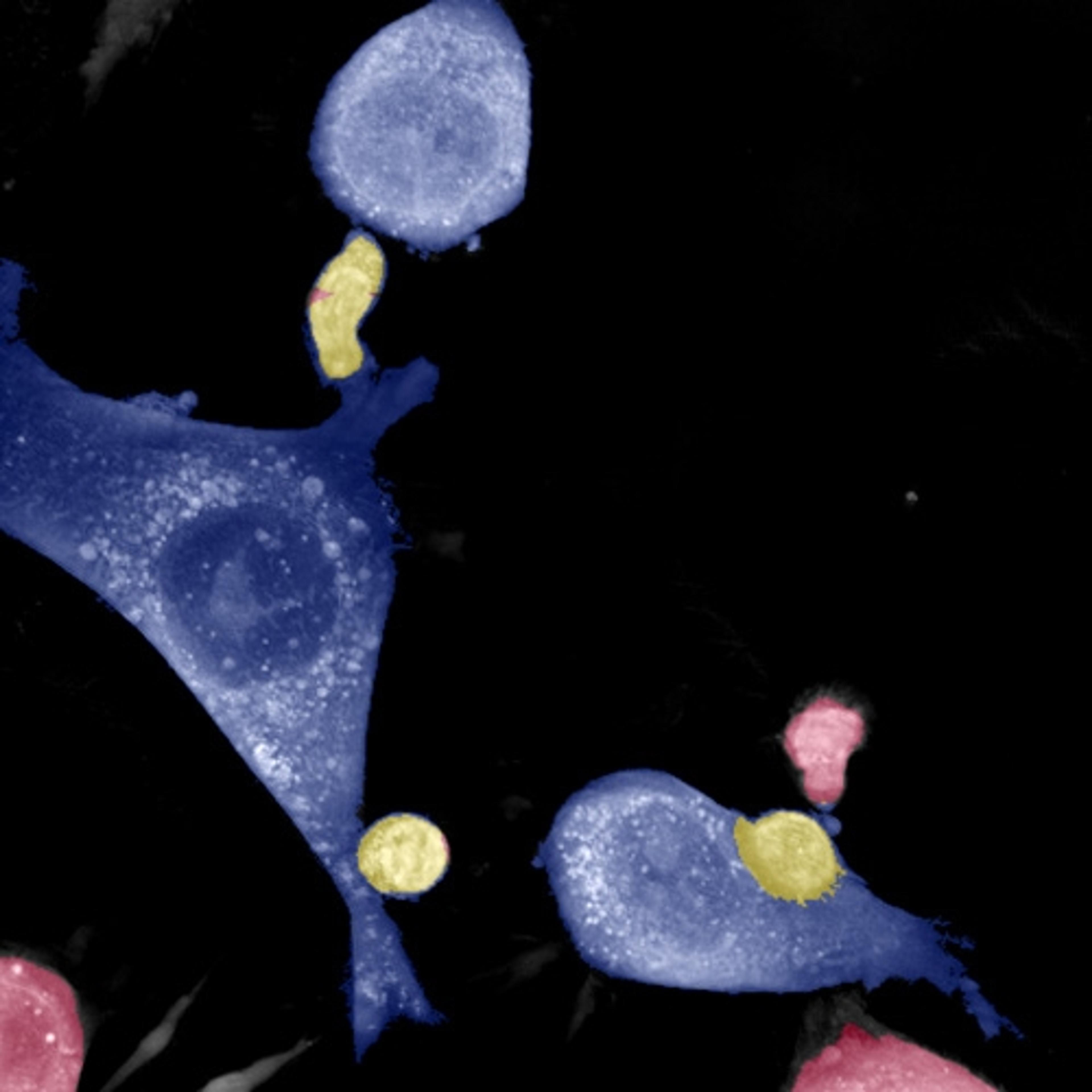
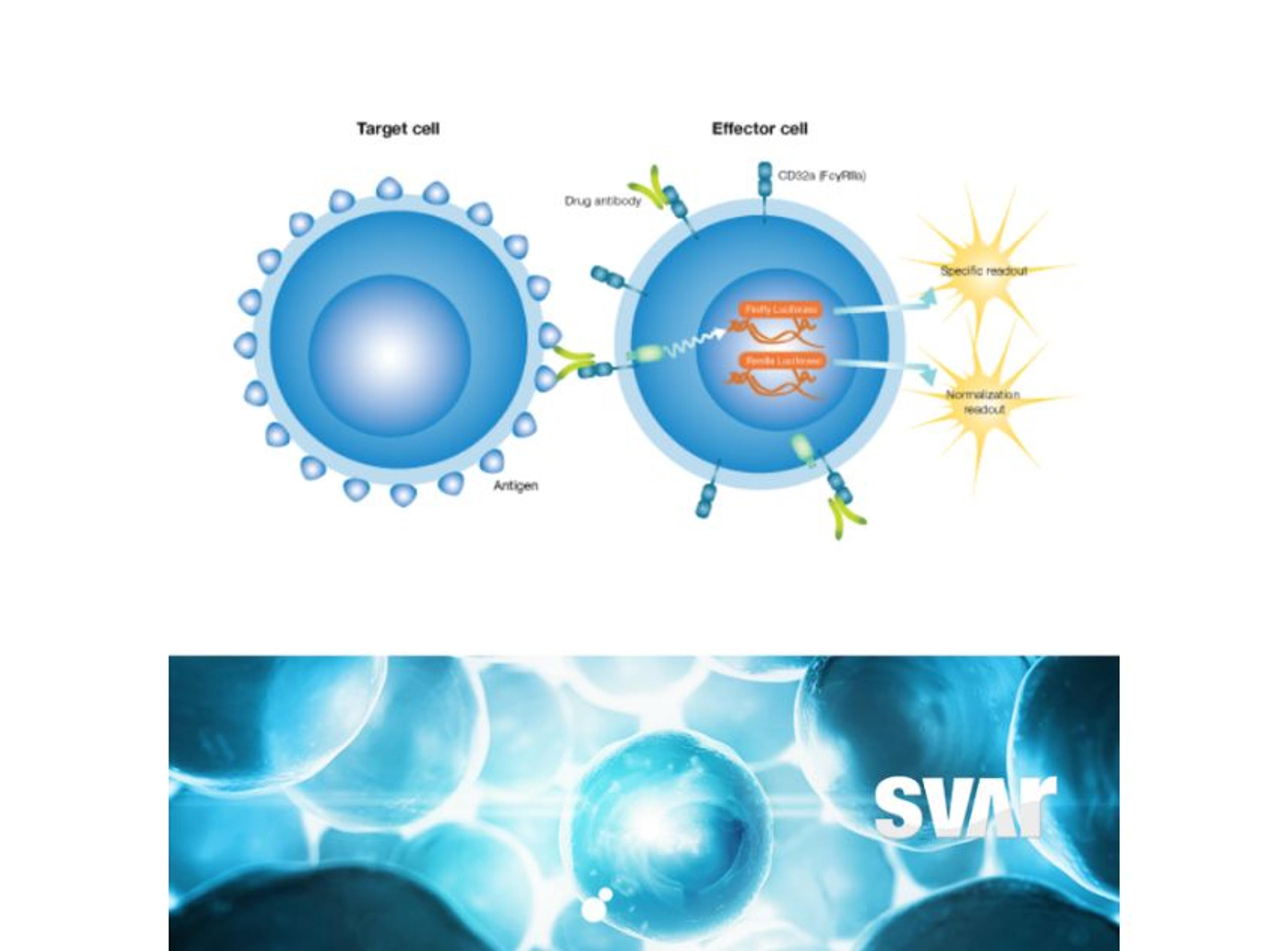
Svar Life Science ABA biologically relevant and powerful way to assess CD32/FcyRlla induced effects.
A biologically relevant and powerful way to assess CD32/FcyRlla induced effects.
A biologically relevant and powerful way to assess CD32/FcyRlla induced effects.
Selvita’s in vivo team has long standing experience in the areas of infection, inflammation and fibrosis and has contributed to numerous preclinical and clinical candidates. Over 60 animal models in mice, rats, and rabbits are fully characterized and validated with clinically relevant pharmacological controls whereas in-depth expertise in translational biomarker selection and validation gives confidence in the smooth transi…
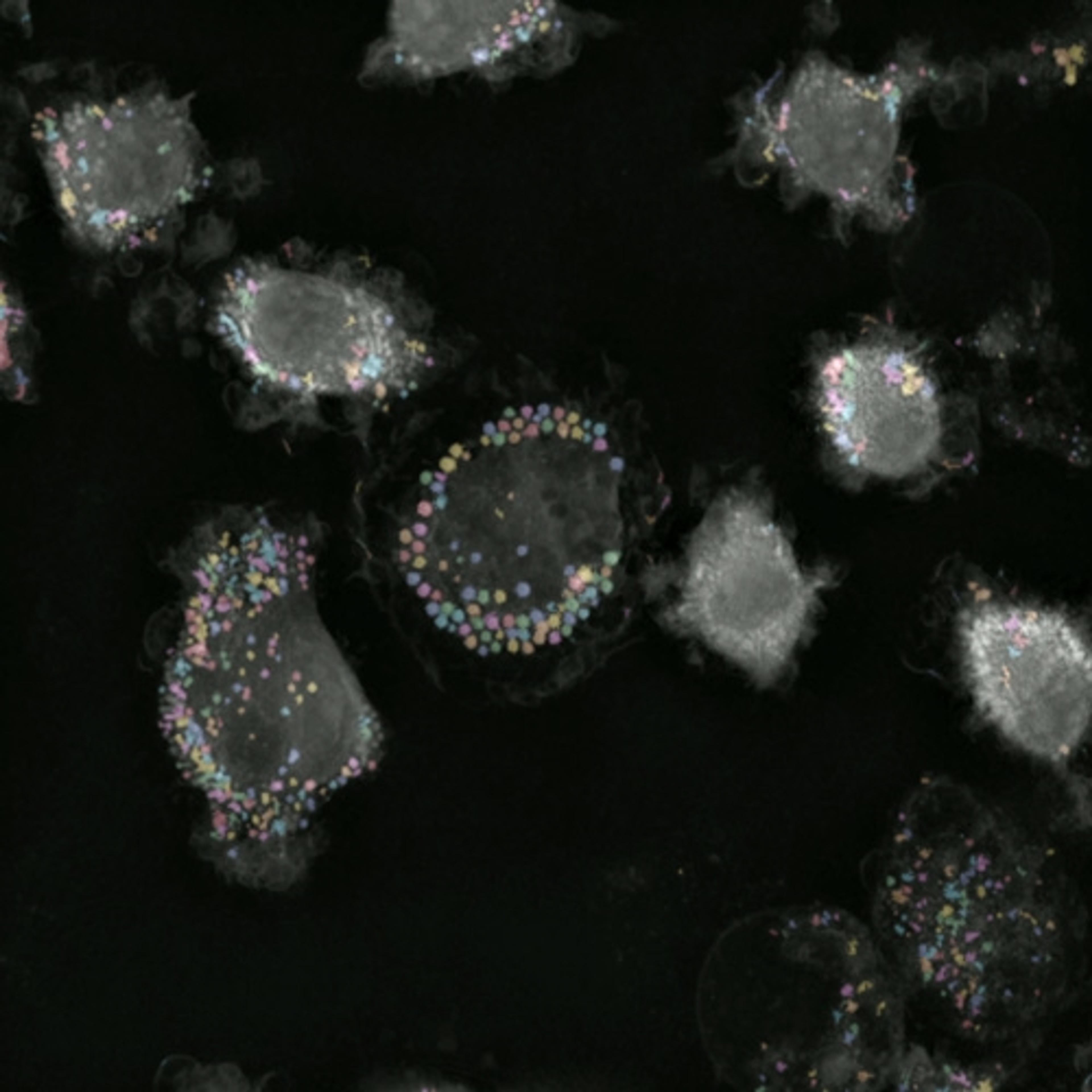
Quantify effector to target cell binding, phenotypic changes, and killing, all in a single label-free assay.
A push-button, automated solution for profiling cell health, death, apoptosis and necrosis, label-free
A push-button solution to analyze lipid droplet dynamics, label-free.
BC001 - 2-channel biochip with narrow cavities for organ-on-chip models by Dynamic42
BC002 - 2-channel biochip with wide cavities for organ-on-chip models by Dynamic42
BC003 - 3-channel biochip with cavities for spheroid/ organoid-on-chip models by Dynamic42
BC005 - 2-channel biochip with thin channels for organ-on-chip models with limited cell material by Dynamic42
Service for toxicology screening, lead identification/ optimization and in vitro model development using organ-on-chip technology by Dynamic42
Apoptosis, or programmed cell death, is a highly regulated pathway that is important in normal developmental processes as well as many diseases. Cells undergoing apoptosis are identifiable by a number of characteristics, including changes in mitochonrial membrane potential, transport of phosphatidylserine (PS) to the membrane surface, activation of caspase proteases, and DNA fragmentation in the nucleus. Guava offers four apop…
Guava Technologies' ViaCount assay is revolutionizing the way cell counting and viability assessment are done in today's laboratories. Fast, automated, and highly reproducible, the Guava ViaCount assay addresses many of the difficulties of conventional cell counting and viability assessment methods. The Guava ViaCount assay provides: Cell counts up to ten times faster than manual counting, in a convenient turnkey assay.…
Cell viability and cytotoxicity detection. Colourimetric microplate assay, one solution type, no washing required, no radioisotopes or organic solvents required.
Gentronix has launched a new, accurate and fast in vitro mammalian cell genotoxicity assay - GreenScreen HC. The TK6 host cells are p53 competent and familiar to most genetic toxicology laboratories. A patented GFP reporter system exploits the proper regulation of the GADD45a gene. The assay delivers both high specificity and high sensitivity and detects all common mechanistic classes of genotoxin. The 96-well microplate for…
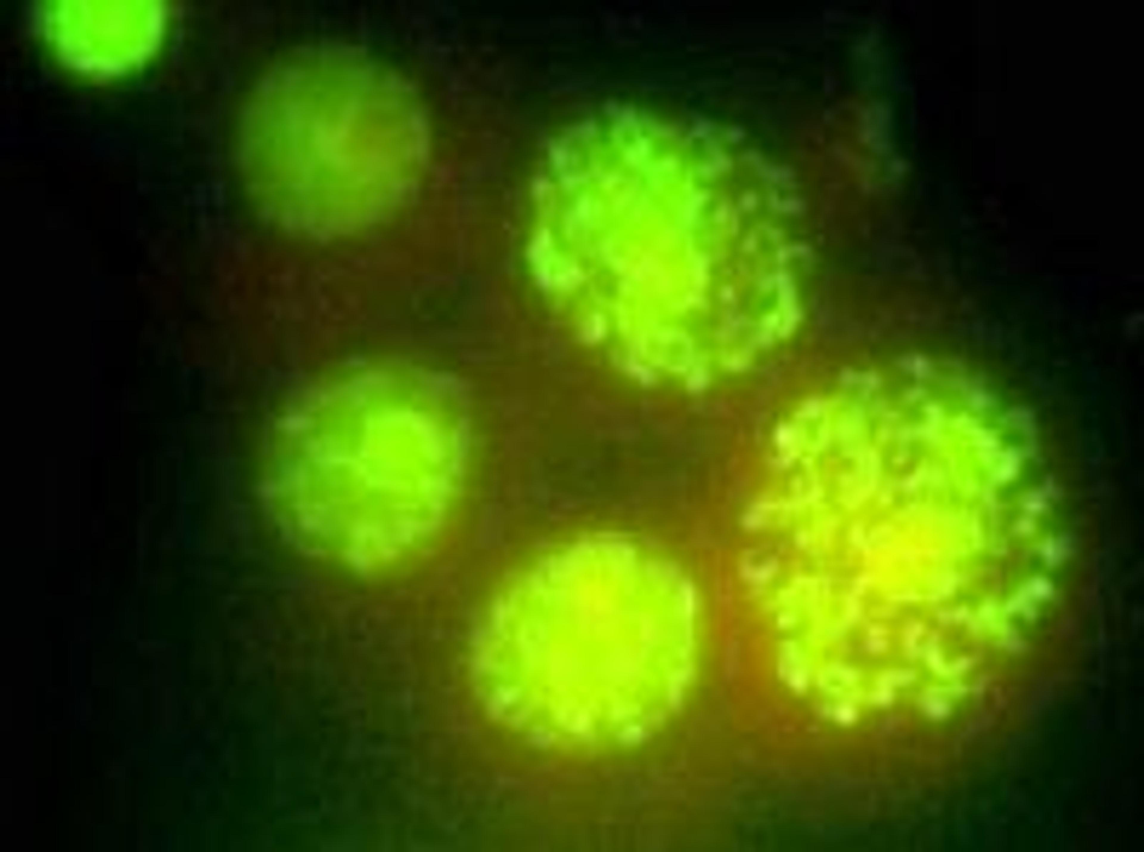
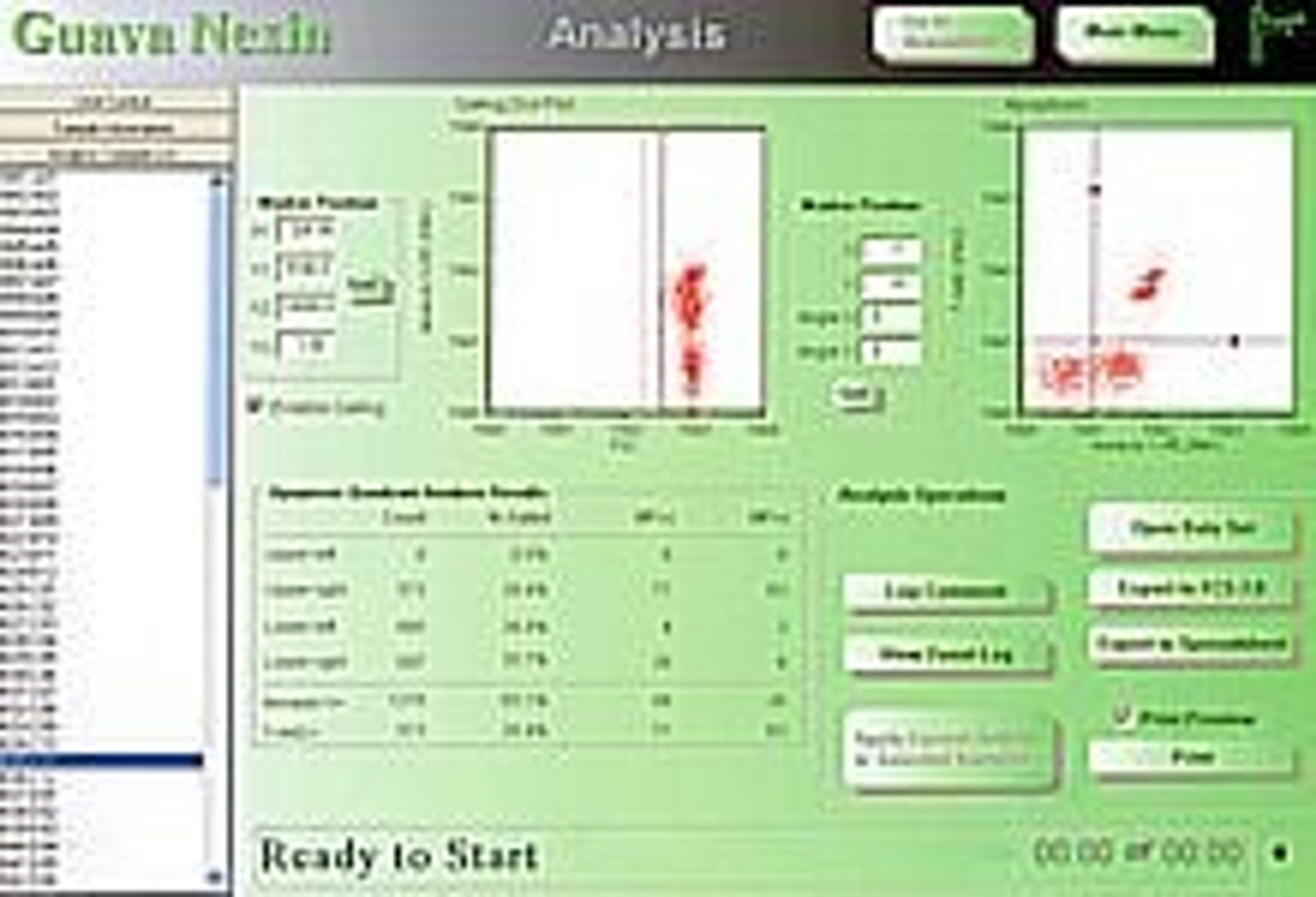
The new Guava Nexin® reagent monitors induction of apoptosis quickly and easily, with minimal chance for contamination. Annexin V is a calcium-dependent phospholipid binding protein with high affinity for phosphatidylserine (PS), a membrane component normally localized to the internal face of the cell membrane. Early in the apoptotic pathway, molecules of PS are translocated to the outer surface of the cell membrane where Ann…
Fluorescein-conjugated anticoagulant for the detection of phosphatidylserine in the outer leaflet of apoptotic cellsProduct Description Detection: Annexin-V-FLUOS can be directly detected in FACS analysis and immunochemistry without a secondary detection system. Sample material: Cell lines and freshly isolated cells. Specificity: Annexin-V binds in a calcium-dependent manner to negatively charged phospholipid surfaces, and sho…
Biotin-conjugated anticoagulant for the detection of phosphatidylserine in the outer leaflet of apoptotic cellsProduct Description Detection: Annexin-V-Biotin can be used in immunohistochemistry with a secondary detection system (e.g., Streptavidin-POD conjugate). Sample material: Cell lines and freshly isolated cells Specificity: Annexin-V binds in a calcium-dependent manner to negatively charged phospholipid surfaces, and…
Alexa 568-conjugated anticoagulant for the detection of phosphatidylserine on the outer leaflet of apoptotic cellsBenefits Detection flexibility: Analysis of results with flow cytometry and/or fluorescence microscopy. Alexa is a red dye, which can be used for double or triple staining with other fluorescent markers (e.g., fluorescein-labeled surface markers). Accuracy: Accurately distinguish apoptotic cells from necrotic…
Kit for the detection and quantification of apoptosis and differentiation from necrosis at the single-cell level, based on Annexin-V-labelingProduct Description Detection: Annexin-V-FLUOS can be directly detected in FACS analysis and immunochemistry without a secondary detection system. Sample material: Cell lines and freshly isolated cells. Specificity: Annexin-V-FLUOS binds in a Ca2+-dependent manner to negatively charged ph…
Kit for the detection and quantification of apoptotic cell death on a single-cell level by light microscopy in immunocyto- and immunohistochemistry Benefits Sensitive: The maximum intensity of labeling (cell staining) of apoptotic cells is higher than the nick translation method. Fast: The use of fluorescein-dUTP allows analysis of the samples directly after the TUNEL reaction, but before the addition of the secondary d…
Kit for the detection and quantification of apoptotic cell death on a single-cell level by flow cytometry and fluorescence microscopy, and for double labeling with fluorescein-labeled cell markers (TMR red)Benefits Convenient: No secondary detection system required. Accurate: Identification of apoptosis at a molecular level (DNA-strand breaks) and identification of cells at the very early stages of apoptosis. Flexible:…
Kit for the detection and quantification of apoptosis (programmed cell death) at the single-cell level, based on labeling of DNA strand breaks (TUNEL technology): Analysis by fluorescence microscopy or flow cytometry Benefits Sensitive: The direct labeling procedure using fluorescein-dUTP reduces background labeling. Fast: The use of fluorescein-dUTP allows analysis of the samples directly after the TUNEL reaction. Co…
ApplicationThe In Situ Cell Proliferation Kit, FLUOS belongs to the second generation of improved kits for the detection of DNA synthesis at the single-cell level (in situ) by immunocyto-/immunohistochemistry. The kit can be used in many different in vitro and in vivo cell systems, for example: Detection and quantification of cells in S-phase Determination of growth fractions for tumors in animal-model systems Detectio…
ApplicationThe immunoreactivity of the M30 antibody is confined to the cytoplasm of apoptotic cells. It can be easily used for: Immunocytochemistry Flow Cytometry Immunohistochemistry: paraffin-embedded tissue sections (secondary enhancer reagents needed for this particular application) and cryostat sections Benefits Determine caspase activity, even in formalin-embedded tissue. Identify the stage of apoptosis (…