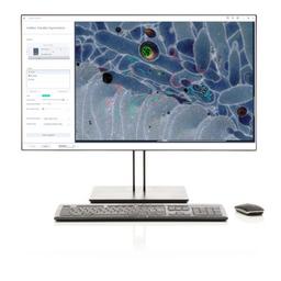
ZEN core AI Toolkit
Advanced Image Processing across Multiple Microscopy Methods


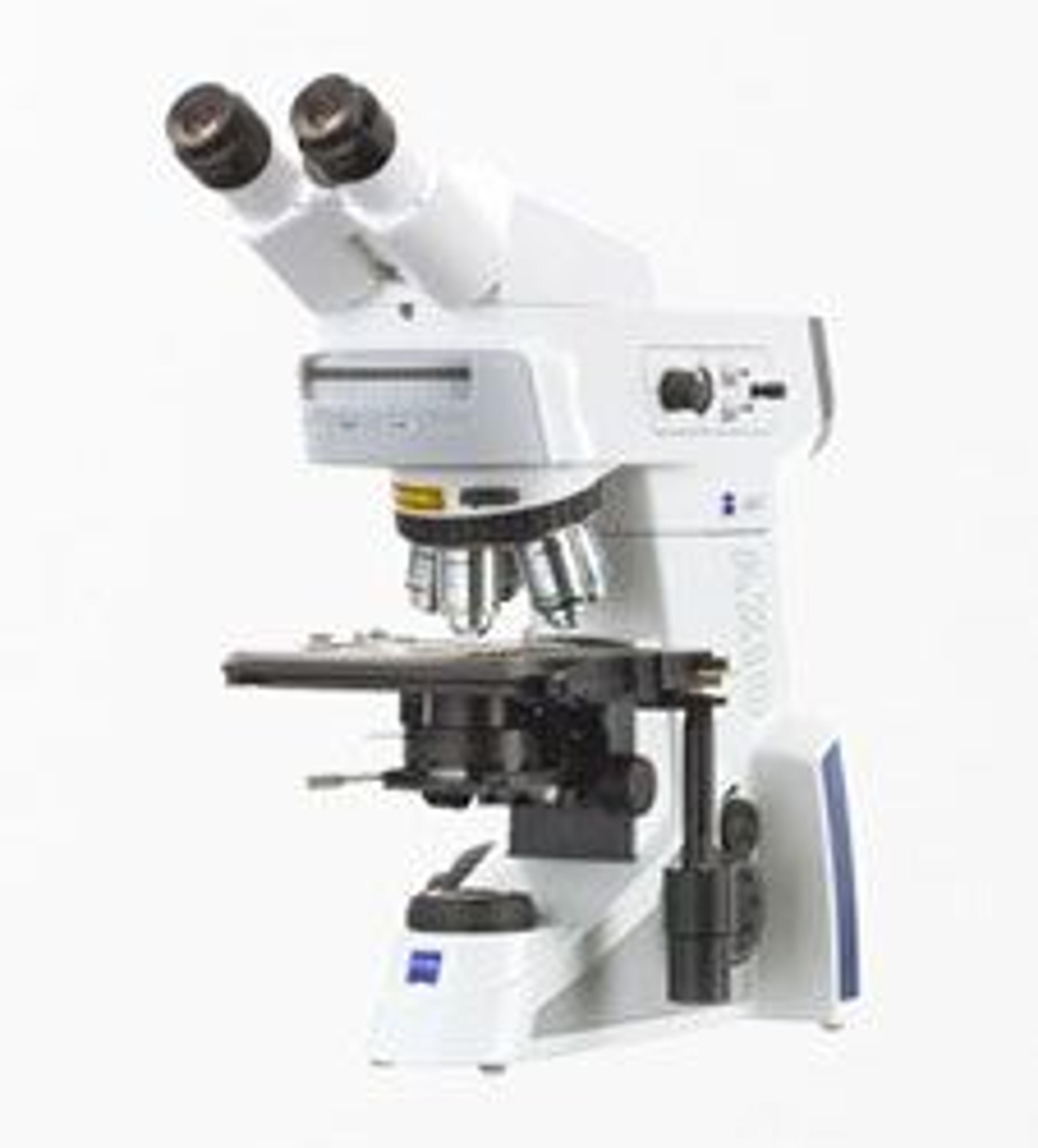
Receive your quote directly from the manufacturer.
The AI Toolkit makes integrated, easy to use, powerful segmentation for 2D and 3D datasets available to routine microscopy users. It is a complete package for AI applications including integrated training interfaces for:
- Automated image segmentation based on machine learning algorithms
- Automated object classification of segmented and analyzed images based on machine learning algorithms
- AI-based denoising of images using noise-2-void algorithms
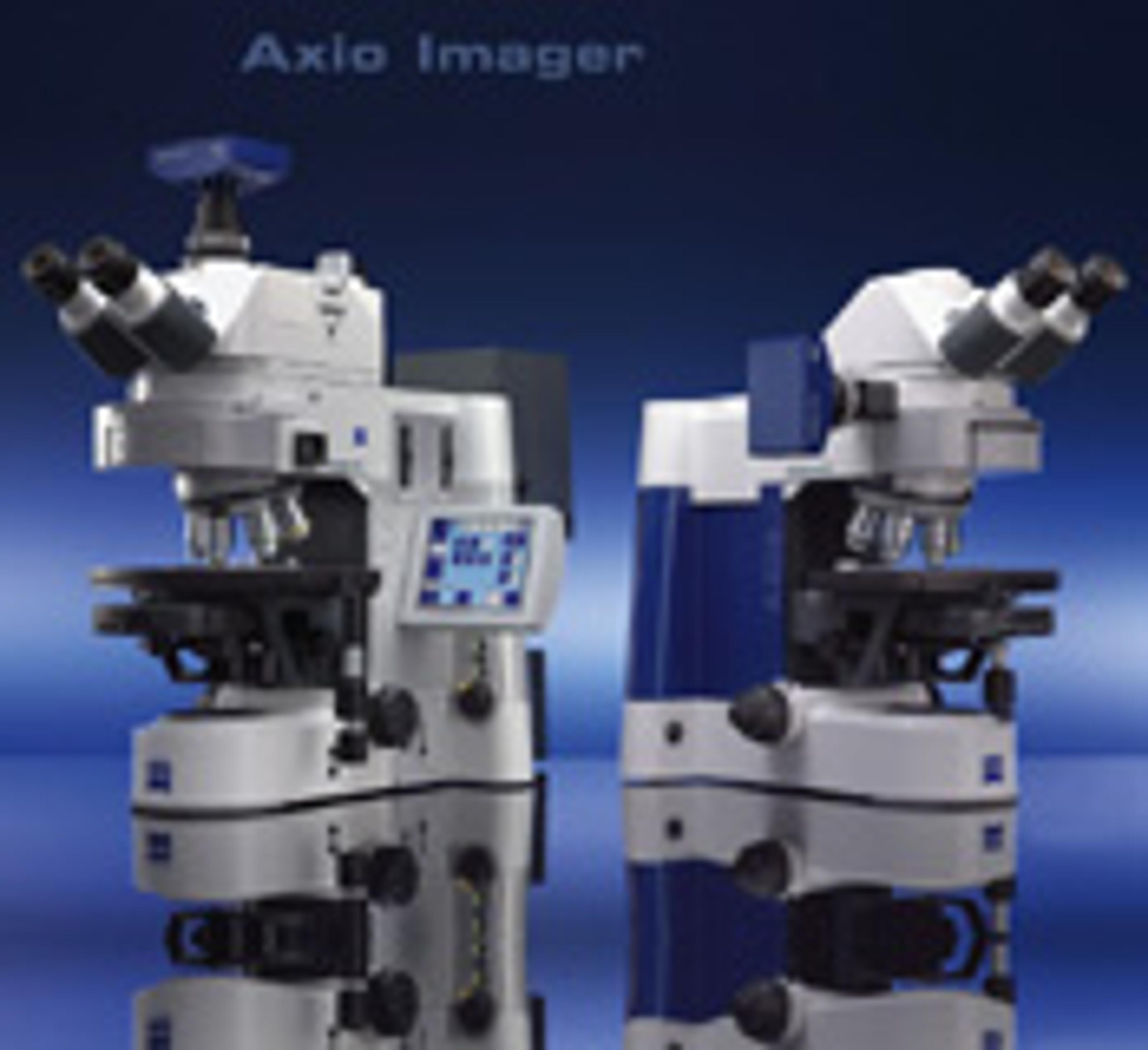
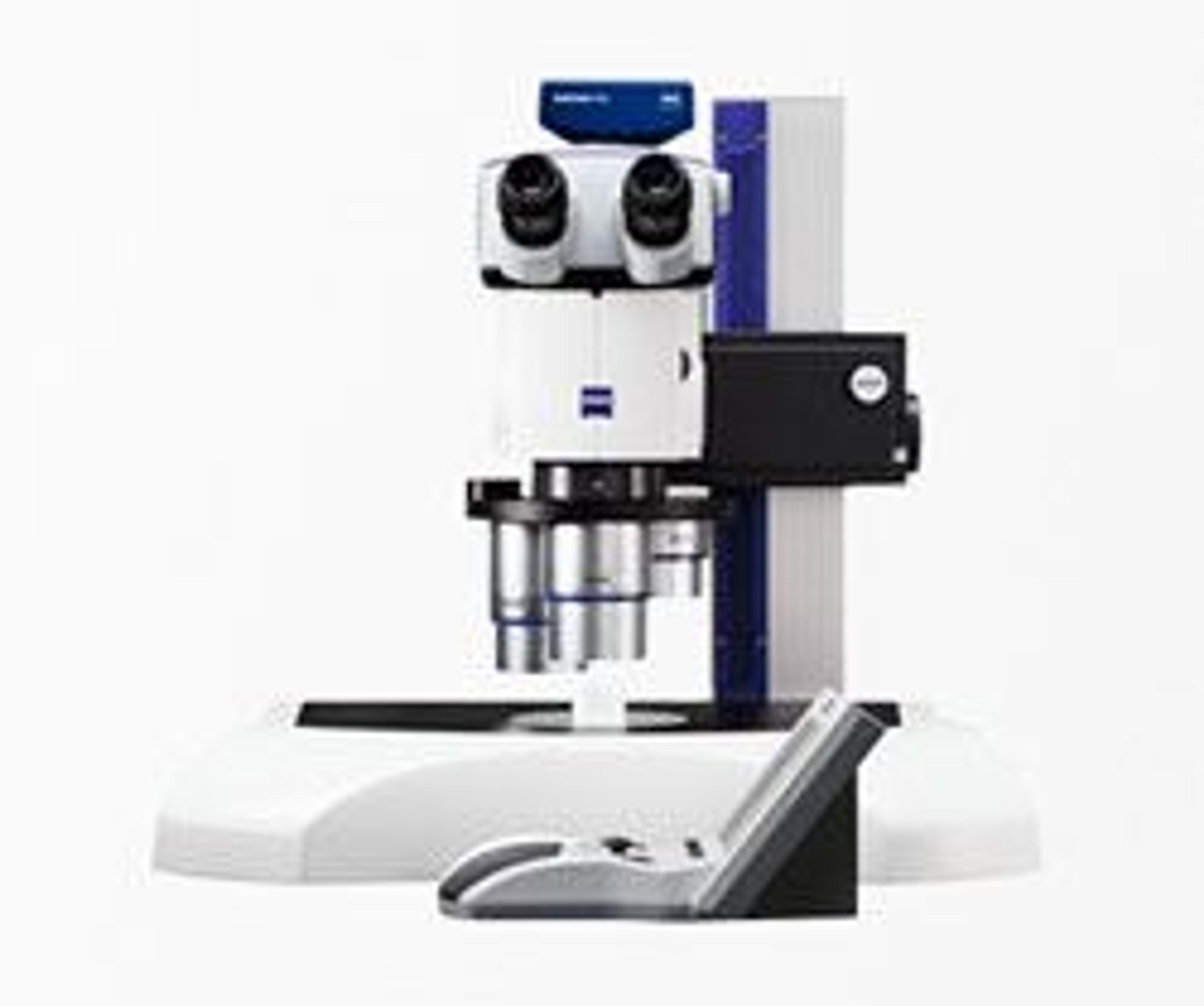
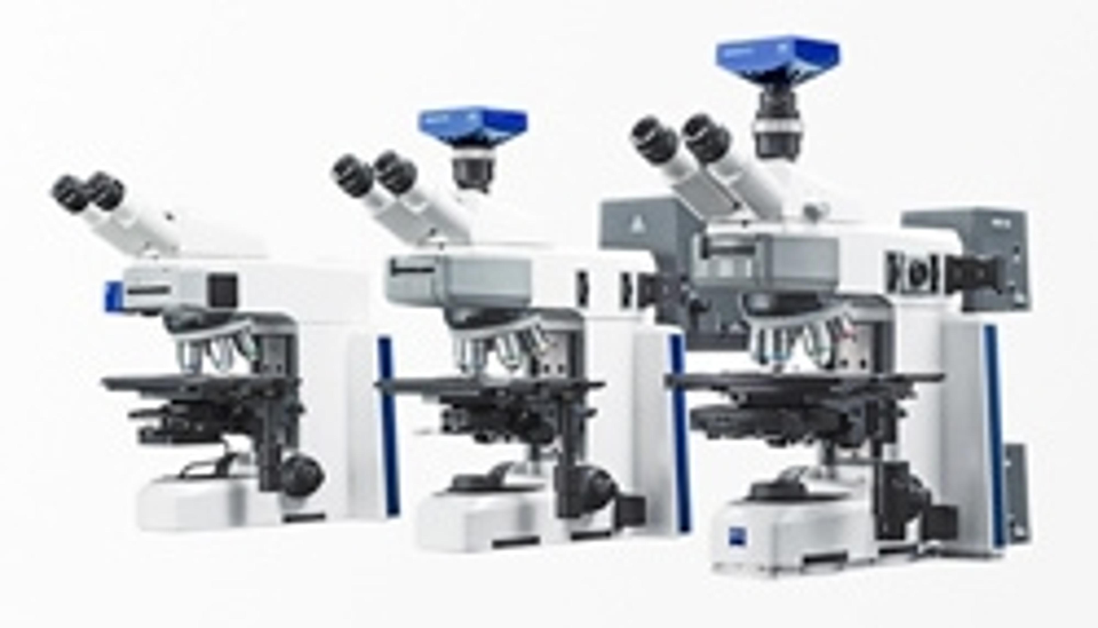
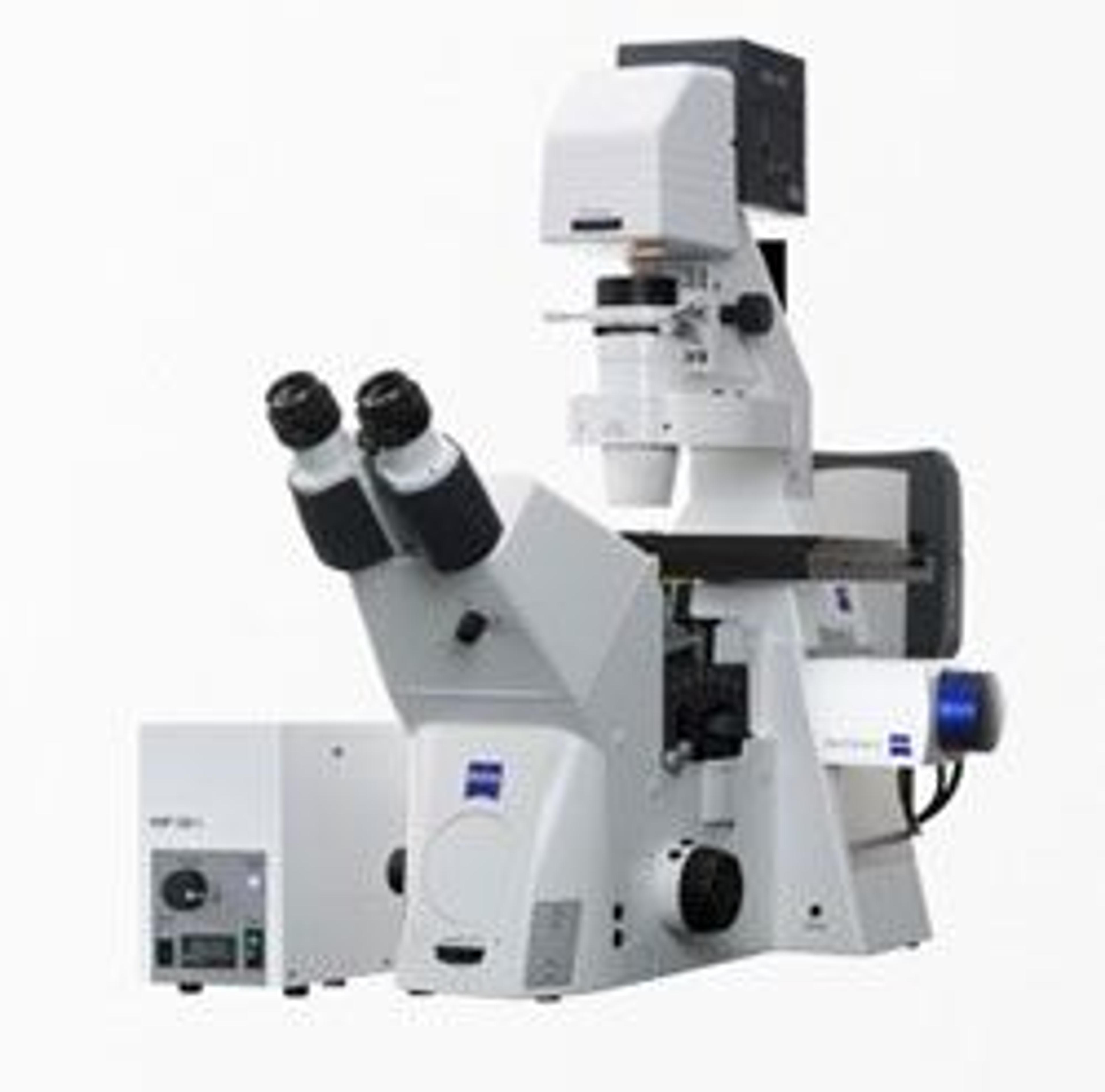
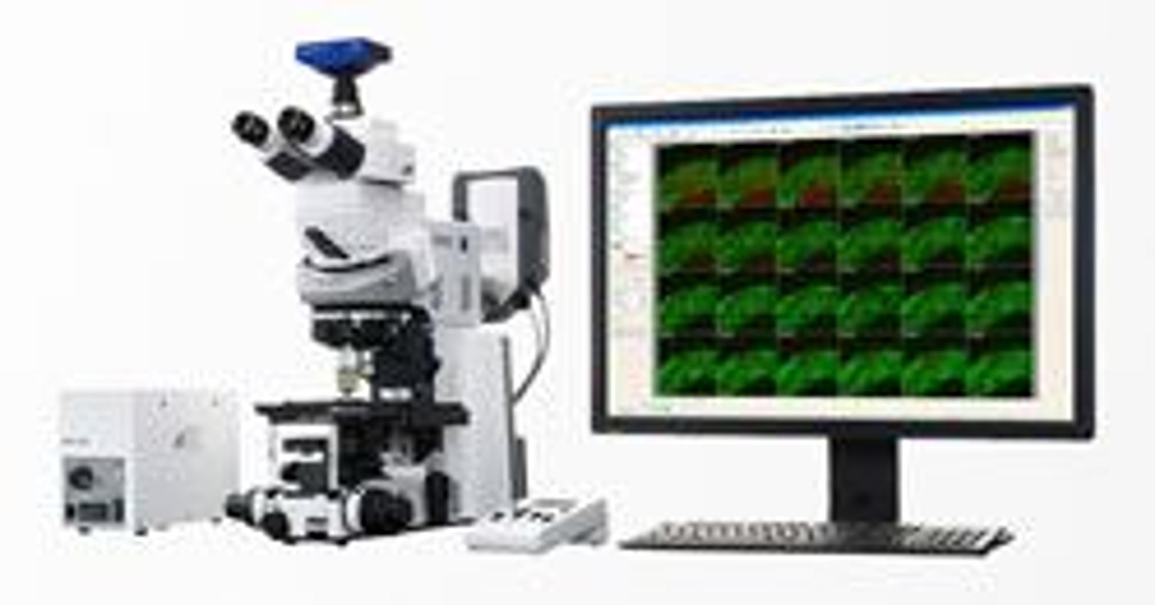
The AI toolkit is a module of ZEN core, a software suite for connected microscopy from the materials lab to production. ZEN core handles more than just microscopy imaging. It is the most comprehensive suite of imaging, segmentation, analysis, and data connectivity tools for multi-modal microscopy in connected material laboratories and lets you benefit from an easy to configure and easy to use adaptive user interface, advanced imaging, and automated analysis.
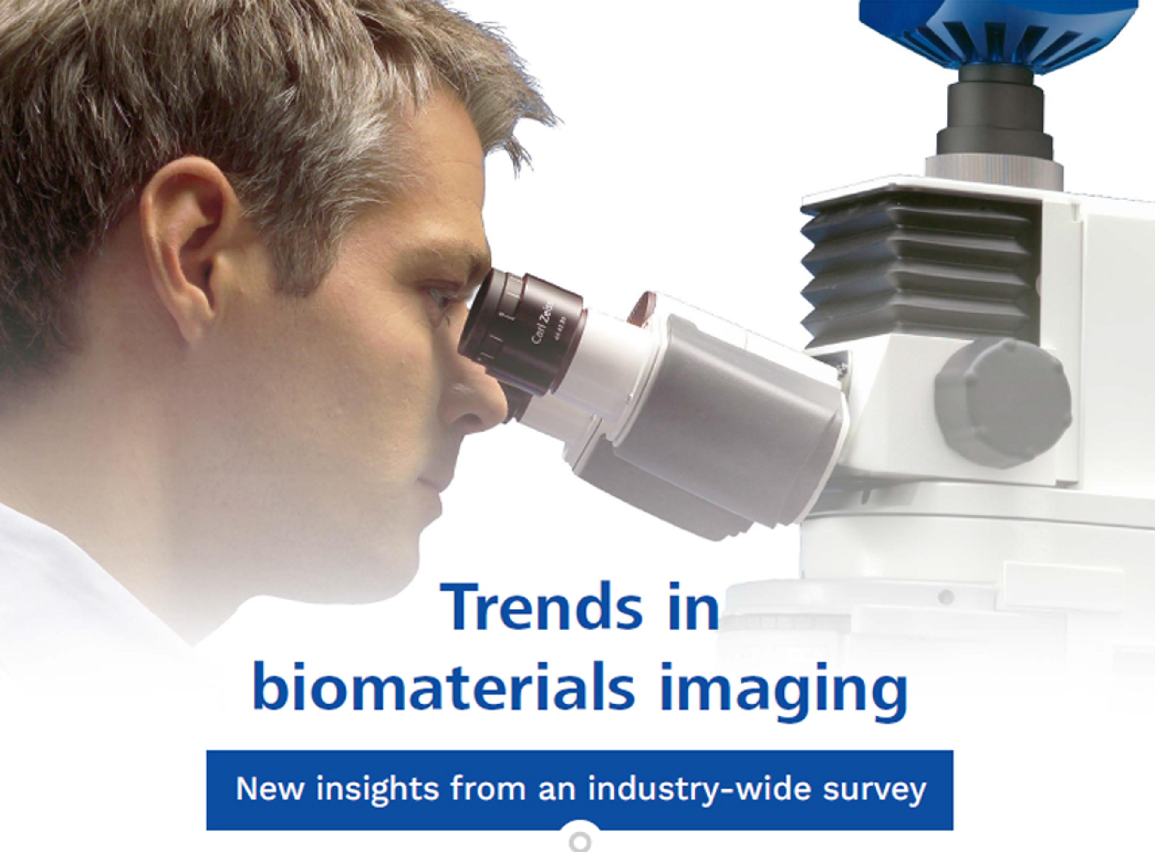
Trends in biomaterials imaging
Explore new insights from an industry-wide survey and discover answers to questions such as:
- What are the key research sectors, applications, and materials being investigated?
- What experimental outcomes are scientists looking for?
- How is constantly developing technology landscape impacting scientists?
Plus, delve into the key challenges in biomaterial analysis and the solutions that can drive your workflows forward, including automation, AI, and beyond.
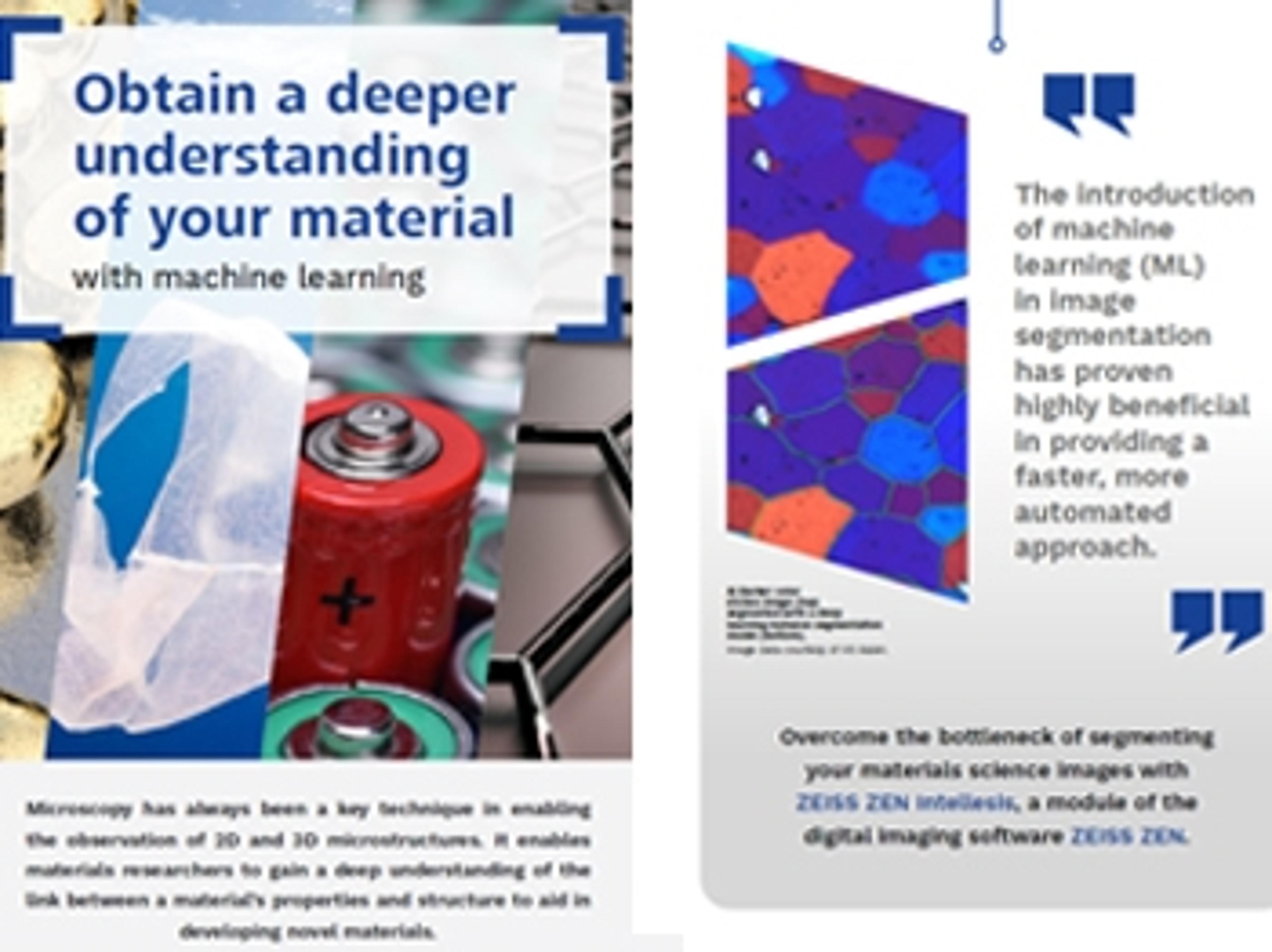
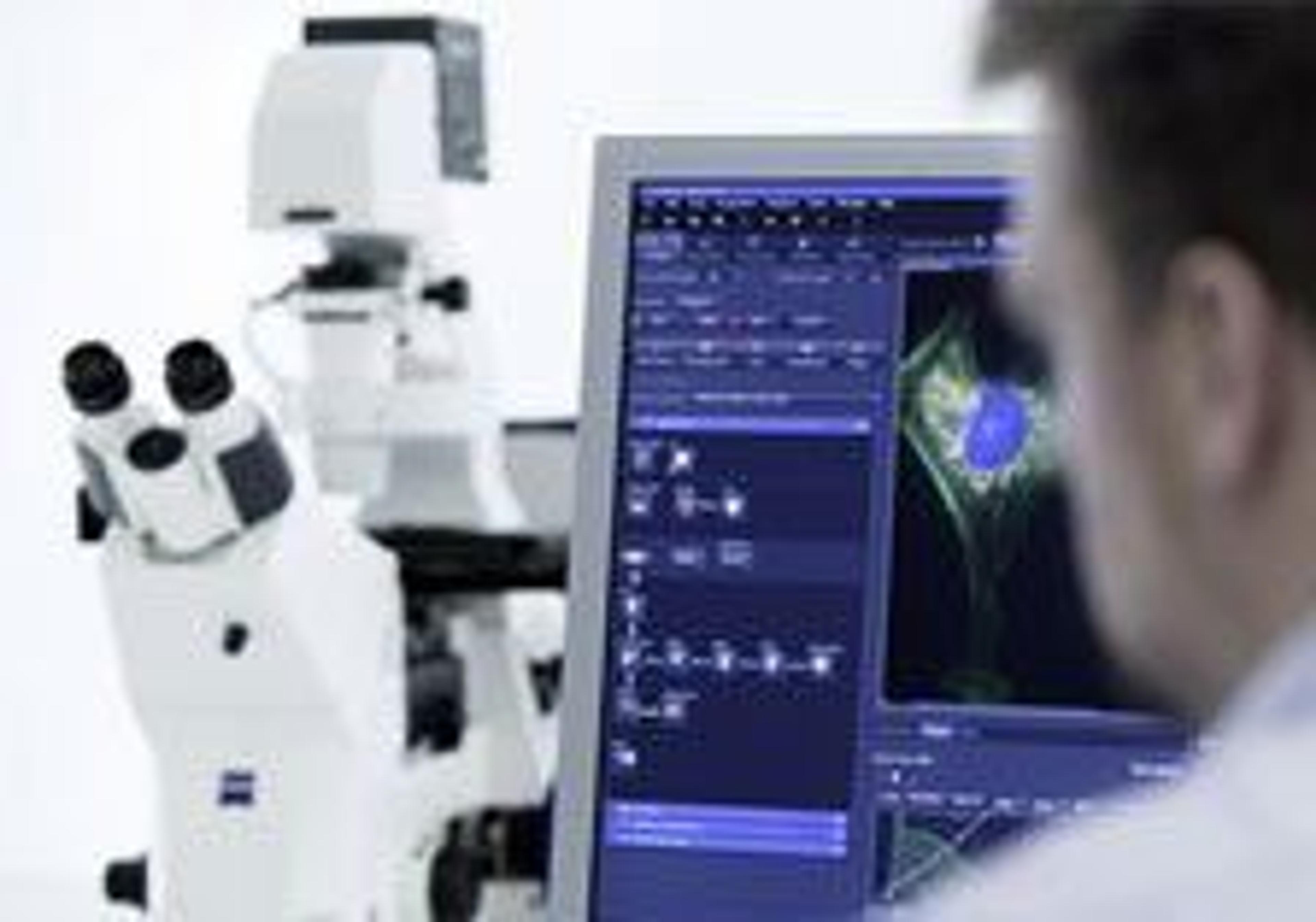
Obtain a deeper understanding of your material with machine learning
Microscopy enables researchers to gain a deep understanding of the link between a material’s properties and structure to aid in developing novel materials. Microscopy image segmentation is currently one of the biggest bottlenecks in quantifying material structure. Traditional microstructure quantification requires manual measurements and can be time-consuming and error-prone.
In this infographic, learn how the introduction of machine learning has proven highly beneficial in providing a faster, more automated approach to image segmentation. Plus, discover artificial intelligence-based software designed to enable superior segmentation and object classification.
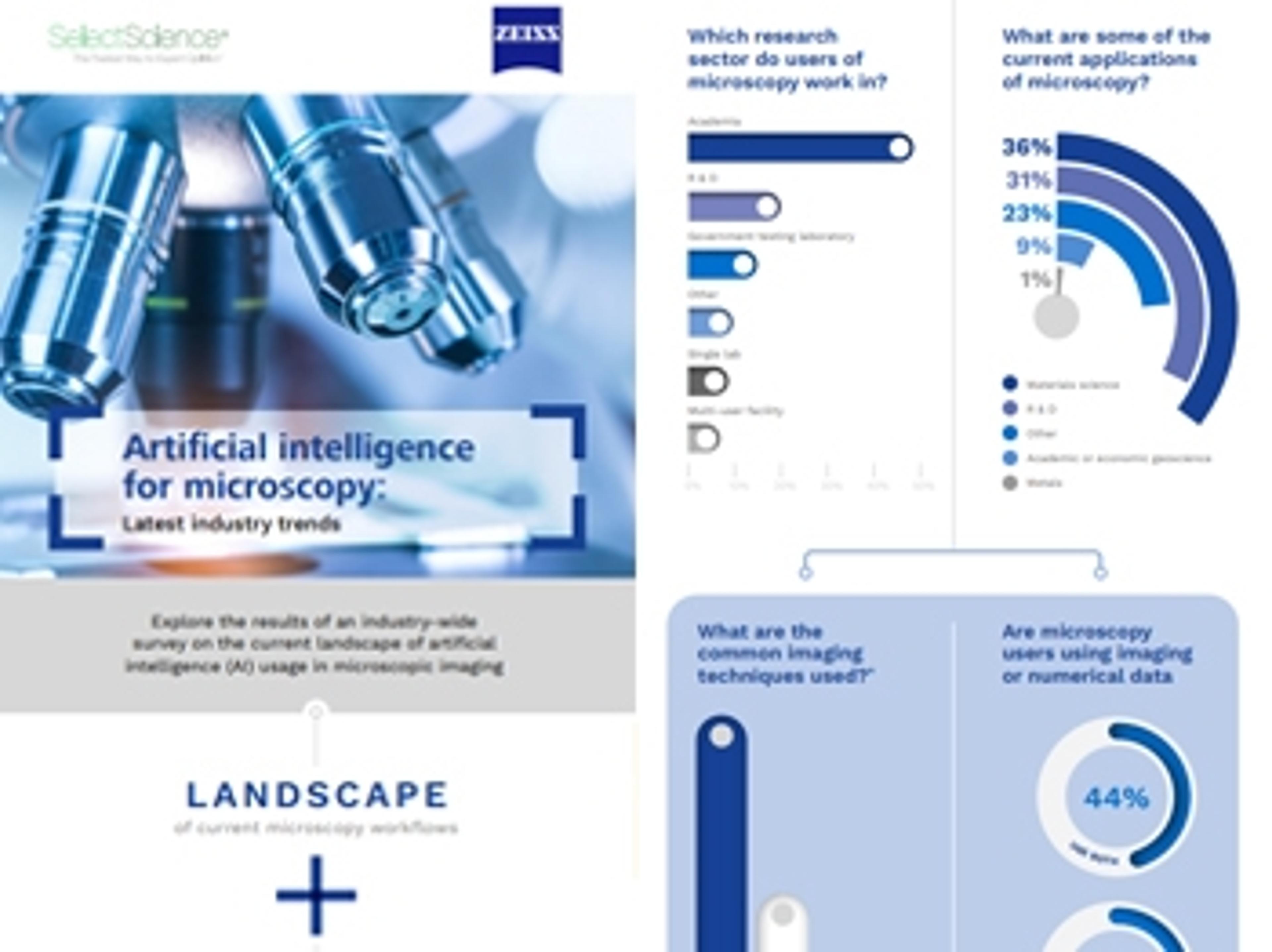
Artificial intelligence for microscopy: Latest industry trends
The introduction of artificial intelligence (AI) in image analysis has proven highly beneficial in providing a faster, more automated approach to quantifying material structure.
In this infographic, explore the results of an industry-wide survey into the current landscape of AI usage in microscopic imaging. Discover the current challenges in microscopy workflows and learn more about the common reasons why labs are choosing to invest in artificial intelligence. Download your free copy and gain insight into AI-based software solutions designed to help improve your image analysis throughput, reproducibility, and more.

Advanced Segmentation for Industrial Materials Using Machine Learning
Every quantitative analysis of a set of micrographs involves some form of segmentation. A region could be a mineral fragment, a grain in a metal, a pore in a composite, an oil contamination on the surface, a blood cell – any area differentiable from a neighboring area. By analysing these regions or the borders between regions, we get useful information. There are several standards for determining useful microstructural properties by image analysis of segmented images – e.g. for grain size, inclusion content, and porosity. These analyses, however, depend on the accuracy and reliability of the segmentation results.
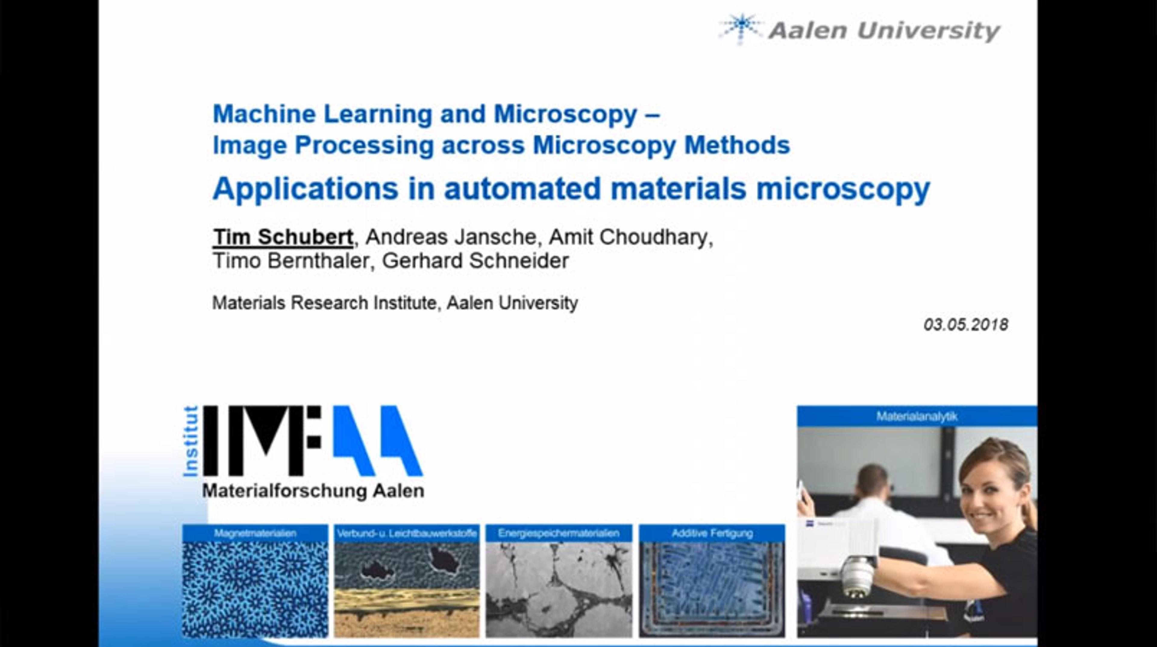
Webinar: How Machine Learning Software Can Accelerate Microscopy Image Analysis
In this webinar, find out how machine learning software, ZEISS ZEN Intellesis, can be applied to speed up and advance image processing capabilities across multiple microscopy methods. Tim Schubert (University Aalen) and Tobias Volkenandt (ZEISS Microscopy) present their latest application examples concerning image analysis and segmentation using machine learning techniques, revealing its excellent application for materials analysis via SEM, FIB-SEM, light microscopy and X-ray microscopy.
Major improvements in workflow offerings for material sciences core facilities
Promise of enhanced analytics, tomography, sample preparation and data integrity