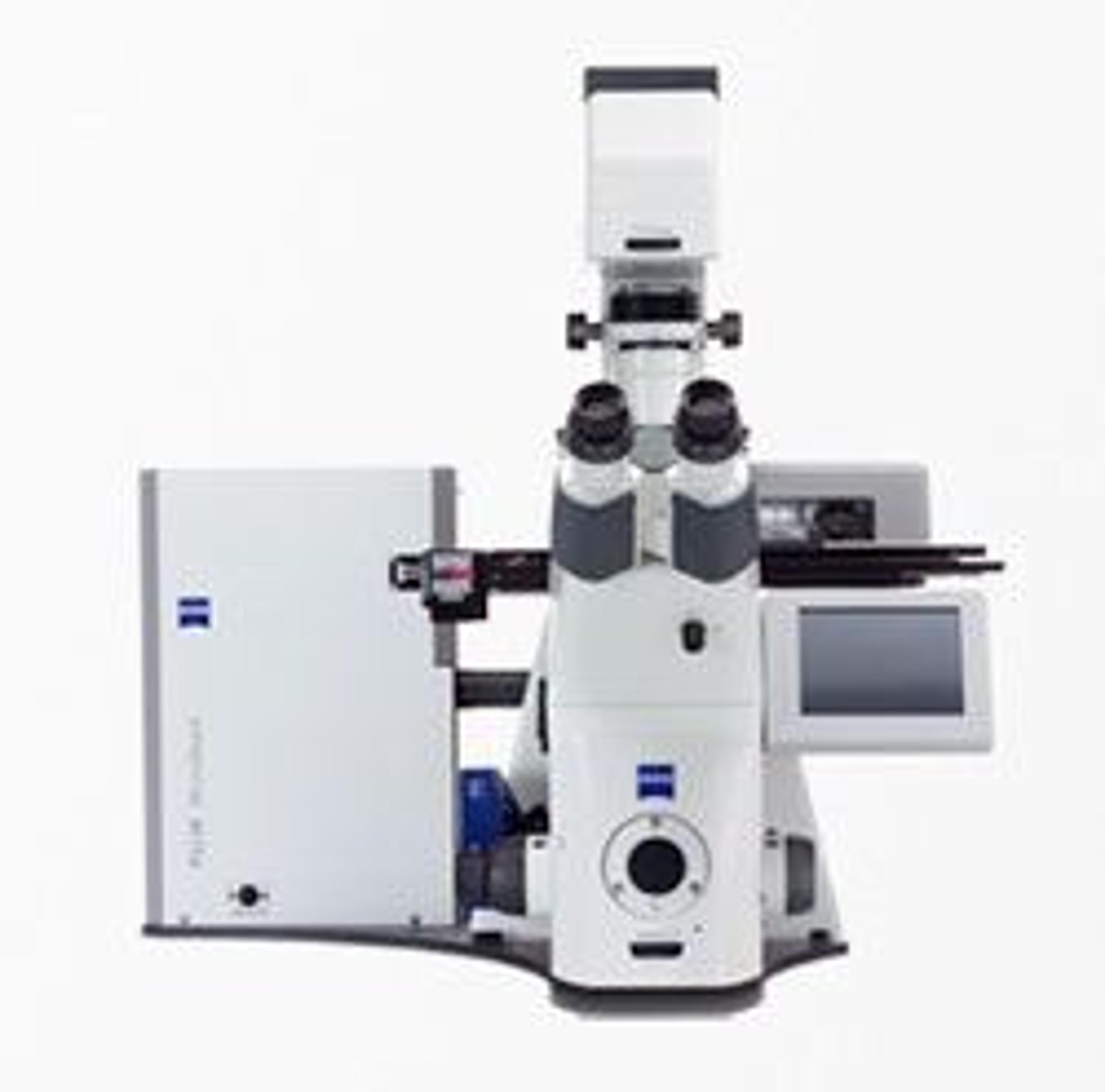
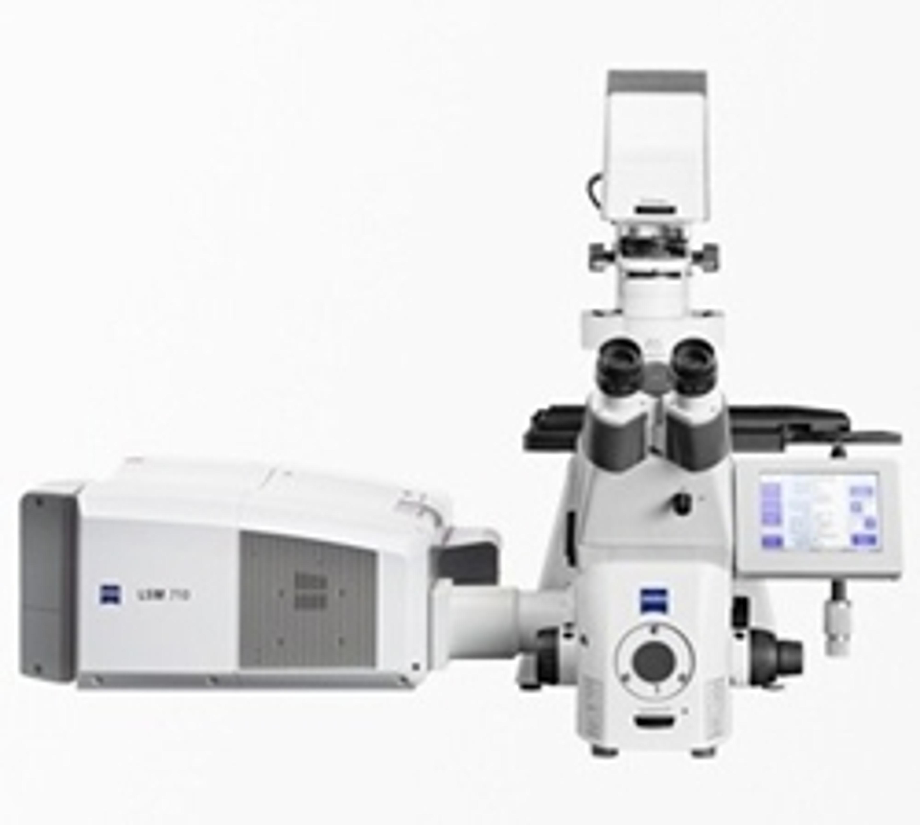
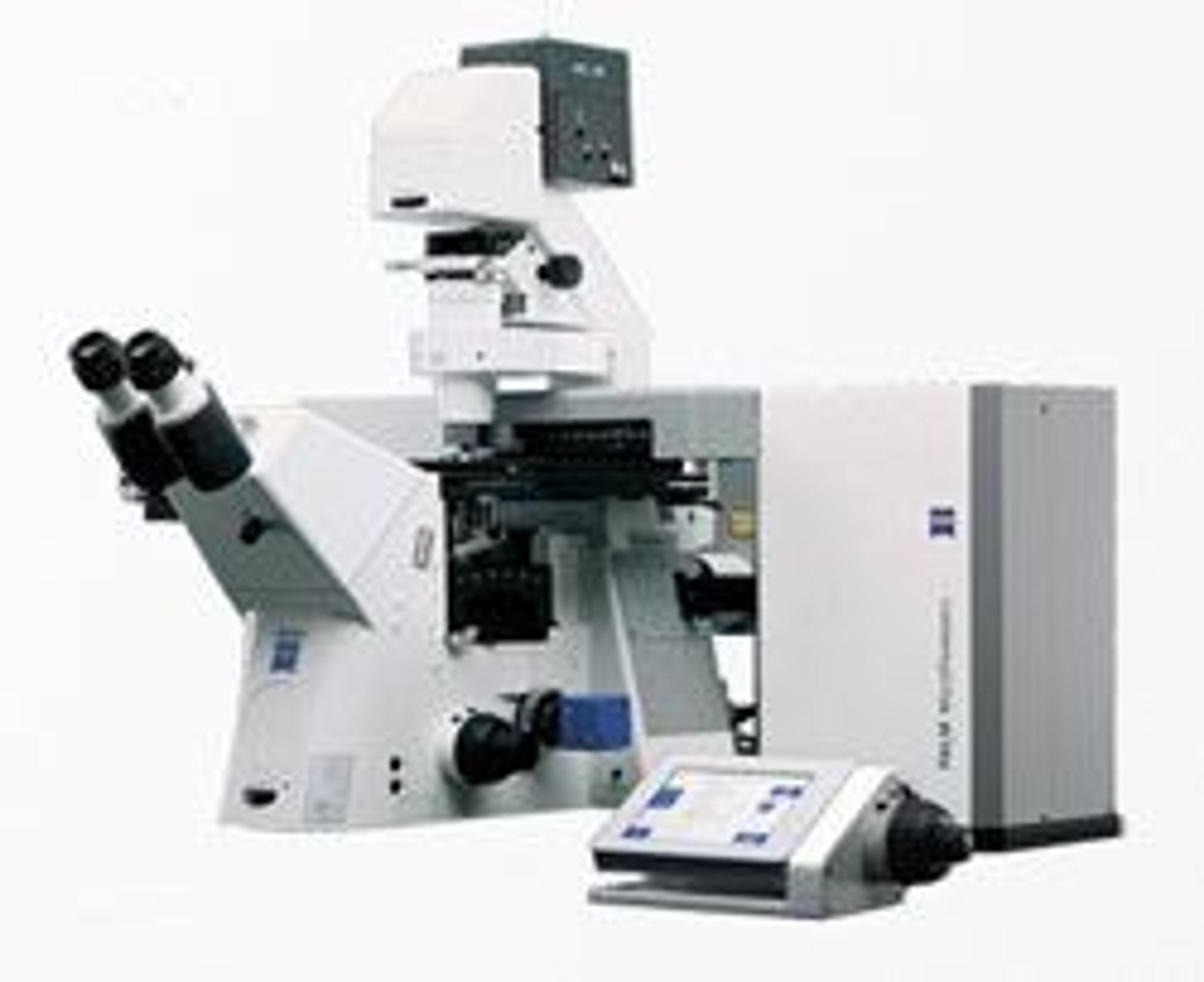
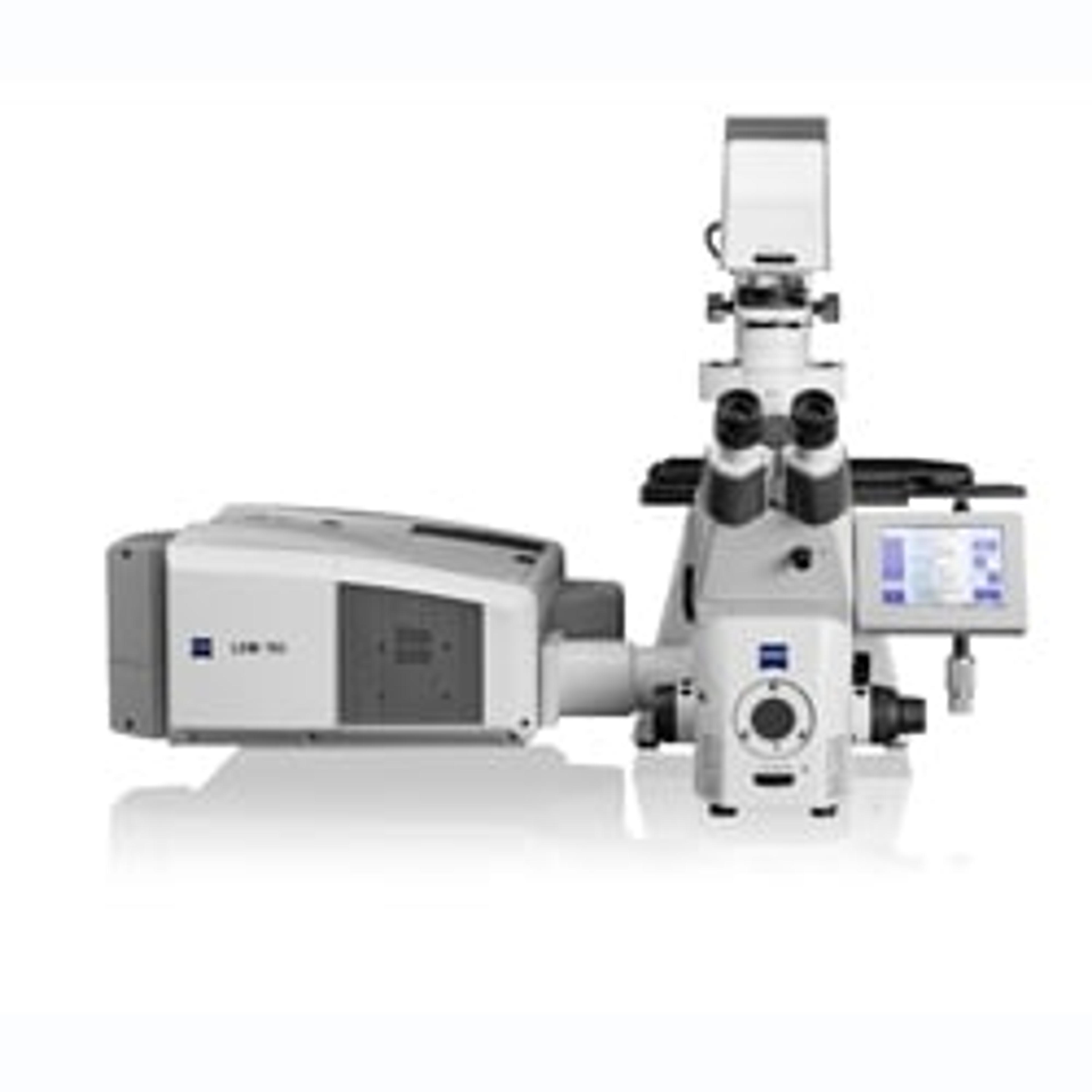
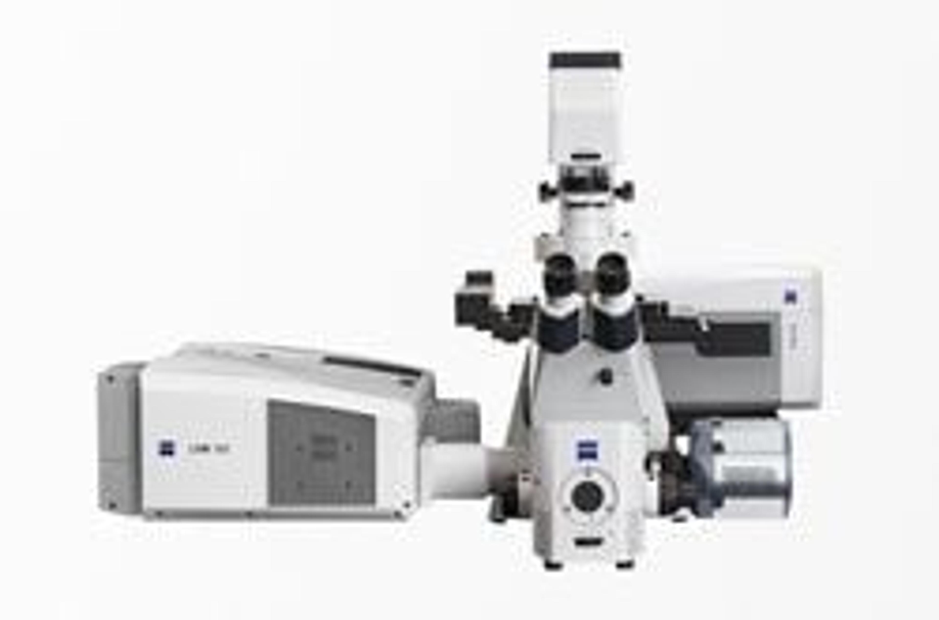
ZEISS Xradia Context microCT
Your X-ray System for Today with Assurance for Tomorrow.
Image large, heavy (25kg), and tall samples in their full 3D context, as well as small samples with high resolution.
Your X-ray System for Today with Assurance for Tomorrow.

ZEISS Xradia Context microCT

Image large, heavy (25kg), and tall samples in their full 3D context, as well as small samples with high resolution.


Axio ImagerAn innovative modular system for digital fluorescence microscopy, featuring advanced flexibility and application versatility. Featuring: New IC2S objectives (Infinity Contrast & Colour Corrected System) - optimise image quality and maximise contrast Special fluorescence filters - reduce exposure and image acquisition time for superior 3D imaging. 'Intelligent Stand' - automatically recognises added components. "Contrast manager" ensures simple changes between contrasting techniques. Integration: standard interfaces permit communication via USB and TCP/IP Vibration-free Imaging Cell - isolated within the stand for stable observation and unparalleled precision. Apochromatic fluorescence beam path - ensures optimum colour correction Active stray light elimination - significantly higher contrast Fast, motorised reflector turrets - hold either six or ten filter modules. HBO lamp: convenient, self-aligning homogeneous illumination LCI Plan-Neofluar objectives - live cell imaging Flexibility - two freely configurable stand variants: Z1 (motorized) and D1(manual) or preconfigured stands M1 (motorized) and A1 (manual). Rapid image acquisition with outstanding quality in up to 6 dimensions means more signal in less time.

PALM MicroBeam uses a patented process to capture even the most demanding specimens – from cryosections, FFPE materials, native tissue like fresh plants, live cells and chromosomes – without contamination. The complete workflow for your laser microdissection research PALM MicroBeam makes isolating uncontaminated source material simple. Precise detection, laser microdissection and patented laser transport allow you to obtain the homogenous analysis material necessary for meaningful scientific results. Because analyses of gene expression patterns rely on exactly-separated analytical material, unwanted cells may alter your results and conceal the signals of the relevant cells. PALM MicroBeam prevents this by allowing you to define cells and tissue regions precisely, ensuring your results are exact and reproducible. Highlights of this technology • Suitability for laser microdissection and analysis for DNA, RNA and protein isolation – from archive material or live cells • A system that completes the step from laser-microdissection to integrated imaging workstation • Applicable to cryosections and FFPE tissue • Performing LCM even from standard glass slides • Extendibility – add digital cameras from the AxioCam camera range • Ability to integrate additional components into MicroBeam and create simple workflows, for individual experiments or automation with flexible collectors • Calibration routines that allow you to easily adjust MicroBeam for laser parameters appropriate to your chosen specimen

Experience freedom with your dyes and applications The new illumination and detection design of LSM 710 brings complete freedom to your fluorescence microscopy. You work with up to ten dyes and use continuous spectral detection across the complete wavelength range. LSM 710 enables confocal microscopy for a wide variety of applications. With the inverse Axio Observer from Carl Zeiss, LSM 710 offers you unrivalled confocal microscopy in cell and developmental biology. Upright stands such as Axio Imager or Axio Examiner offer you have all the equipment you need to record neurobiological, physiological and developmental relationships to an exceptional standard. LSM 710 gives you flexibility, allowing you to perform confocal microscopy in exactly the way you need. Features: Oversampling with 30 percent longer sampling time per pixel for low noise Excellent contrast characteristics, even with strongly reflective samples – thanks to laser suppression that has been improved by 100-1000x A huge range of QUASAR detector configuration options – to meet your requirements The innovative grid and spectral recycling loop design ensures that not even a single valuable photon will be lost from your sample. Up to 80 percent dark noise reduction – thanks to the QUASAR array detector by Carl Zeiss 34-channel parallel imaging – across the complete wavelength range APD imaging with photon counting Low noise electronics and lasers

PALM MicroTweezers guarantee simple and precise micro-manipulation With PALM MicroTweezers you manipulate cells and particles in the micro- and sub-micrometer range without contact. The highly focused laser beam allows you to trap, move and sort live cells, organelles and other large biomolecules. A simple mouse click on your target is sufficient to trap it. You are able to complement PALM MicroTweezers with the Force Measurement software module and with high-end imaging and supplementary technologies from ZEISS, such as live cell imaging systems or PALM MicroBeam for laser microdissection. • Contact-free, non-invasive sample manipulation in 3D • Trap, position or sort microscopic objects • Intuitive dual-beam operation with click & trap functionality • Combined micromanipulation and high end imaging workstation • Compatible with other Zeiss solutions such as PALM Microbeam • Applications range from micromanipulation, cell-cell interactions, cell sorting to quantitative force measurements

LSM 780 - Obtain Quantitative Information About Your Cells and Individual Molecules. The sensitivity of LSM 780 is quite simply outstanding. The GaAsP detector achieves 45 percent quantum efficiency compared to 25 percent typically by conventional PMT detectors. This results in accurate details and contrast-rich images of the challenging specimens you encounter in your live cell imaging.The system’s illumination and detection design allows you to simultaneously acquire up to ten dyes. You excite any common fluorophore with up to eight different lasers, detecting the signals with the 32 channel GaAsP detector. LSM 780 is so sensitive, the system even allows photon counting.The maximum possible acquisition speed available to you is determined by the sensitivity of your detection system. When you choose LSM 780, you benefit from new GaAsP detectors that bring a significant improvement on signal-to-noise ratio, allowing you to acquire your image sequences at least twice as fast.Parallel spectral detection also reads 34 channels simultaneously in Lambda mode: you acquire and separate up to ten colors simultaneously.LSM 780 allows you to achieve images of unparalleled detail, even from the most challenging specimens. The superior quality and sensitivity has been recognized by a distinguished R&D 100 Award in 2011.LSM 780 Laser Scanning Microscope Features: 32-channel GaAsP detector with a typical quantum efficiency of 45% – a significant improvement on the 25% typically achieved by classic PMT detectors. Potential for photon counting on GaAsP and the new side PMT. With lasers from 355 nm true UV to 660 nm far red, any dye will be excited optimally. Simple adaptation to changing acquisition strategies via the ZEN software’s Smart Setup. Improved ZEN software with modules for e.g. correlative microscopy, deconvolution and time lapse. Improved dynamics for better visualization of weak signals. Active cooling together with oversampling and photon counting modes deliver improved signal-to-noise ratio.

Your Flexible Imaging System for 3D Superresolution Microscopy.

Your modular stereo microscope with motorized 20x zoom.

Your modular microscope for research and advanced routine.

Examine and Evaluate Living Cells - Fast and Efficiently.

Colibri.2 The LED Light Source for Fluorescence Microscopy that is Gentle on Your Samples With the Colibri.2 choose out of 13 LEDs to cover the complete standard spectrum from UV to dark red. Adjust each illumination intensity depending on the sensitivity of the sample. The narrow-band illumination reduces crossstimulation, increasing the sensitivity of your images. There’s no UV leakage of the sort you encounter with filter-based systems with and conventional white light sources like HBOs. Profit from an excellent signal-background ratio for imaging. Thanks to the high dynamic range of the images you detect the finest details. Colibri.2 Highlights • Complex Images – Adjusted in Microseconds: Switch on or off each LED. Continuously adjust their intensity in just microseconds. Direct the process via a control panel or the system control software. Or use an integrated trigger function to synchronize the imaging process for real-time applications. Achieve complex images for investigating the fastest processes in a way that’s simple and easy to reproduce. • Optimum Results - LED-optimized Filters and Direct Coupling Increase Sensitivity: Colibri.2 uses a coupled light source that ensures homogeneous illumination at the same time. Achieve the desired narrow spectral bandwidth for any examination using additional filters. Profit from the ideal match of the LED spectrum to the filter set. From single band to quad band filters. Fibre optics that are susceptible to wear are now a thing of the past. • Low Cost Light Source – Reliable Long Life LEDs: Long-life LEDs keep running costs low. Do away with the tiresome job of changing and adjusting high-pressure lamps (HBO/XBO/).

LSM 700 rivals high end confocals with superb image quality Perform fluorescence measurements, create optical sections of your samples and combine them into 3D image stacks. ZEN imaging software shortens the training period and stores the settings for all users in your team. Smart Setup lets you configure new experiments easily with optimized recording settings. The compact design of LSM 700 means you will always have room for it in your work environment. Combine your LSM 700 with the upright microscope stand Axio Imager 2, inverted Axio Observer or the fixed stage microscope Axio Examiner for complete flexibility in your live cell imaging or photomanipulation applications.

ZEISS Axio Observer for Life Science Research (eom)

Your fixed stage research microscope.
