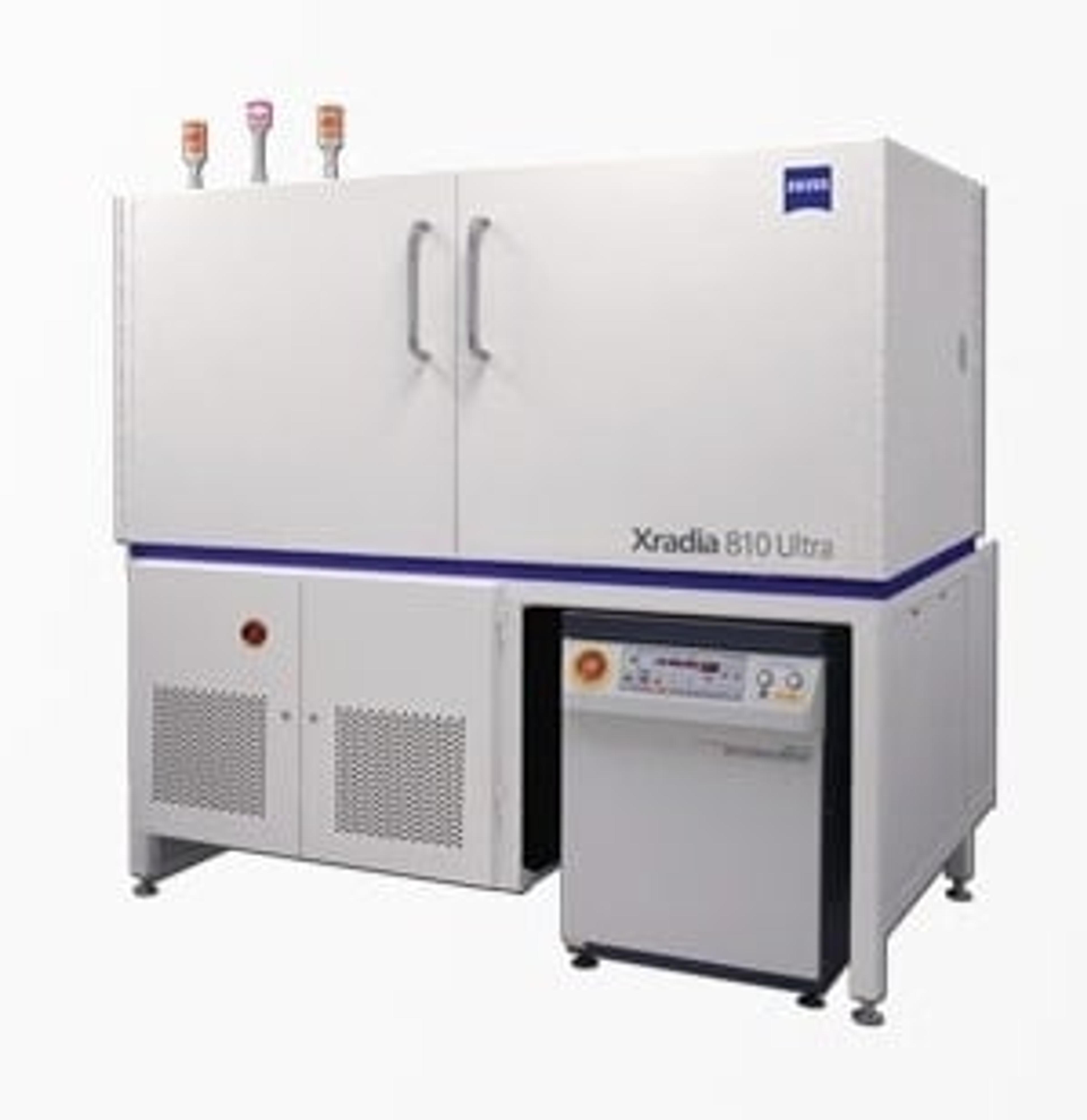
ZEISS Xradia 810 Ultra
Extending the reach and value of non-destructive nanoscale imaging


Receive your quote directly from the manufacturer.
Achieve spatial resolution down to 50 nm with ZEISS Xradia 810 Ultra X-ray microscope, the highest among lab-based X-ray imaging systems.
Experience unparalleled performance and flexibility with the non-destructive 3D imaging that plays a vital role in today’s breakthrough research. The innovative Xradia Ultra architecture, with unique X-ray optics adapted from synchrotron technology, features absorption and phase contrast. Now with energy at 5.4 keV you can increase the throughput of your nanoscale imaging by up to a factor of 10. Achieve even better contrast and image quality for medium to low Z samples with the lower energy of Xradia 810 Ultra.
Expect to accomplish advanced in situ and 4D capabilities for studying structural evolution over time and under varying conditions. Extend the limits of exploration with 3D X-ray imaging for materials research, life sciences, natural resources, and diverse industrial applications.

Characterization of the 3D microstructure of nanofibrous scaffolds for tissue engineering
In this application note, ZEISS presents X-ray microscopy (XRM) characterization of cross-linked electrospun gelatin nanofibers. These nanofibers are potential candidates for applications like nanofibrous scaffolds for skin regeneration and wound dressings in medical care. The note delves into the unique phase-contrast techniques, specifically using the Zernike phase plate, applicable to low atomic number materials using the laboratory-based X-ray microscope ZEISS Xradia 810 Ultra.
In situ 3D imaging of crack growth in dentin at the nanoscale
Dentin is a nano-composite material which forms the majority of the mineralised tissue in teeth. A better understanding of fracture in dentin is important to develop a framework for failure prediction, not only for clinical understanding but also for developing biomimetic restorative materials that are able to mimic the tissue’s mechanical response. ZEISS shows how a novel in situ nanomechanical test stage for the ZEISS Xradia Ultra nanoscale X-ray microscope could be used to initiate and propagate cracks in elephant dentin (tusk) during tomography. This enabled the progressive crack growth to be studied in 3D and in situ (under load) at 150 nm resolution for the first time. The results can provide new insights into anisotropic fracture behavior and crack shielding mechanisms.
Microscopic characterization of polymer fibers with mechanical properties similar to dragline spider silk
In this application note, ZEISS presents the microscopic characterization of synthetic fibers using ZEISS field emission scanning electron microscopy (FESEM) and X-ray microscopy (XRM). This analysis aids in optimizing the manufacturing process of strong and tough polymer fibers. Spider dragline silk, known for its exceptional mechanical properties, is explored. While it surpasses steel in strength and toughness on an equal weight basis, it is also lighter, thinner, and more flexible. If commercially harvested, spider silk could revolutionize various high-tech, commercial, and consumer applications, including bridge cables, aerospace components, biodegradable water bottles, and antimicrobial medical devices due to its superior properties.
Diffraction contrast tomography
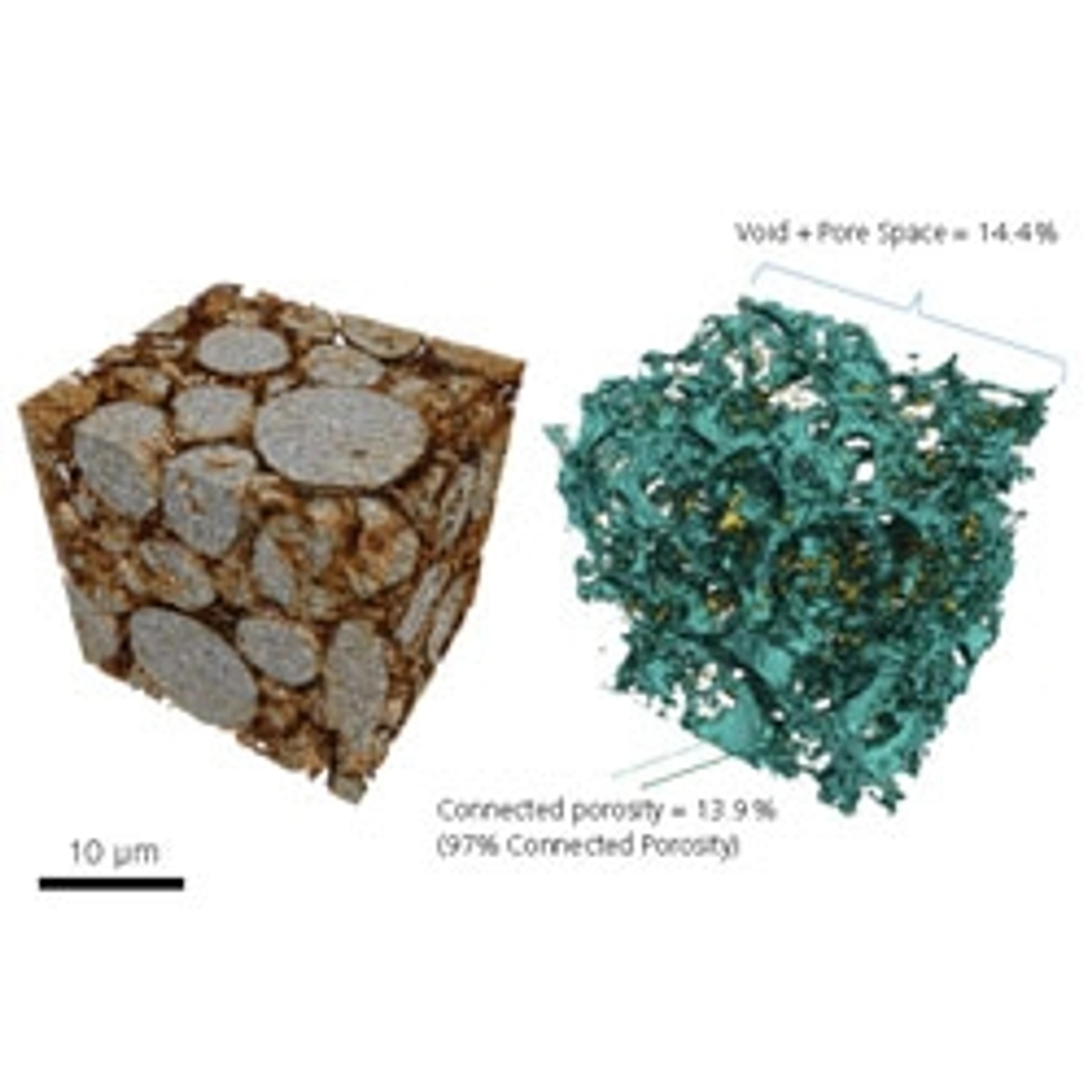
In this application note, ZEISS provides an overview with experimental data on diffraction contrast tomography. X-ray tomography has operated under two primary contrast mechanisms for some time: X-ray absorption and phase contrast, which both rely on material density differences within the sample. However, single-phase polycrystalline materials (e.g. steels, alloys, etc.) do not exhibit any significant contrast using absorption or phase mechanisms. Synchrotron-based X-ray microscopy (XRM) has demonstrated results in this area for about a decade with diffraction contrast tomography (DCT), which provides crystallographic/diffraction information from poly-crystalline samples, non-destructively, in 3D.
Microstructural Investigation of Austempered Ductile Iron (ADI) with "Shuttle & Find"
Austempered ductile iron (ADI) excels through strength, wear resistance and toughness – characteristics that make ADI the material of choice for use in combustion engines and gear box components. This means that safety aspects are also involved in addition to purely functional aspects. For this reason, changes in the ADI production process need to be monitored with respect to the material's characteristics and must be optimized systematically. For the micro- and nanoscopic analysis of the structure and precipitations, scientists typically use both light and electron microscopes. To date, however, there has been no possibility of relocating regions of interest without doubt when transferring the sample from the light to the electron microscope or vice versa. "Shuttle&Find" – the interface for correlative microscopy in materials analysis — offers an easy-to-use solution, enabling seamless integration of these two complementary technologies for the first time.
Biological Sample XRM Imaging from Selected Publications
The new field of 3D X-ray microscopy (XRM) brings dramatic resolution and contrast improvements to X-ray tomographic imaging of biological specimens for correlative studies and hierarchical structure investigations of hard and soft tissue. This document is a compendium sampling the vast diversity of biological samples and preparation methods published in literature by researchers utilizing ZEISS X-ray microscopes for their research.

Non-destructive characterization of electrospun fibers with 3D high-resolution X-ray microscopy
Watch this on-demand webinar to gain an understanding of the benefits of phase-contrast imaging for characterization of biomaterials

Analyzing Catastrophe: Why Lithium Ion Batteries Explode [and How to Prevent it]
The insightful work of Professor Paul Shearing
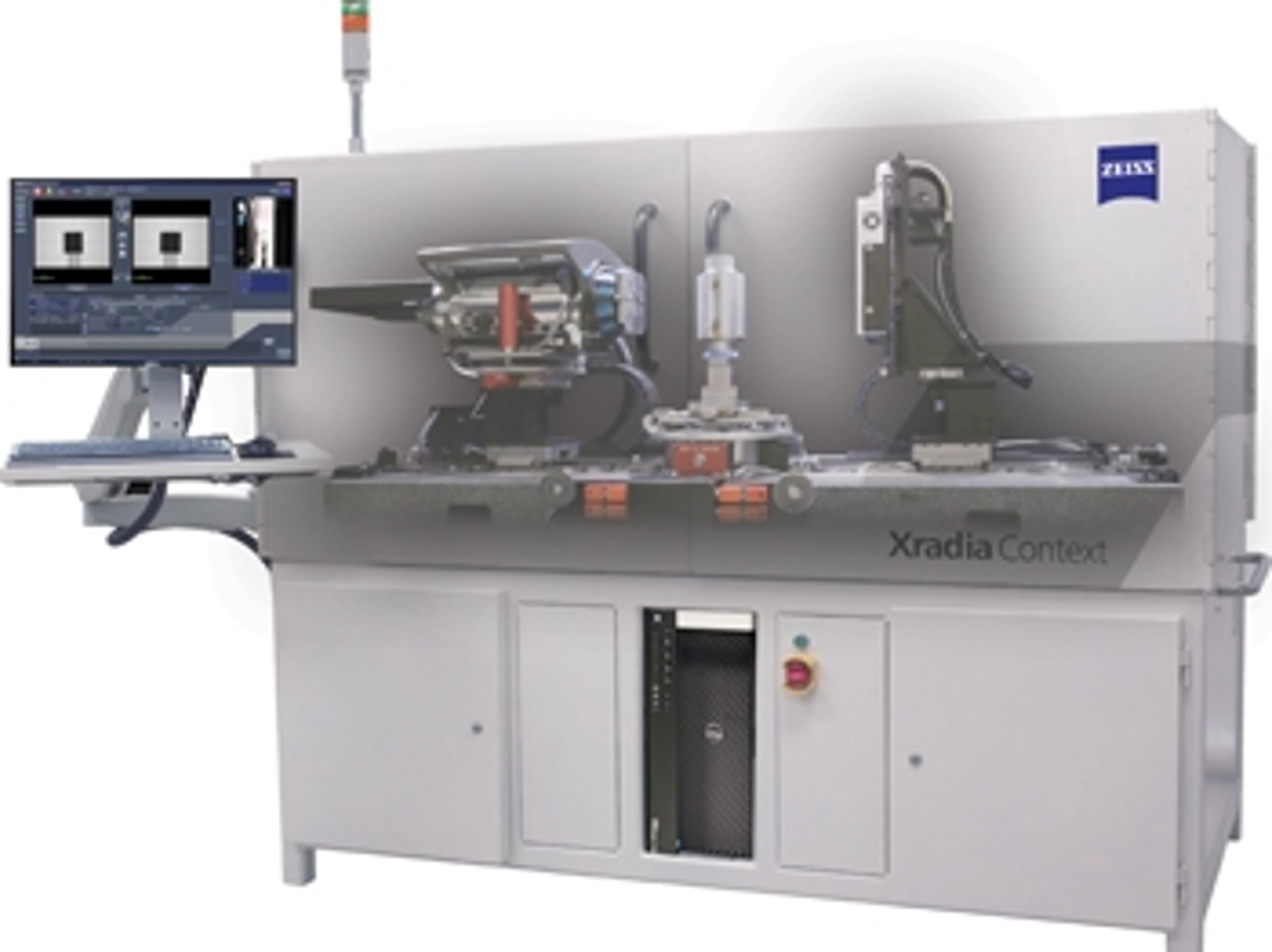
ZEISS Reveals New X-ray Imaging Instrument: ZEISS Xradia Context microCT
X-ray imaging instrument is built on renowned ZEISS Xradia platform and field-convertible to ZEISS Xradia Versa X-ray microscope
Advancing the Next Generation of Batteries
University College London research team uses ZEISS Xradia 810 Ultra X-ray microscope to study batteries and fuel cells