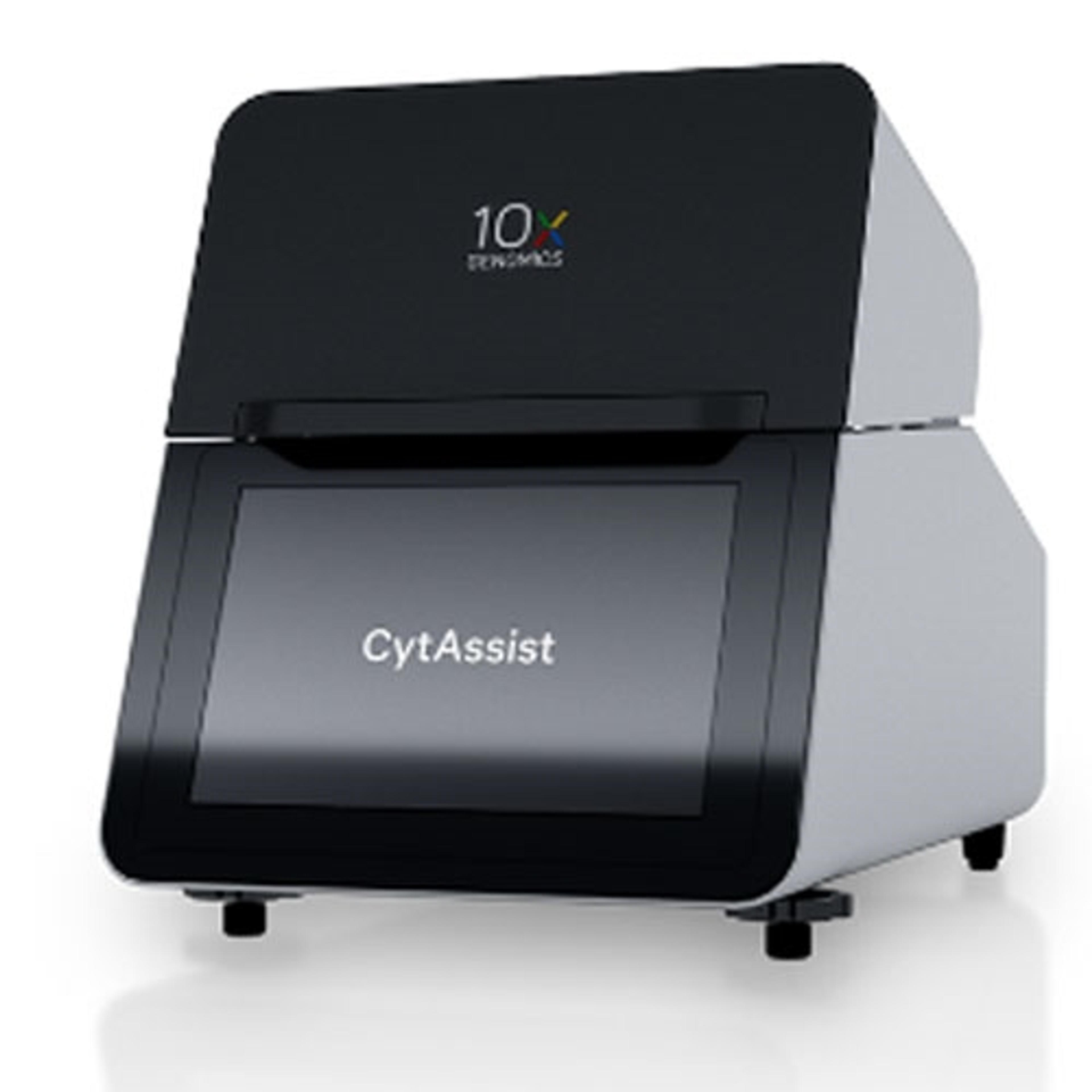
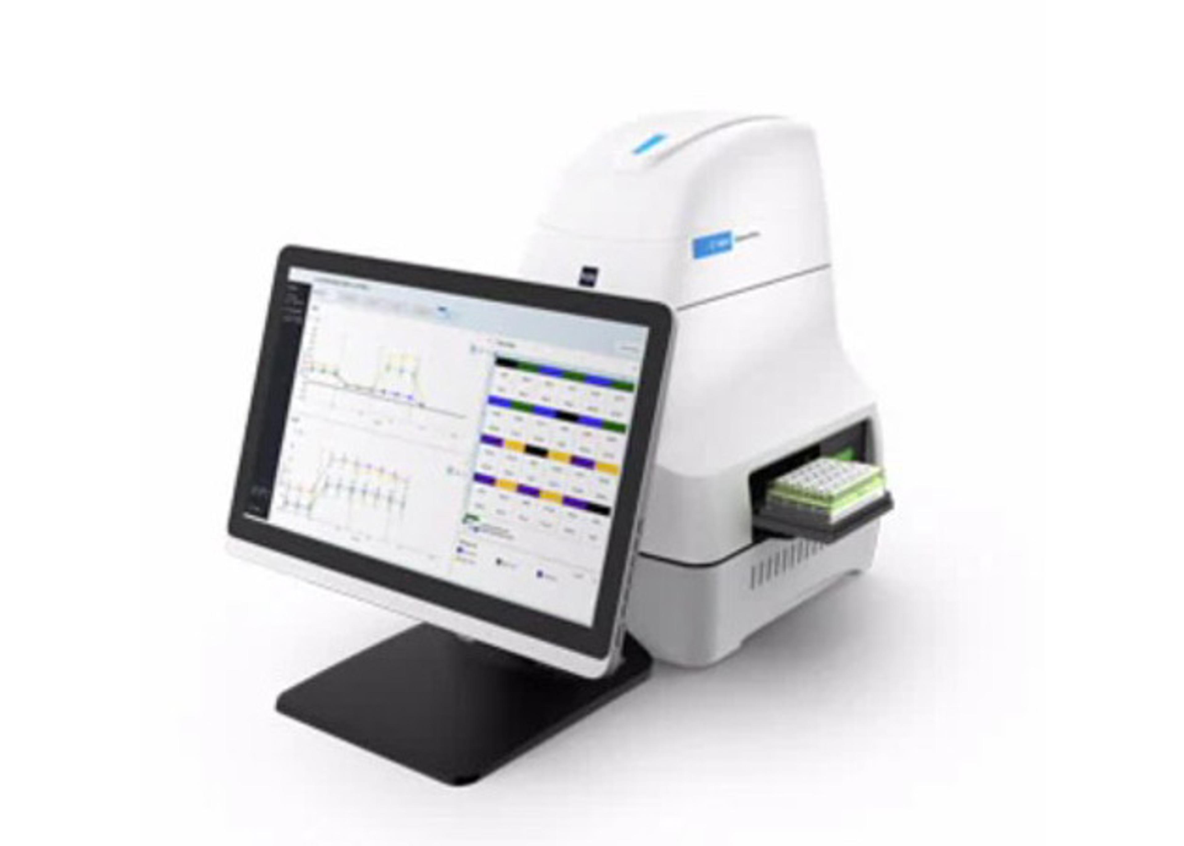
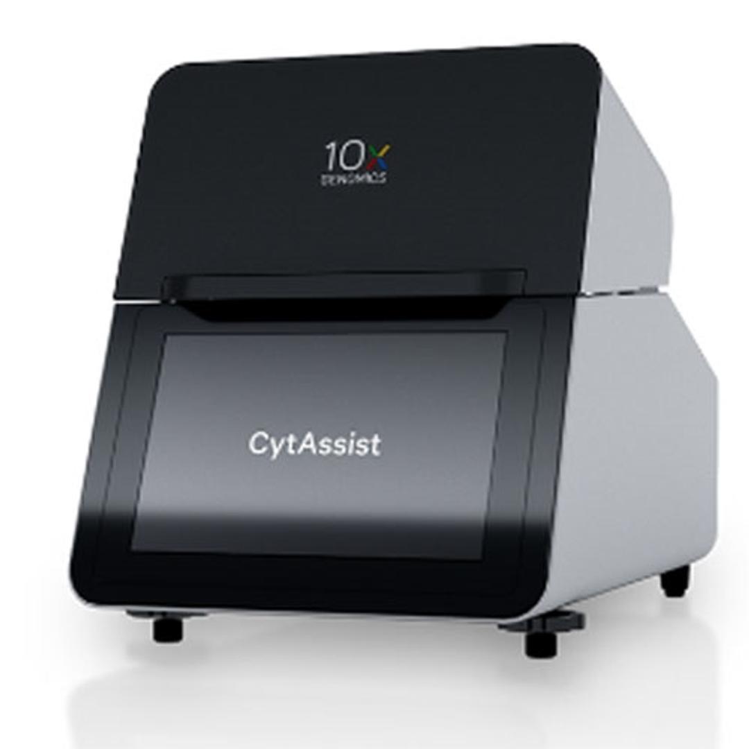
Visium CytAssist
Bridging histology and genomics

The supplier does not provide quotations for this product through SelectScience. You can search for similar products in our Product Directory.
A major step forward in spatial omics
Another major step forward
A major step forward in the field of spatial omics. Can allow users to submit samples in the form of slides.
Review Date: 29 Sept 2023 | 10x Genomics
The Visium CytAssist is a compact instrument designed to simplify the Visium workflow by facilitating the transfer of transcriptomic probes from standard glass slides to Visium slides, enabling spatial profiling insights to be gained from an expanded range of FFPE samples.
Simplified sample management
It all starts with a standard glass slide. The Visium CytAssist enables users to start with tissue on standard glass slides, allowing input tissue to be prepared, stained, and imaged using standard histology workflows.
Expanded sample access
Compatible with H&E or immunofluorescently stained FFPE tissue sections, the CytAssist instrument allows you to use pre-sectioned and pre-stained tissues with 6.5 x 6.5 mm or 11 x 11 mm Visium Capture Area slides.
Maximized spatial insights
Pre-screen tissue sections and then use the instrument for precise manual alignment of tissue section to Visium Capture Areas making the most of your spatial discovery studies.
Brochures

Bridging histology and genomics: Introducing the Visium CytAssist
In this product brochure, explore the features of the Visium CytAssist a compact, benchtop instrument that enables the transfer of transcriptomic probes from standard glass slides to Visium slides, designed to enable spatial profiling insights to be gained from even more samples.
Guide to novel approaches in spatial biology: Gain deeper insights into tissue heterogeneity
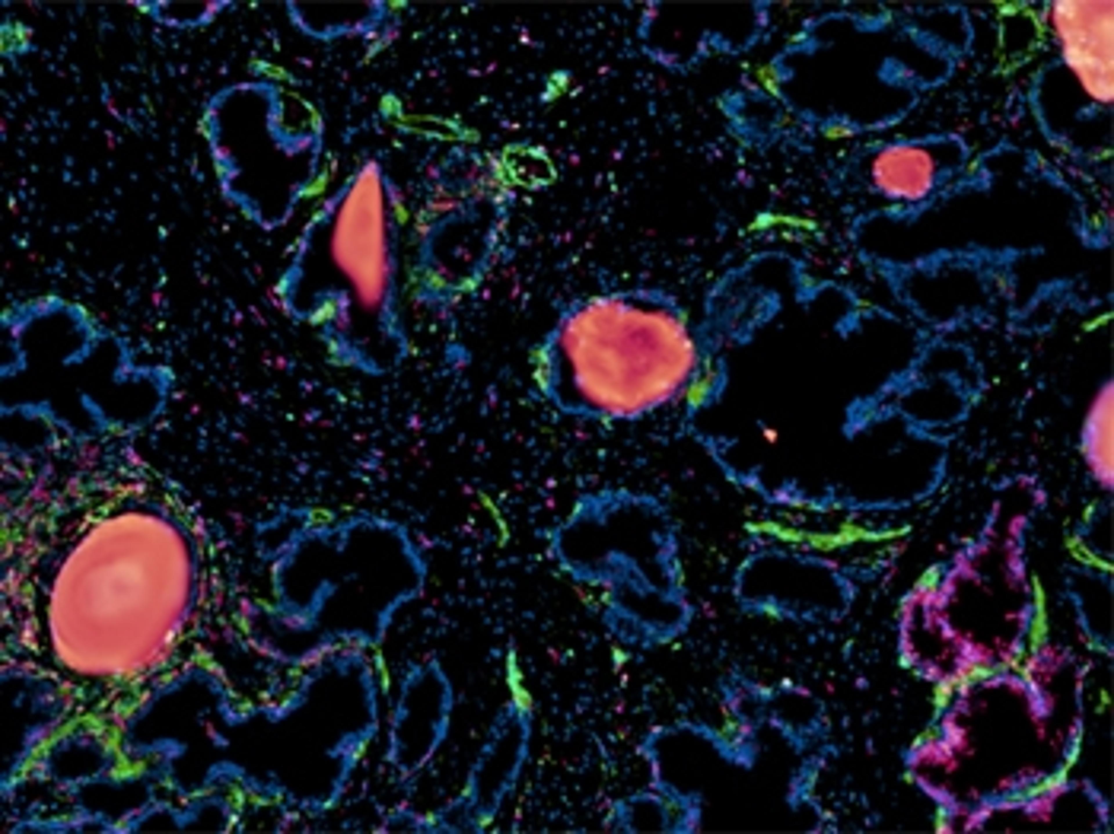
Spatial biology techniques are expanding our understanding of biological architecture with ever greater resolution in the context of tissue microenvironment. Next-generation molecular profiling solutions for the analysis of spatial gene expression look set to be game-changers, allowing us, for example, to classify tissue based on total mRNA, and enabling advances in spatially resolved gene expression that tell us where in a cell that expression is changing.
In this eBook, learn how coupling single-cell approaches with new spatial analyses of gene expression is enabling researchers to see biology in new ways through significant shifts in spatial resolution and scale, whether for cancer cell profiling or greater basic understanding in neuroscience.
Download the eBook to learn how the latest spatial biology profiling platforms from 10x Genomics can be used to resolve cancer tissue types and brain architecture in normal and diseased states, including case studies of the following tissue types:
- FFPE prostate cancer tumor samples
- Triple negative breast cancer
- Human squamous cell carcinoma
- Brain architecture
- Alzheimer’s disease markers
Introduction to Spatial Transcriptomic Data Analysis - A Case Study of Renal carcinoma
Are you interested in how spatial transcriptomics allows researchers to answer biological questions? Dr. Isaias Hernandez, postdoctoral researcher, Centre de Recherche des Cordeliers and Paris Brain Institute, will demonstrate how his team were able to unravel the differences in tertiary lymphoid structures (TLS) of responding patients to immune checkpoint inhibitors.
Key learning objectives
- Learn in practice how to analyse spatial transcriptomic data from your experiments and which pitfalls to avoid
- Discover how information from multiple Visium slides can be integrated to reveal shared and distinct features in the TLS of responding and non-responding patients
- Learn how to obtain tumoral-immune hubs that provide biologically relevant information
- Understand how to apply spatial analysis to characterize cellular heterogeneity inside and around TLS
Who should attend?
Researchers and bioinformaticians working in areas such as:
- Spatial biology
- Immuno-oncology
- Spatial transcriptomics
- Renal carcinoma
- Tertiary lymphoid structures
- Tumor microenvironment
- Cancer
Certificate of attendance
All webinar participants can request a certificate of attendance, including a learning outcomes summary, for continuing education purposes.