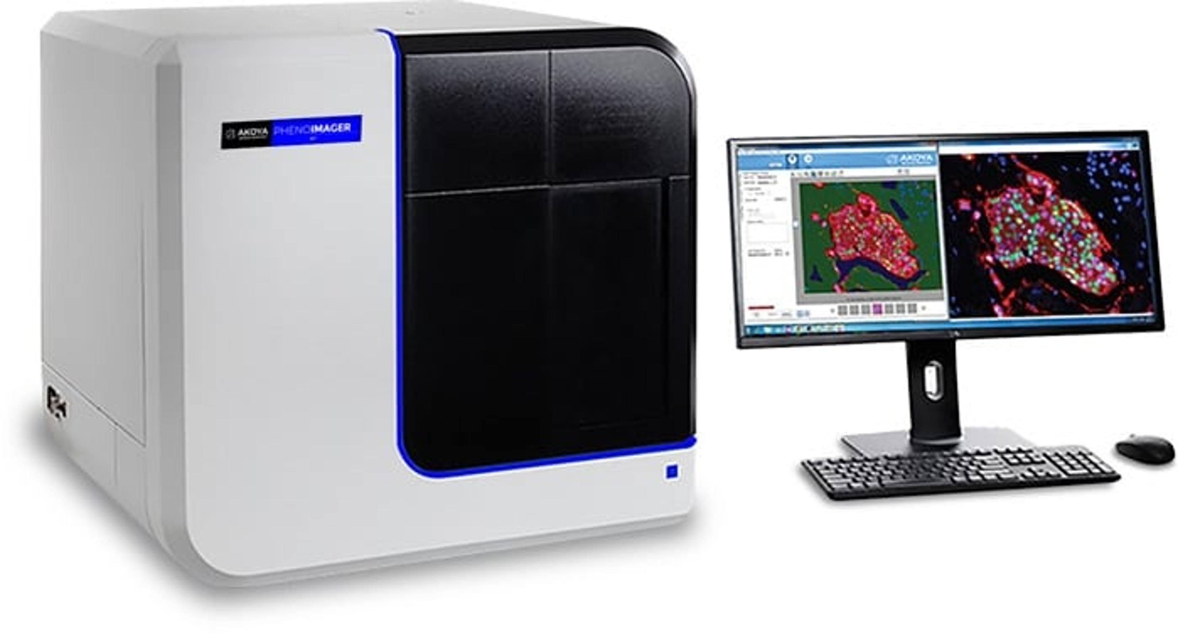
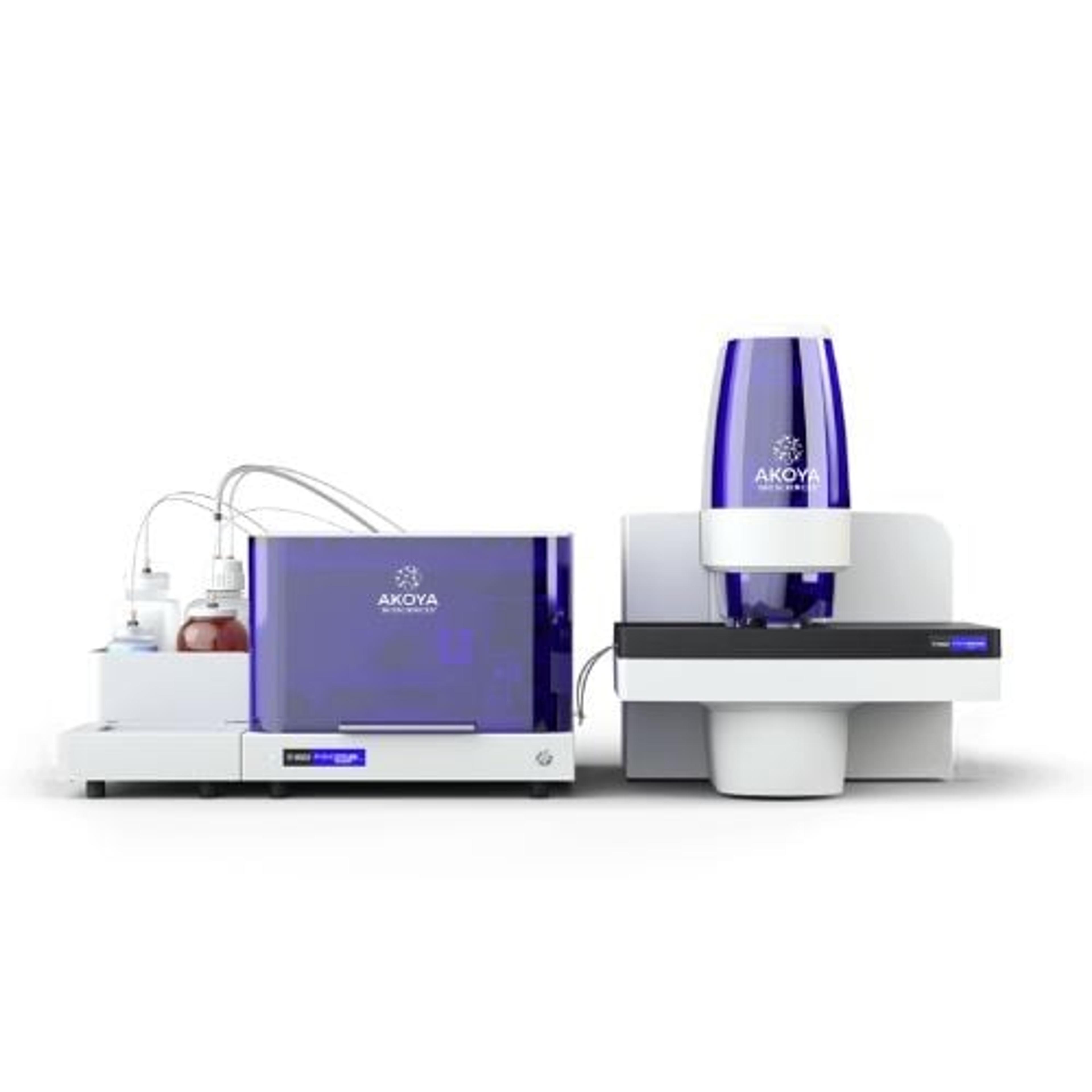
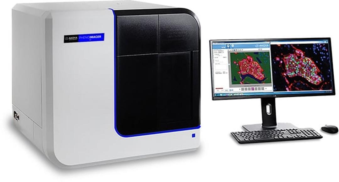
PhenoImager® HT
PhenoImager ® HT (formerly Vectra ® Polaris™) is the fastest and most highly cited whole-slide, single-cell resolution imaging platform for spatial phenotyping and the development of spatial signatures. Featuring Akoya’s patented Multispectral Imaging (MSI) and spectral unmixing technologies, this platform can be easily integrated into high-throughput workflows to accommodate projects regardless of your scale.
PhenoImager™ HT


Receive your quote directly from the manufacturer.
Key to complete studies successfully
In the field of cancer research studies
The product is quiet good and user friendly. It helps imaging and also analysis as it has built in image analysis facility. Cool machine
Review Date: 20 Apr 2023 | Akoya Biosciences
A game-changer in immunohistochemistry-based research of tumor microenvironments.
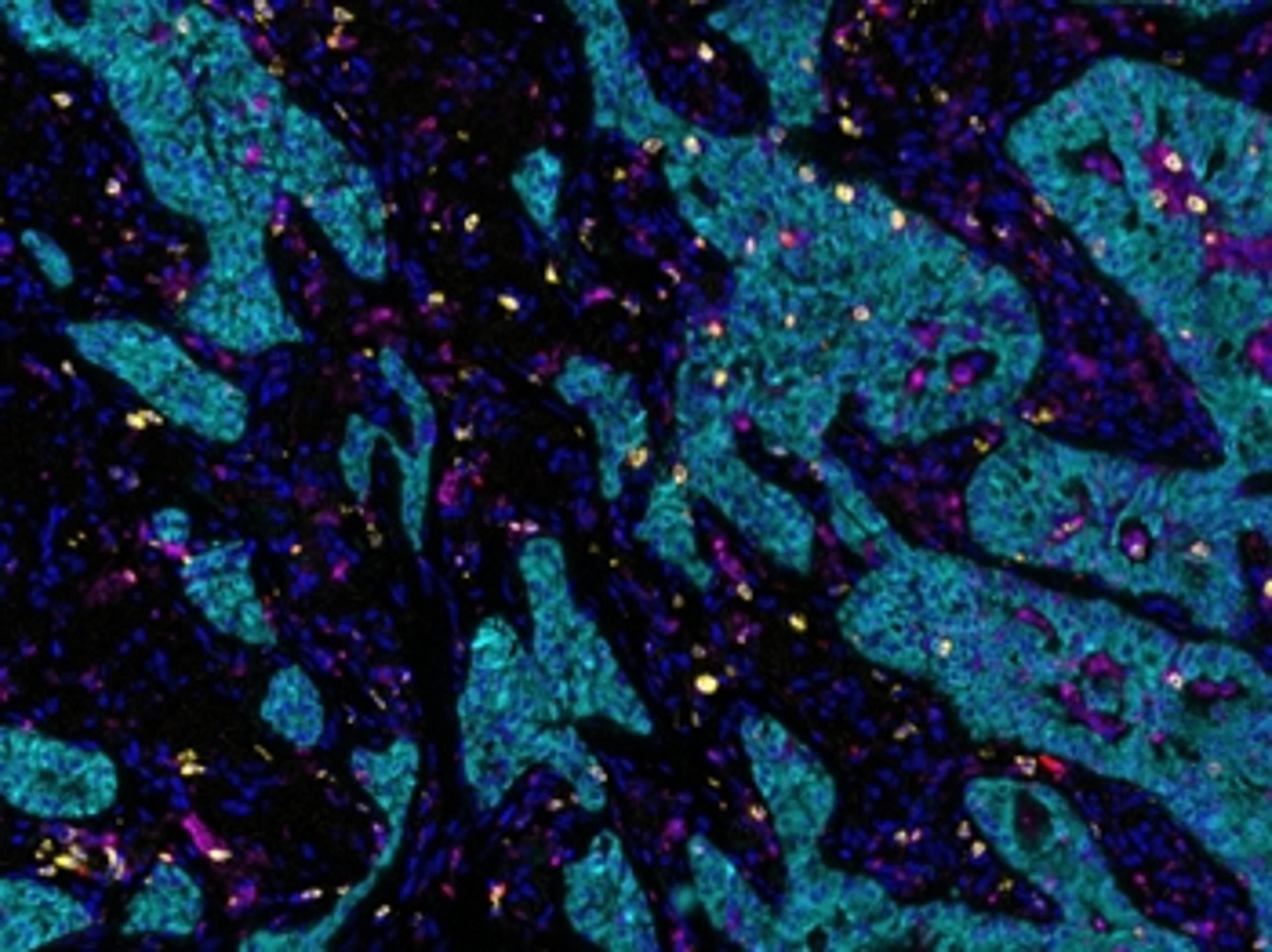
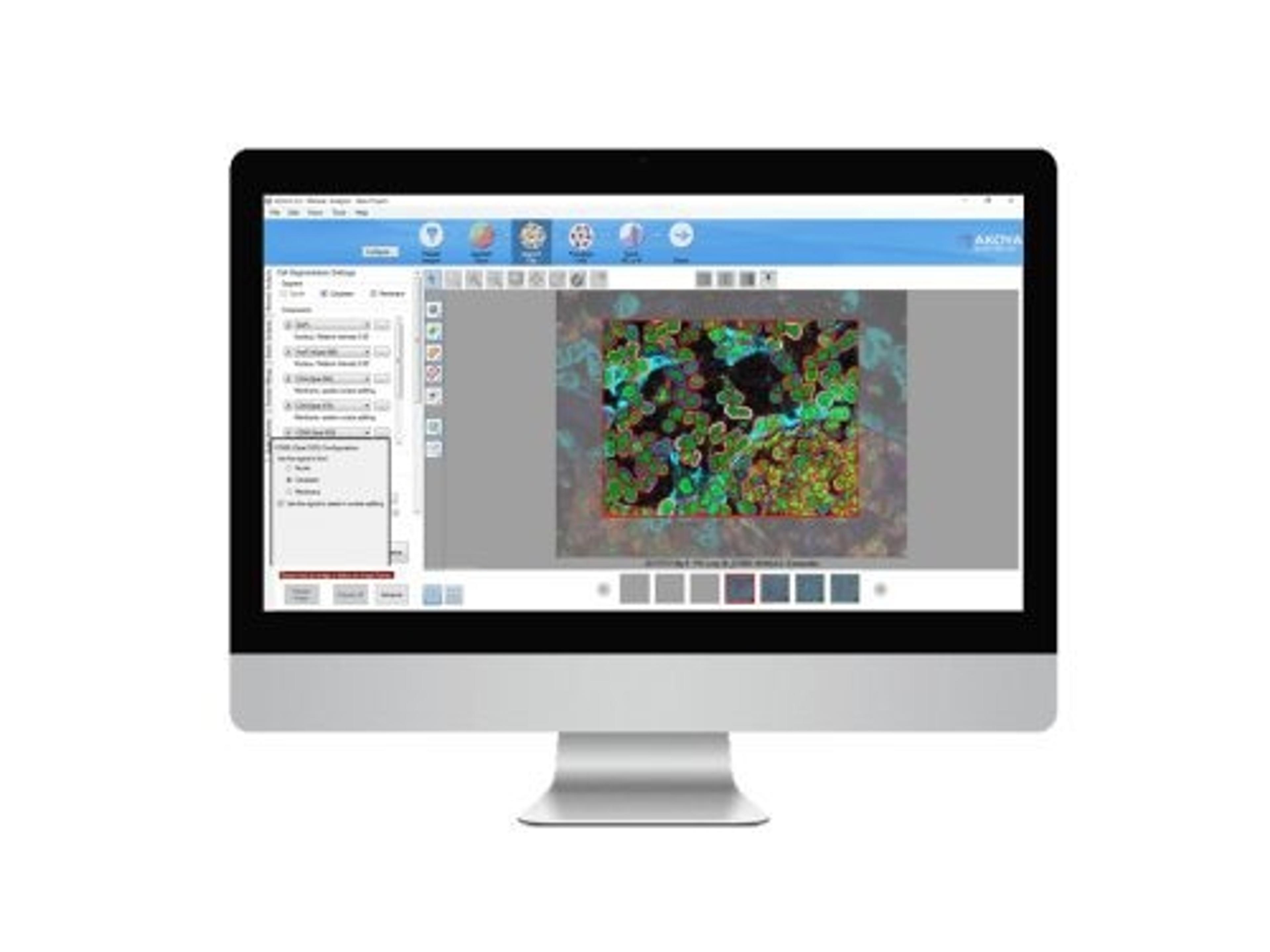
Analysis of immune markers in tissue - multiplex IHC
Having the Vectra Polaris in our lab has transformed our ability to analyze and understand the tumor immune microenvironment. Within a year of having this technology we have developed novel 9-plex immune marker panels and applied them to endometrial, bladder and esophageal cancer tissues. PhD students with little to no experience of IHC have quickly mastered the ability to perform multiplex IHC and are also now fully competent at using the software needed to analyze their samples. Before obtaining this technology, we were unable to carry out equivalent immunohistochemistry analyses, both in terms of multiplex staining capability and also quantitative regional analysis of multiplex-stained tissue sections. The increased information and thus understanding of the tumor microenvironments this technology is revealing to us will greatly help inform us in our development of more effective combination immunotherapies.
Review Date: 3 Dec 2020 | Akoya Biosciences
PhenoImager HT running the newly released 2.0 software provides researchers with a unique technology stack combining onboard spectral unmixing, rapid imaging and manageable data outputs.
Key Benefits:
- Speed: The fastest imager for spatial phenotyping and signature development, PhenoImager HT supports walk-away automation with a straightforward and user-friendly workflow enabling you to scale projects effortlessly.
- Accuracy: A new onboard spectral unmixing process that runs seamlessly in parallel with imaging means never having to compromise on data quality, regardless of your project size.
- Throughput: PhenoImager HT is ideally suited for an automated mIF staining and imaging workflow, providing the capacity to image over 400+ multiplex stained slides per week to fit any project of your scale.
- Proven: Akoya’s technologies have been cited more than a 1,000 times. With the installation of our 1000th instrument, we are proud to be one of the most trusted partner in the field of spatial biology.
Key Features:
- Whole-slide scanning: 10X-40X in brightfield or fluorescence
- Multispectral Range: 440 – 780 nm
- Multiplexing Capability: Separates up to 9 colors, even if overlapping (integrated spectral unmixing)
- Slide Capacity: 80 slides with continuous loading technology
- Automation: Touchless, with walk-away image acquisition
- Scan Speeds: Generate whole slide scans of up to 7 colors in less than 12 minutes (15mm x 15mm)
- Sample Types: Tissue microarrays and tissue sections
- Data Output: Akoya Biosciences’ whole slide scan image compatible with 3rd party analysis software packages (.qptiff); Multispectral field format (.iM3); monochrome or color images (JPEG, single-layer TIFF, BMP, or PNG)
- Analysis Software: Integrated inForm and phenoptr tissue analysis software packages support configurable projects for biomarker quantification and spatial analysis.