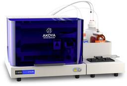
PhenoCycler®
The PhenoCycler ® is a cutting-edge fluidics control unit that integrates with the PhenoImager Fusion imaging platform creating the fastest spatial biology solution with unparalleled throughput.

Akoya’s PhenoCycler™ System

The supplier does not provide quotations for this product through SelectScience. You can search for similar products in our Product Directory.
Best in multiplex image quality!
Multiplex Multiomics
Phantastic looking images, reliable data and great scientific support!
Review Date: 23 Mar 2023 | Akoya Biosciences
Still trying to unlock the magic.
Phenotype rare cells in tissue
Value for money: Staining reagents can only be sold as a full kit... makes troubleshooting prohibitively expensive and wasteful. After sales care: One-on-one troubleshooting help for computer troubleshooting and image segmentation. Would like help tailoring panels/exposure times, analysis for research questions (ie. it currently feels very clinically-oriented). Ease of use: Clear instructions if using standard workflow.
Review Date: 3 Dec 2020 | Akoya Biosciences
Great instrument!
High parameter imaging of immune cells
Great machine, great service, dedicated team. Needs experience in panel design and high parameter imaging.
Review Date: 3 Dec 2020 | Akoya Biosciences
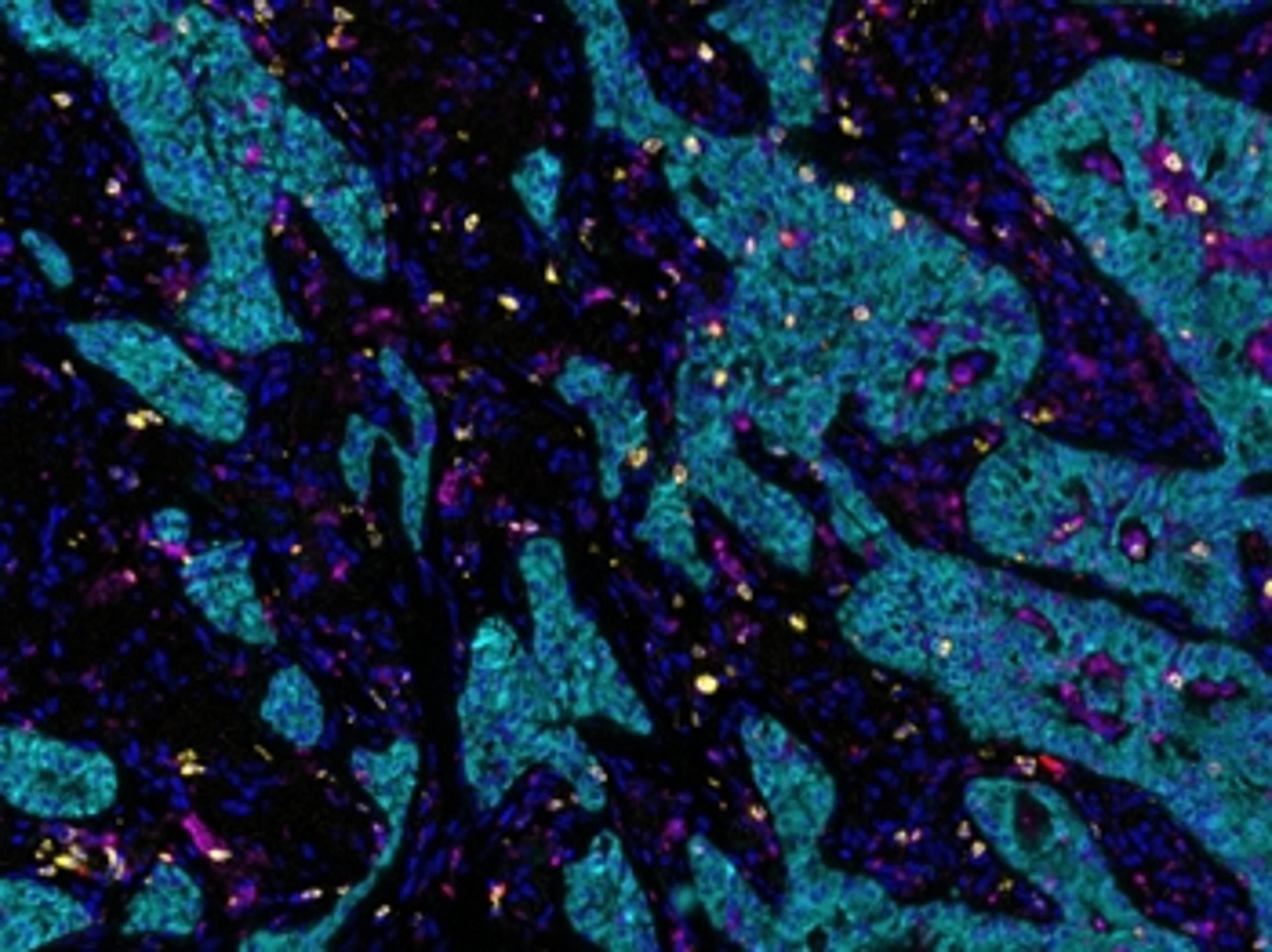
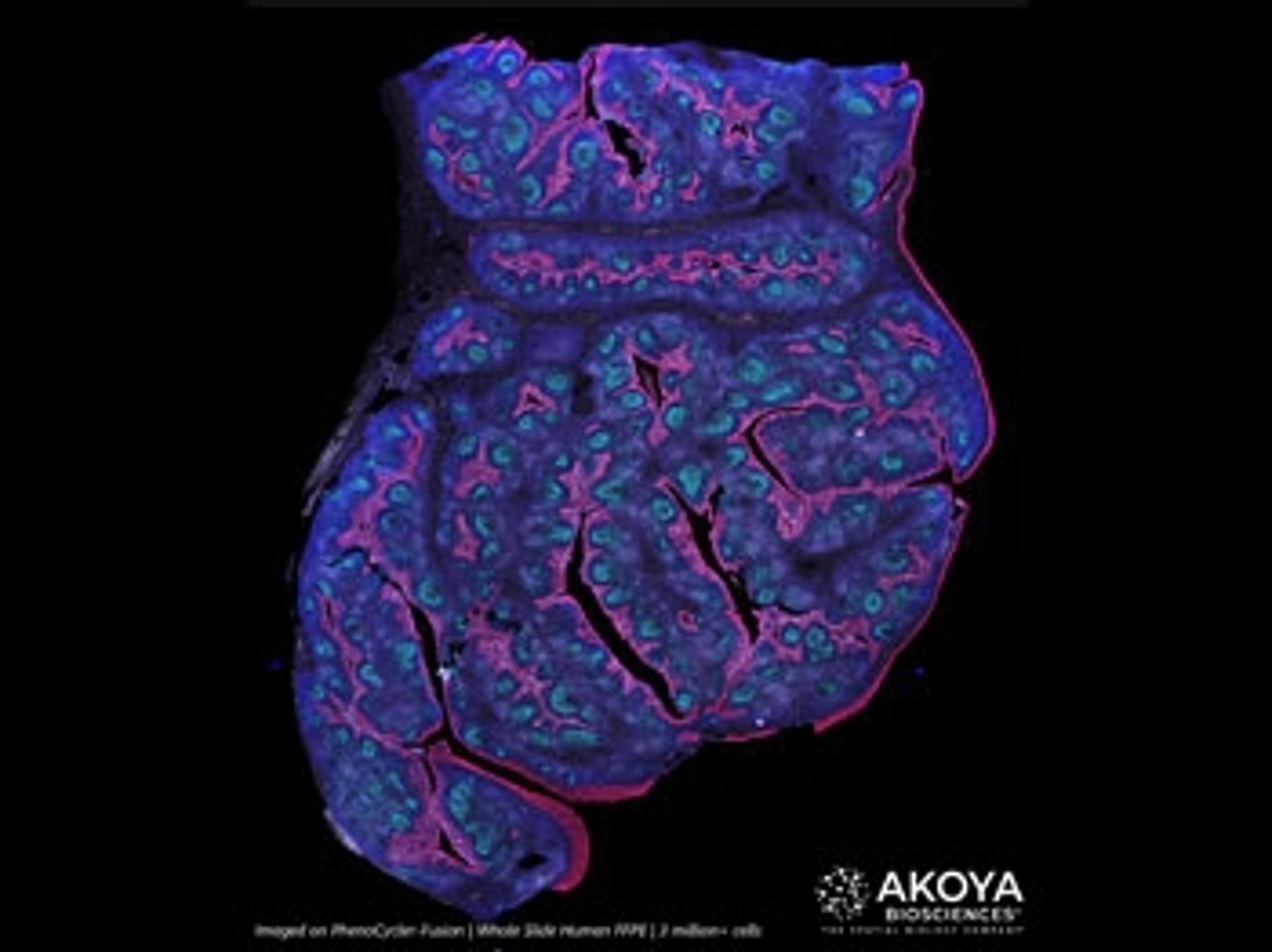
The PhenoCycler®-Fusion system is the fastest spatial biology solution that enables ultrahigh-plex spatial phenotyping of whole slides at single-cell resolution by integrating automated fluidics and iterative imaging. Capable of imaging 1 million cells in 10 minutes, the system allows unbiased spatial phenotyping of tissues, accounting for every single cell across the whole slide. Moreover, it is high-throughput and the only system that enables translation of spatial discoveries into actionable biomarkers.

Spatial phenotyping without compromise in plex, resolution, or throughput
Biological systems are complex, and full spatial understanding of tissue heterogeneity and cellular microenvironments has been hampered by trade-offs that are forced by the available analytical technologies.
All of which has given rise to the concept of the ‘iron triangle’ – the hard problem of improving resolution, plex and throughput simultaneously in one analytical solution.
But the world is set to change with the advent of new spatial phenotyping approaches that enable viewing, characterization and quantification of cells by lineage and variant with single-cell resolution, all in the context of an intact tissue sample.
From capturing tumor heterogeneity and validating cancer models, to teasing out how the cellular microenvironment impacts disease progression and treatment response, this guide will explain how spatial phenotyping can deliver advances in automation, efficiency, tunability, and speed that are set to transform how we study tissue biology.
Transform your discovery research with spatial phenotyping
Spatial phenotyping of whole tissue sections enables the study of different cell phenotypes and how they interact in situ to impact disease pathology and progression. In areas such as oncology, immunology, and neurodegenerative disease, this is rapidly increasing our understanding of underlying cell-level biology and holds the potential to drive a new wave of therapeutic discovery.
In this new eBook, we provide a user guide to spatial phenotyping and highlight a range of its benefits and applications, including how it can enable researchers to:
- Unlock single-cell discoveries in FFPE tissues
- Explore tumor heterogeneity
- Visualize tumor-infiltrating lymphocytes
- Perform highly multiplexed hypothesis-free biomarker discovery
- Conduct hypothesis-driven biomarker discovery and high-throughput translational research
How spatial phenotyping can uncover novel insights into tissue biology
Spatial phenotyping allows a researcher to view, characterize, and quantify cells by lineage and variant with single-cell resolution in the context of an intact tissue sample. In areas such as oncology, immunology, and neurodegenerative disease it has opened new insights into the interplay between different cellular actors in promoting or suppressing disease.
This white paper offers examples of how spatial phenotyping can impact your research tool kit.
Using spatial phenotypic signatures to predict immunotherapy response
In this infographic, discover how spatial phenotypic signatures can act as invaluable whole slide image-based quantitative biomarkers to predict immunotherapy response, and learn more about Akoya Biosciences’ comprehensive multiplex immunofluorescence solutions for spatial phenotyping.
Study shows spatial biology is essential for predicting response to immuno-oncology treatment
This application note outlines the results of an in-depth study comparing immuno-oncology biomarker types by scientists at Johns Hopkins University, Yale University, and other institutions. Here, Akoya Biosciences shows that the study determined that multiplex immunofluorescence with spatial characterization significantly outperformed other biomarker testing approaches for predicting patient response to treatments targeting PD-1/PD-L1.

Akoya Biosciences unveils comprehensive roadmap of innovations at inaugural spatial day event
Innovations include a novel spatial transcriptomics chemistry and universal protein chemistry for accelerated biomarker discovery and validation

Akoya Biosciences announces a groundbreaking collaboration with PathAI to combine spatial biology with AI-powered tools to facilitate discovery of novel predictive biomarkers
The partnership pairs Akoya’s spatial phenotyping solutions and PathAI’s AI-based algorithms to elucidate spatial biomarker signatures for biopharma partners
CrestOptics selected by Akoya Biosciences for spatial biology collaboration
Collaboration aims to provide new workflows, faster acquisition speeds and deeper resolution


Akoya Biosciences unveils spatial biology platform to enable rapid image analysis and secure data sharing
The Proxima spatial analysis software solution integrates image acquisition to data analysis in the secure cloud for rapid access by any user


Study Shows Spatial Biology Is Essential for Predicting Response to Immuno-Oncology Treatment
Akoya Biosciences determines that multiplex immunofluorescence technology preserves critical data missed by other biomarker strategies