Spatial phenotyping without compromise in plex, resolution, or throughput
How ground-breaking innovative approaches in spatial phenotyping can deliver high-throughput single-cell resolution within the context of fully intact tissues samples
5 Mar 2024
Biological systems are complex, and full spatial understanding of tissue heterogeneity and cellular microenvironments has been hampered by trade-offs that are forced by the available analytical technologies.
All of which has given rise to the concept of the ‘iron triangle’ – the hard problem of improving resolution, plex and throughput simultaneously in one analytical solution.
But the world is set to change with the advent of new spatial phenotyping approaches that enable viewing, characterization and quantification of cells by lineage and variant with single-cell resolution, all in the context of an intact tissue sample.
From capturing tumor heterogeneity and validating cancer models, to teasing out how the cellular microenvironment impacts disease progression and treatment response, this guide will explain how spatial phenotyping can deliver advances in automation, efficiency, tunability, and speed that are set to transform how we study tissue biology.
Related products
Request Quote for All Products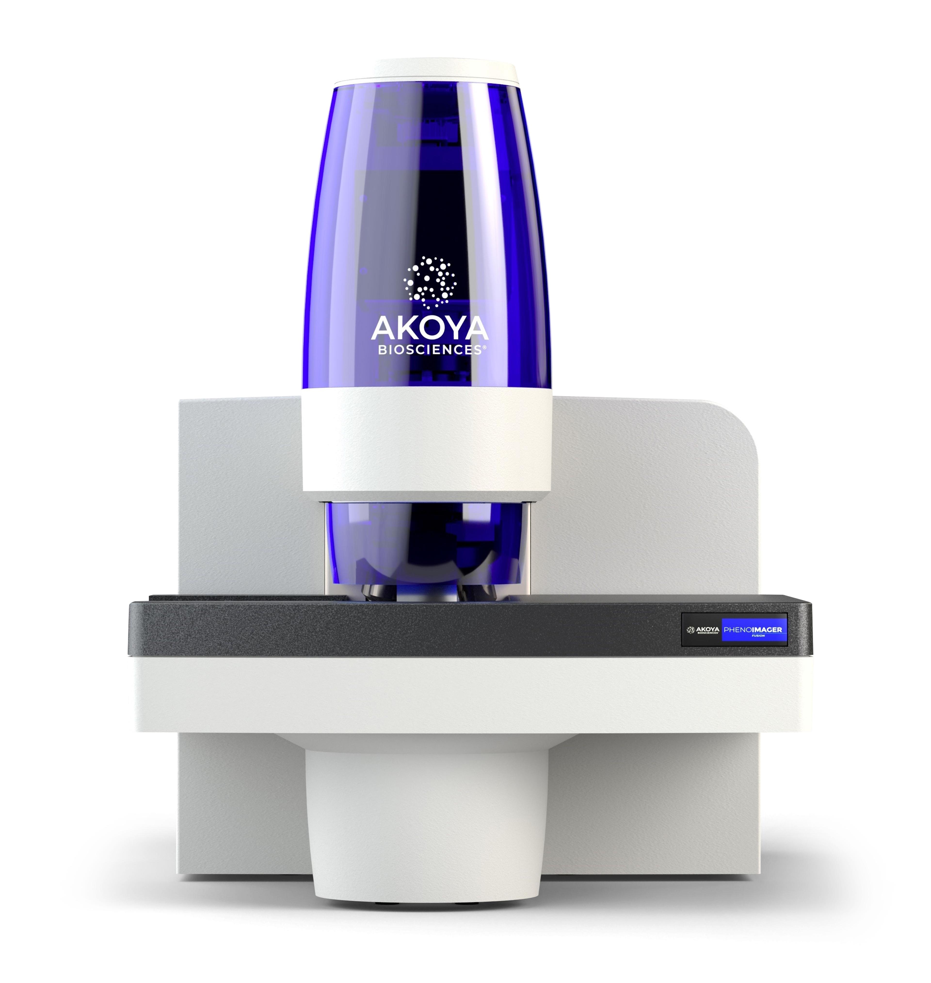
PhenoImager® Fusion
Akoya BiosciencesThe PhenoImager ® ; Fusion system is a whole-slide, single-cell resolution, ultrafast multispectral imager ideal for standard throughput brightfield and fluorescence imaging applications. This imaging platform can be integrated with the PhenoCycler ® ; system (formerly CODEX ® ) for ultra high-plex spatial discovery at scale.
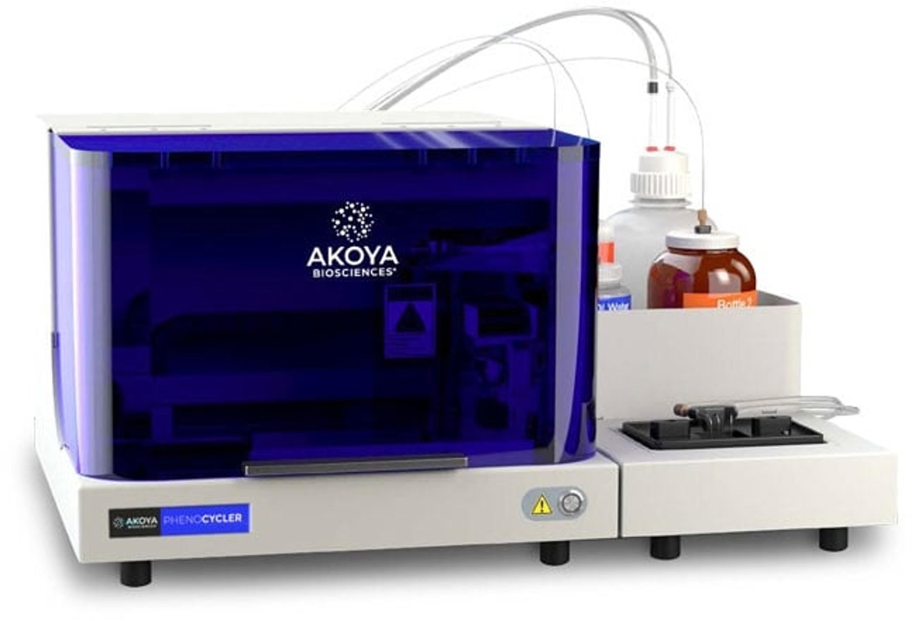
PhenoCycler®
Akoya BiosciencesThe PhenoCycler ® is a cutting-edge fluidics control unit that integrates with the PhenoImager Fusion imaging platform creating the fastest spatial biology solution with unparalleled throughput.