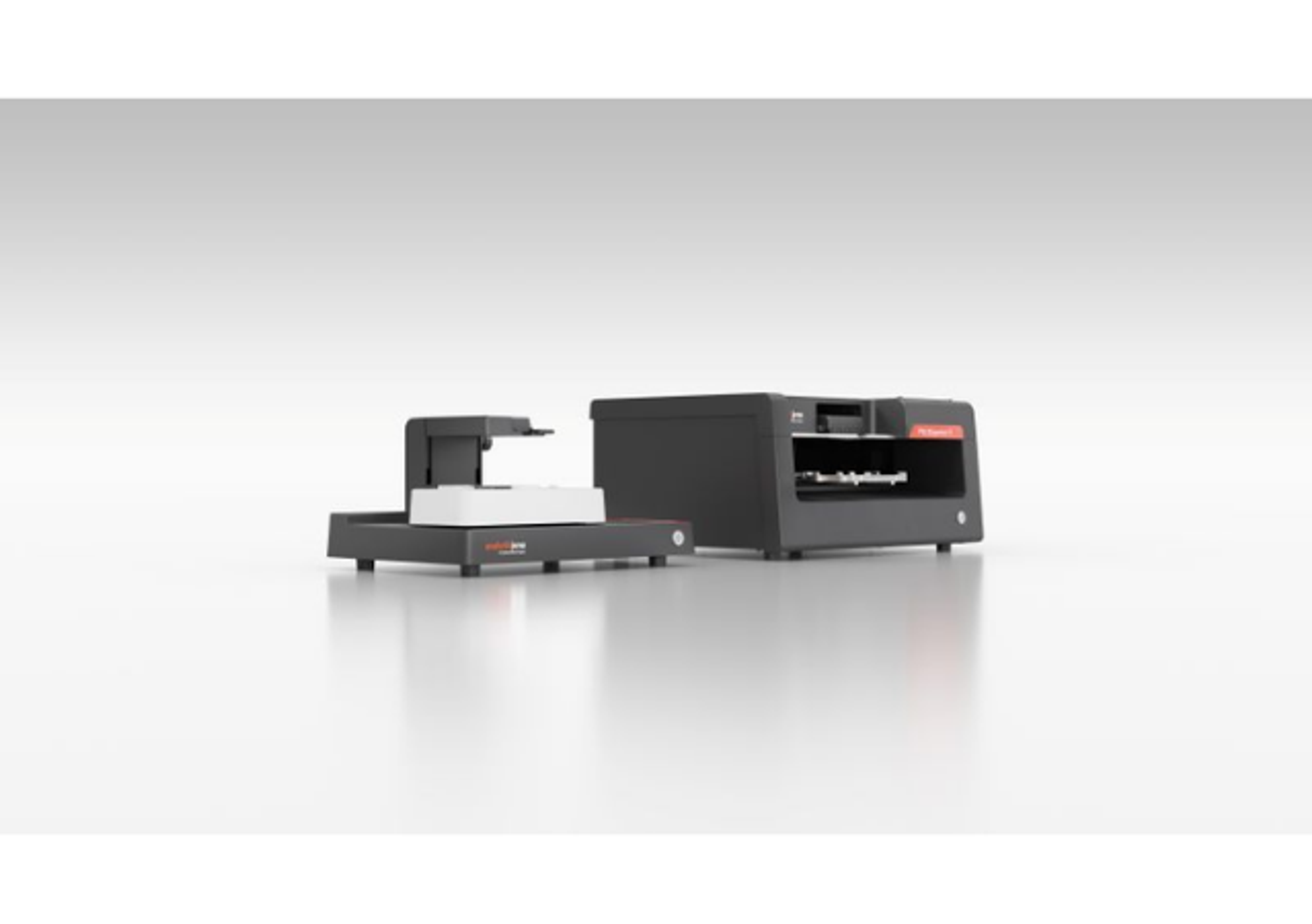
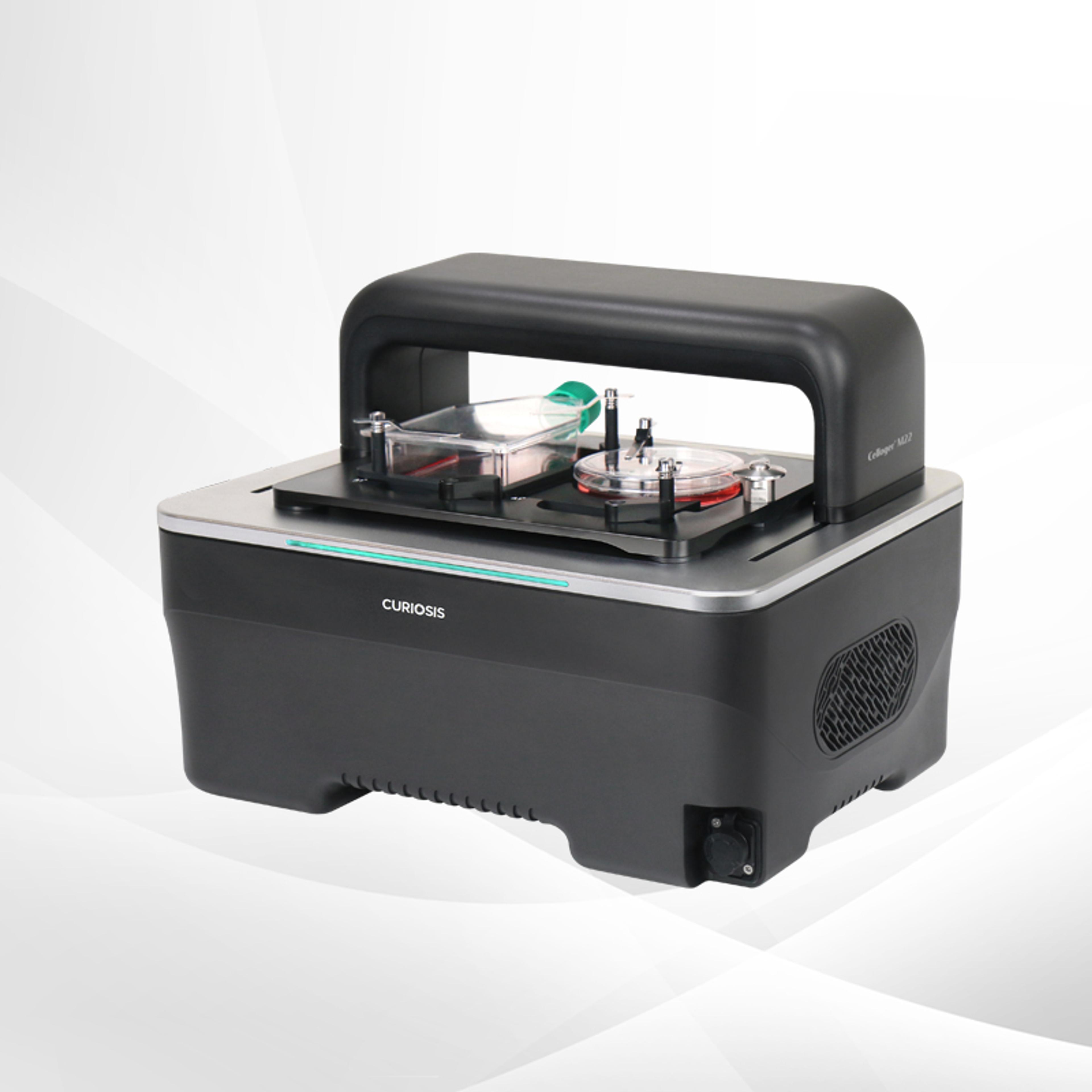
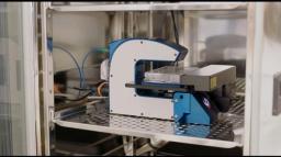
HoloMonitor® M4 Live Cell Analysis System
The HoloMonitor ® live cell imaging system enables long-term non-invasive analysis of cell cultures within your standard incubator. Cell biologists worldwide use PHI’s label-free cell imager to automatically obtain accurate live-cell morphology, migration, and proliferation data as well as multi-well time-lapse videos, down to a single-cell level in real-time.

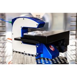
HoloMonitor fits your standard incubator
Both single-cell & cell population data
Get multiple results from 1 sample


The supplier does not provide quotations for this product through SelectScience. You can search for similar products in our Product Directory.
This instrument enables us to obtain novel data that nobody knows
Cell statement evaluation (especially macrophage)
We usually use it without any problems and make great use of it in our research. There are few devices in the world that can acquire three-dimensional information of cells while they are alive. This device helps us to make new discoveries by capturing the characteristics of cells that have not been focused on before. To be honest, automatic tracking of migrating cells is not 100% accurate. We have reduced the density of cells, still some tracking mistakes occur. But don't worry, because the accompanying software, SINGLE CELL TRACKING, has enough tools for manual correction, and once corrected, useful data can be obtained. In this regard, we look forward to technological advances.
Review Date: 20 Sept 2024 | Phase Holographic Imaging (PHI)
Manufacturer's Response
Thank you for your insightful feedback! We're thrilled to hear HoloMonitor is helping drive new discoveries in your research. We appreciate your feedback on the automatic tracking feature and are happy to share that improvements for the single cell tracking app are already in the works. Your input helps us continue enhancing the system—thank you for your support!
High-quality images with associated data that can be re-analyzed in endless ways!
Real-time proliferation, motility and dose-response assays, imaging, single-cell analysis of 48 morp
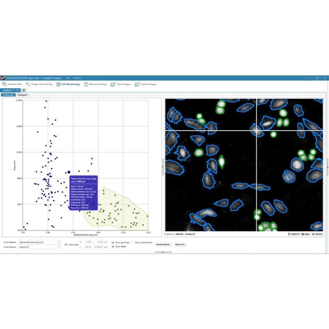
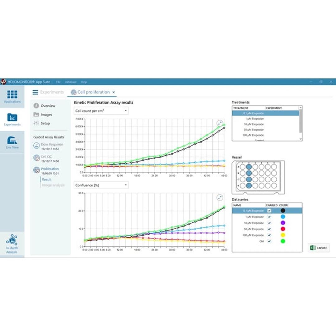
The system is very easy to use. You set up an experiment, place the cell culture vessel under the microscope, and select the experiment of interest (dose-response). The system auto-calibrates itself and collects data in real time. Sometimes external factors such as condensation on cell culture flask interfere with single-cell detection. This is visible in the images, moreover, the system flags low-quality images, which you can exclude from the data analysis. At the end of the experiments, the system generates all the figures for you (graphs, images, videos). You can then use the collected data and have them re-analyzed by the software using a different application e.g. proliferation, motility, etc. All in one, performing one experiment allows you to collect data using multiple applications. The PHI lab team are friendly, approachable and caring. The team always tried their best to help us solve issues, guide us through applications we were not familiar with or provide training where needed. At times, I needed help with the calibration of the laser, solving the accumulation of condensation on my cell culture flask. Equally, the system generates so much data that sometimes I needed help with analysis. The PHI lab team were always happy to help, responding quickly to all queries, and having video calls to navigate us through the process. The system generated reproducible cell proliferation data. I have run two proliferation assays in parallel. One is kinetic proliferation using HoloMonitor (real-time analysis) the other is tritiated thymidine incorporation proliferation assays (end-point analysis). The assays generated reproducible data. What I liked about the system is that you select how many positions within your cell culture flask the laser monitors and how often the laser monitors those positions. This is very useful if you are expecting the effect of the treatment to manifest within a short period or to study apoptosis. There is an additional “in-depth analysis” function which gives endless ways of analyzing your data. In addition to the applications that detect single-cell automatically and generate data automatically (wound-healing assay, proliferation, motility, dose-response, cell quality, etc.) with ready-to-export graphs, there is an “in-depth analysis” function. This enables you to look at cell tree plots, 48 morphological features collected for every single cell at the selected position, like roughness, and eccentricity, which you can then use to investigate the effects of the treatment on your cell morphology. The “in-depth analysis” provides infinite options to analyze your cells. This application, however, is less intuitive to use. The single-cells are detected automatically by software, but I recommend double-checking the single-cell selection manually (note: automatically selected borders can be manually adjusted here). I found this function extremely helpful when studying cytotoxic vs cytostatic cellular responses to treatments. After publishing my results, PHI lab contacted me to hear about my user experience. They were particularly interested to see where the system could do better and were very responsive to feedback. The only drawback of the system: -An in-depth analysis of 48 morphological features does not provide mean values. Let’s say you want to see the mean cell volume from three wells in your 6 well-plate. To do this, you would need to export the Excel data and analyse them yourself. -Statistical analysis is not incorporated within the software, so you have to manually export the data in Excel format and re-analyse them. All in all, I would highly recommend this product if you want to get a comprehensive understanding of your cell morphology and how it changes to stimuli (of your interest). I think it is good value for money because you only need to set up one experiment, which can be re-analysed in many ways.
Review Date: 5 Mar 2024 | Phase Holographic Imaging (PHI)
Manufacturer's Response
Hi Zuzana! Thank you for your detailed review and invaluable feedback. It was great speaking with you and learning about your positive experiences and insights and the areas where you see room for improvement. Your suggestions are crucial as we strive to make HoloMonitor even more user-friendly. Thank you for recommending HoloMonitor, Your PHI team
Small, very effective, time saving
Live imaging of the cells
Easy to use system placed directly in the cell culture incubator; support various cell culture vessels including small flasks, 6-well plates, 24-well plates and other; definetly time and resource saving, once the experiment is started you get your ready results; possibility to upgrade and extend your analysis software
Review Date: 6 Jun 2023 | Phase Holographic Imaging (PHI)
Manufacturer's Response
Thank you, Bogusz for your kind feedback! We’re thrilled to hear that you and your cells are happy! Best wishes from your PHI team.
Big bang for buck, but can be quite labor-intensive.
Analyse cells morphologically in adherent cultures at single cell level
A high value for money product. Easy to use and gives nice insights into adherent cell cultures and how they develop. The technological approach is very advanced and offers a high potential for the future. However, the device is very sensible to vibrations and generates often artifacts, so many images must be excluded manually. An advanced algorithm which can detect such artifacts and could automatically exclude them from analysis, as opposed to manually selecting the ones which should be analyzed, would be preferred to reduce user workload. Also a better algorithm for image segmentation (perhaps based on machine learning) would be highly preferred (also for later analysis based on image parameters).
Review Date: 20 Sept 2022 | Phase Holographic Imaging (PHI)
Manufacturer's Response
Hey, Martin! Thanks for providing us with detailed feedback about your experience with HoloMonitor. We take every opportunity to improve our product to fit customers' needs. HoloMonitor analyzes over 30 different parameters for every single cell and therefore generates tons of data which sometimes can be labor-intensive. Our team will continue to improve the algorithms to provide our users with a better experience. Let us know if we can assist you in any way.
The results add great value to all our toxicity studies!
Proliferation analysis for toxicity testing
The system is very easy to use and generates high-quality, reproducible results. Dedicated data analysis offers detailed information on cell proliferation frequency, cell division duration, cell morphology and cell motility to precisely assess additive toxicity.
Review Date: 1 Aug 2022 | Phase Holographic Imaging (PHI)
Manufacturer's Response
Hallo, Jessica! We are happy to hear that you get the high-quality and reproducible results you want with your HoloMonitor system. Thank you for sharing your feedback with other scientists.
HoloMonitor is an excellent microscope
Analyze the morphology and behavior of cancer cells under anti-cancer treatment.
The HoloMonitor machine is efficient for operating up to 96 samples simultaneously. Holomonitor can provide high-quality images and time-lapse movies. We always get professional operation instructions and data analysis suggestions from the service team.
Review Date: 2 Jul 2022 | Phase Holographic Imaging (PHI)
Manufacturer's Response
Thanks for sharing your experience with us, Tianyan! HoloMonitor is developed by and for biologists, and our team is always happy to provide fast and professional support to our customers.
Great live cell imaging system with a wide range of data and parameters!
Analyze migration, morphology and cell growth
I am using HoloMonitor system for analyzing migration, morphology, and cell growth of cells from cell lines and primary cells, and I am very happy with the imaging system and using it a lot in my research. It is easy to use and you can get amazing data: videos/images of the cells (2D, 3D, magnifications, etc) and a range of different parameters from one single experiment. I believe it is a big advantage that it is label-free and inside the incubator, making it a more stable, "natural" milieu and less interference with the cells, and also it enables long experiments. Great microscope!
Review Date: 23 Nov 2021 | Phase Holographic Imaging (PHI)
Manufacturer's Response
Thanks a million for sharing your experience, Frida. When you and your cells are happy with HoloMonitor, we are as well! Let us know whenever we can support you further in your research.
Best microscope I've tried so far - worth every penny
Vascular cell biology
This microscope is perfectly suited for our cell culture under flow application as well as many other types of experiments where tracking cell behaviour and motility in real-time is a must. Because it even provides morphology data in 3D we now have insights into what the cells are doing at a level which we simply could not achieve before. Highly recommended.
Review Date: 18 Nov 2021 | Phase Holographic Imaging (PHI)
Manufacturer's Response
Thank you for sharing your feedback, Markus. Our team is happy to see that HoloMonitor is what you have been looking for in your cell culture under flow application.
I like and recommend this device.
Differential cytotoxicity screening
The HoloMonitor M4 is a beneficial device for live-cell imaging, providing a lot of data. The device does not require large space and the software application is user-friendly, while recorded pictures look great. The good thing is that you can analyze your experiment using one software assay, and later, you can rerun the recorded data in another assay, which allows you to get the maximum from your experiment. I like it and would recommend it to all people doing a cytotoxicity assessment based on cell proliferation and optical thickness of the adherent unstained cells. Cell migration tracing is another application I like and recommend.
Review Date: 3 Nov 2021 | Phase Holographic Imaging (PHI)
Manufacturer's Response
Hello Sasa! We are glad to hear that you easily get the data you are after. And that you can maximize the results from every experiment setup by combining the different HoloMonitor live cell assays! Thank you for sharing your experience.
An affordable, powerful, indispensable part of our research program.
Imaging heart muscle cell responses
I'm the principal investigator of a small research group at a public primarily undergraduate institution. Even considering our limited resources, I believe that the Holomonitor M4 is a fairly priced system and is definitely worth the investment. It has truly been and will remain a critical component of our research program. Our microscope lives directly in our cell culture incubator and allows us to see how various treatments affect our cells in real-time with three-dimensional single-cell resolution - it's amazing! The Holomonitor App Suite software is particularly powerful allowing us to quantify changes in cell morphology and movement. Importantly, I also think this instrument is great for education and my students are absolutely fascinated by the quality and beauty of the videos produced. This system is intuitive to operate and it is very effective in generating label-free long duration time-lapse videos. We dissociate complex tissues containing a mixed population of cells for culture, and with the Holomonitor M4 we are able to easily distinguish different cell types by their unique size, shape, and movement patterns. We can't wait to see what new biology we will discover with this innovative approach! I highly recommend it!
Review Date: 20 Oct 2021 | Phase Holographic Imaging (PHI)
Manufacturer's Response
Our team is excited to hear that your students cannot get enough of watching their beautiful cells! We thrive that HoloMonitor is an incubator microscope for any cell lab by making it high-quality but affordable and easy-to-use everyday lab equipment. Thank you, Alexander!
A Live Cell Analysis Tool for everyone in your lab.
- Non-invasive: Detailed kinetic cell morphology data without fluorescence.
- Compact: Small footprint to fit your standard incubator.
- Affordable: Not all live cell imaging systems have to be expensive.
- Automated: Automatic single-cell tracking of all individual cells in real-time.
- Easy-to-use: Intuitive software to guide you from imaging to analysis.
- Time-efficient: Multiple results from one sample with this 8-in-1 live cell analysis tool.
The compact HoloMonitor cell culture microscope facilitates label-free single-cell analysis on a population level. With this quantitative live-cell imaging system, any cell research lab is equipped for long-term non-invasive analysis of their cell cultures within a standard incubator.
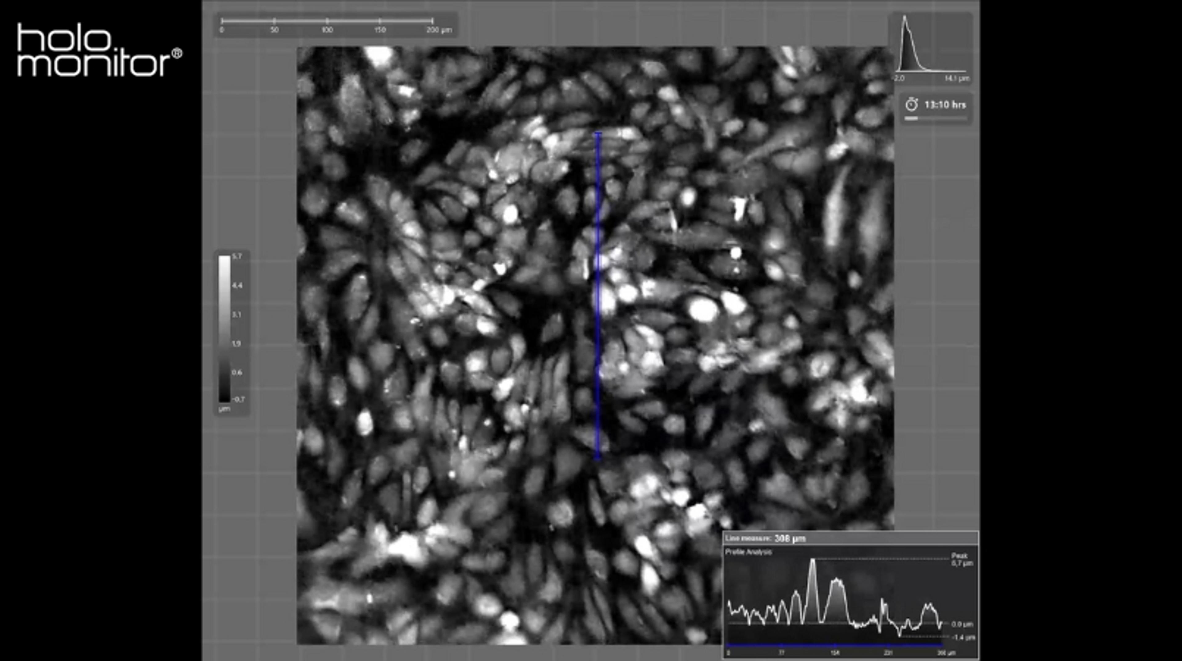
Without the need for any cellular labels or stains, HoloMonitor uses digital holographic microscopy (DHM) with quantitative phase imaging (QPI) to record cells in real-time. The result is real 3D images & videos and accurate quantitative data on your cultures, all the way down to single-cell level – collected in a completely non-invasive way.
HoloMonitor is ideal for long-term kinetic studies. Images can be recorded with high temporal resolution (maximum image rate: 1 image/s), ensuring that important cellular events are not overlooked.
All HoloMonitor live cell assays are non-destructive. Label-free assays not only reduce the risk of unwanted toxicity. They also allow cell samples to be reused, as no reagents are added to the sample. Hence, they save time and money.
Furthermore, recorded time-lapse images can at any time be reanalyzed using other HoloMonitor live cell assays. In that way, multiple results are obtained from the same cell sample and experiment. HoloMonitor is designed for and by biologists to meet the demand of researchers to increase assay reproducibility, analysis efficiency, and productivity in the cell lab.
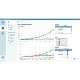
The HoloMonitor software offers a variety of live cell assays – all having the same straight-forward assay setup and following you through automated image analysis, result presentation, and data export.
HoloMonitor Key Applications – as seen in 270+ peer-reviewed publications worldwide:
- Kinetic Cell Morphology (with data on 30+ parameters)
- In-depth Single Cell Tracking & Analysis
- Kinetic Cell Motility & Migration
- Wound Healing (Scratch assay) | Chemotaxis
- Kinetic Cell Proliferation
- Cell Growth | Cell Division | Cell Cycle
- Cell Differentiation
- Kinetic Drug Dose-Response
- Cytotoxicity | Cell Death
- Cell Counter
- Cell Quality Control (QC)
Why use HoloMonitor?
- Run automated live-cell assays 24/7 directly inside your standard incubator
- Image your cells non-invasively – no labels or toxic stains needed
- Get real-time quantitative data on both single cells & cell populations
- Generate multiple results from one experiment setup using the re-analysis functionality
- Study cell proliferation, morphology, and movement behavior at the same time
- Save time with intuitive software & guided workflow developed by biologists
Brochures

HoloMonitor M4 product brochure
In this product brochure, Phase Holographic Imaging (PHI) presents the HoloMonitor® M4 live cell imaging system, offering non-invasive, long-term analysis of cell cultures within standard incubators. The system enables the automatic capture of label-free live-cell morphology, migration, and proliferation data in real-time, down to a single-cell level. HoloMonitor is designed for the visualization and quantification of adherent cells over time, suitable for various cell types, including delicate ones like induced pluripotent stem cells and primary human cell cultures. The label-free imaging preserves cell viability for subsequent studies. The HoloMonitor software is a user-friendly tool that extracts quantitative data from holographic images, guiding users through experiment setup and result analysis. The software also allows easy creation of visual content for publications, and data can be exported to Excel for further analysis.
Non-invasive live cell analysis with HoloMonitor M4
In this application note, Phase Holographic Imaging presents HoloMonitor® M4, a non-invasive live cell imaging system enabling long-term analysis of cell cultures within standard incubators. It offers label-free data on live-cell morphology, migration, and proliferation down to the single-cell level in real-time. The user-friendly software provides easy experiment setup and result analysis, making it suitable for cancer research, drug discovery, and toxicology studies.
Exploiting the potential of HoloMonitor digital holographic microscopy by combining it with 3D matrix cell culture assays
In this application note, a different type of microscope, HoloMonitor M4, based on digital holography, has demonstrated high compatibility with 3D Matrigel cell cultures. PHI researchers were able to quantify wound closure and cell movement in Matrigel by single-cell tracking. Researchers at PHI also found possibilities for further applications, such as analysis of drug response, tumor growth, metastasis, and invasion, in our 3D preparations.
Evaluation of HoloMonitor M4 cell motility applications as compared to standard methods
In this application note, Phase Holographic Imaging evaluates the HoloMonitor M4 software modules for cell tracking, wound healing, and population motility based on outcome and reproducibility. Additionally, the results were compared to the standard methods - Boyden transwell assays.
Imaging living cells without compromising cell integrity
The HoloMonitor® label-free live cell imaging system is based on the principle of quantitative phase imaging, enabling non-invasive visualization and quantification of living cells without compromising cell integrity. In this application note, Phase Holographic Imaging describes the rationale and advantages of using quantitative phase imaging for live cell kinetic analysis of cellular events — explaining the power of live cell time-lapse cytometry and how cells are made visible without labels or stains.
Comparison of the effects of pharmaceutical compounds on tumor cells in 2D and 3D in vitro models using label-free, quantitative 4 dimensional holographic imaging
Development of in vitro models for the evaluation of drugs represents a useful approach as in vivo studies may be costly and time consuming. Ideal models should take into account the effects of the cellular microenvironment, which includes the extra-cellular matrix, stroma and neighboring cells. In this poster, the HoloMonitor® M4 from Phase Holographic Imaging was used for label-free long-term kinetic cellular analysis, in order to compare the effects of pharmaceutical compounds on tumor cells.
Non-invasive DHM and its unique advantages for cell phenotype measurements
In this video, Dr. Kersti Alm, CSO, Phase Holographic Imaging, presents a talk that she gave at the National Institute of Standards and Technology (NIST) 2022 workshop on measurement needs for the biofabrication of tissue-engineered medical products. During the presentation, she introduces the label-free HoloMonitor® technology for digital holographic microscopy and provides an overview of its unique advantages for minimally destructive cell phenotype measurements.
Endothelial response to flow 67 hours: HoloMonitor
In this video, Phase Holographic Imaging shows endothelial response to flow, imaged for 67 hours with the HoloMonitor® M4 by Markus Bosteen, a Senior Scientist at the Department of Vascular Biology at Novo Nordisk.
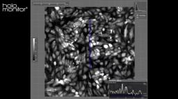
Automatic single cell tracking with HoloMonitor App Suite: Accurate, real-time live cell analysis
The cell-friendly HoloMonitor® cell culture microscope lets you study your cells directly inside a normal incubator, completely label-free. The HoloMonitor Single Cell Tracking assay automatically tracks the cells in your culture over time. It provides you with comprehensive data on, for example, cell movement, cell division, and cell death, both for every single cell but also for the whole cell population.
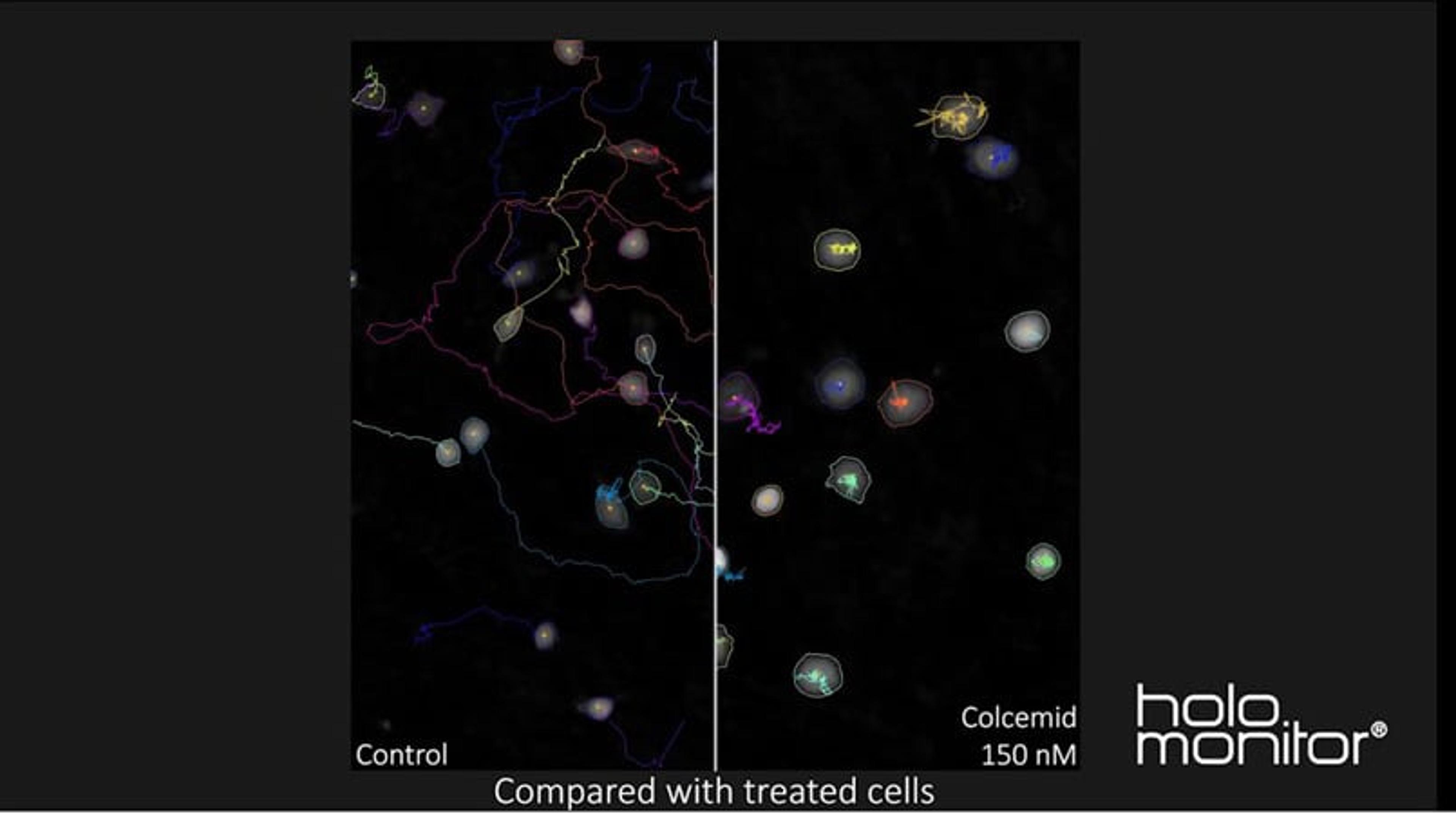
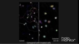
This video shows a 48-hour time-lapse with L929 mouse fibroblasts. All cells in the field of view are automatically tracked, and non-treated cells are compared with the same cells treated with the chemotherapeutic drug colcemid. It is evident that the drug not only prevents the cells from dividing but also lowers their movement to a minimum.
HoloMonitor live cell imaging system in 1 minute
The HoloMonitor® live cell imaging system enables long-term non-invasive analysis of cell cultures within a normal incubator.
In this video, Phase Holographic Imaging describes how the HoloMonitor® live cell imaging system obtains beautiful 3D images of your cells. The HoloMonitor® promises to collect real-time, quantitative data on many cellular events such as cell differentiation, migration, and cell death.
Automatic single-cell tracking assay
In this video, Phase Holographic Imaging provides insight into the benefits of using the HoloMonitor® Single-Cell Tracking Assay. The HoloMonitor allows you to study individual cells non-invasively, increase your analysis efficiency with automatic single cell tracking, simultaneously monitor cell movement and cell morphology changes, and explore both individual cell and cell population behavior over time.
Spatial tracking of mouse fibroblasts using HoloMonitor
This video, by Phase Holographic Imaging, demonstrates spatial tracking of mouse fibroblasts (cell line: L929). Control cells and cells treated with the anti-cancer drug colcemid are tracked using HoloMonitor and then compared.
Live-cell imaging made simple with the HoloMonitor App Suite
In this video, Phase Holographic Imaging illustrates how the HoloMonitor App Suite software is designed to make live cell imaging and analysis easier than ever before, with a simple step-by-step workflow, from the set-up of the experiment to the automatic presentation of results.