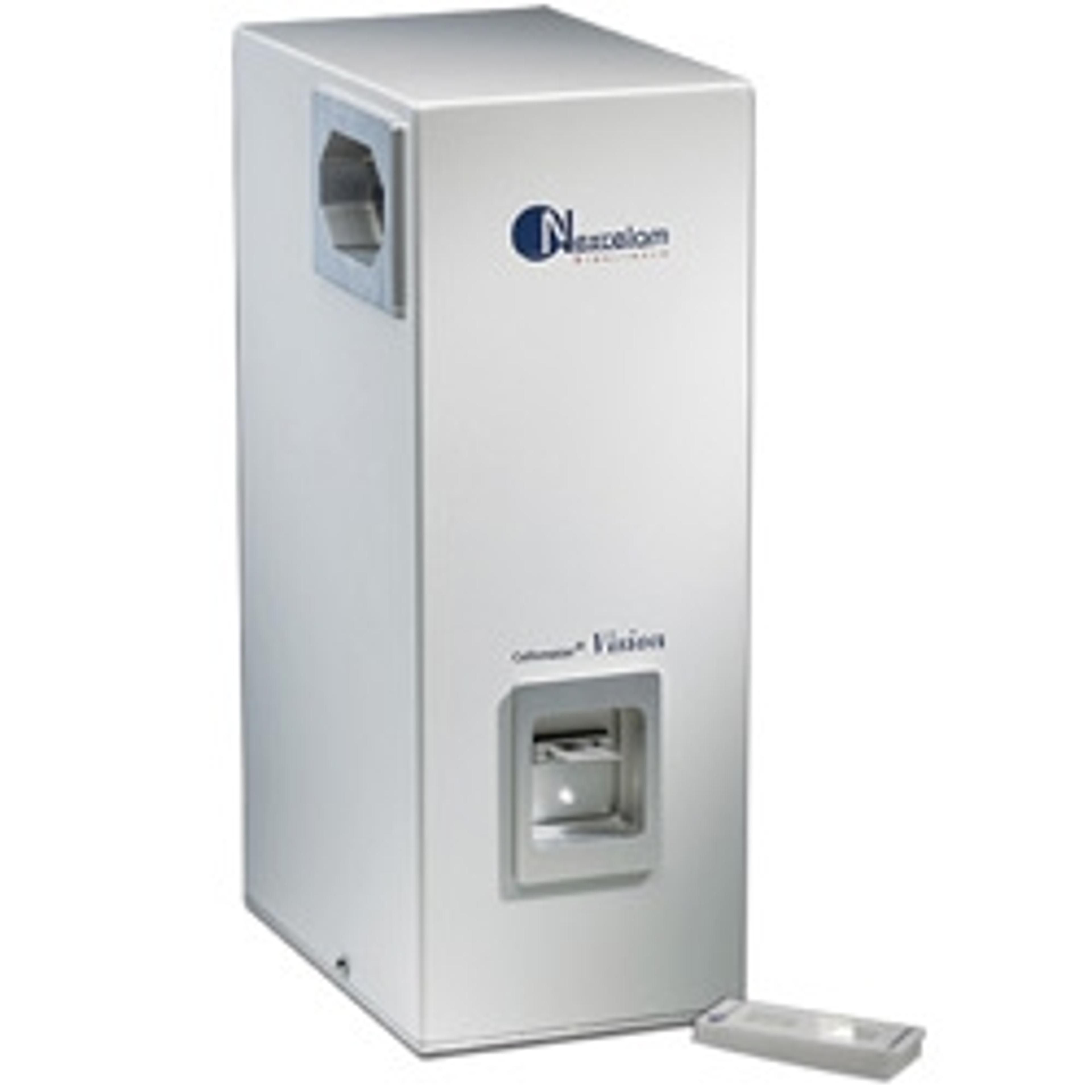
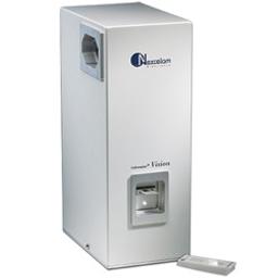
Cellometer Vision CBA Image Cytometer
Image cytometer with two interchangable fluorescent optic modules for simple, single-sample 20 uL cell-based assays with flow-like data output.

Great instrument, using everyday with no problems!
Counting cells, measuring viability
It is the most frequently used equipment in the lab by far. We are very happy overall and I recommend this product to everyone.
Review Date: 4 Jun 2019
The Cellometer Vision CBA combines the simplicity of image cytometry with the power of flow analysis software to offer user-friendly cell based assays. The Vision CBA offers both bright field imaging and dual, user-changable fluorescent optics modules, covering a spectrum from UV to far-red, enabling analysis of a wide range of fluorescent proteins, dyes and fluorophores. The Cellometer software captures and analyzes images in less than 30 seconds to provide count, concentration and viability measurements. Data and images are auto-saved and can be auto-printed.

Novel method to assess primary hepatocyte concentration and viability
In this application note, Nexcelom outlines its new method for use in complex cell population characterization assays, such as the reliable counting and viability determination of primary hepatocytes. With dual fluorescence detection capabilities, the Cellometer® Vision Cell Analyzer incorporates image-based cell counting and fluorescence detection in a compact and easy-to-use instrument.
Comparison of Trypan Blue and Fluorescence-Based Viability Detection Methods Via Morphological Observation
All cell-based biological experiments, from standard cell culture to primary cells for evaluating pharmacological agents, require accurate determination of cell viability. The Trypan Blue (TB) exclusion assay is one of the oldest and most common methods by which to assess cell viability. Fluorescent-based dyes now provide an alternative to this traditional method.
Accurately Count PBMC and Measure Viability in Presence of Residual RBC
One of the major issues of PBMC isolation is RBC contamination, which can be sampledependent. RBC contamination in a PBMC sample can introduce error to the measured concentration and viability, which can pass down to future experimental assays performed on these cells. To resolve this issue, RBC lysing protocol can be used to eliminate potential error caused by RBC contamination.