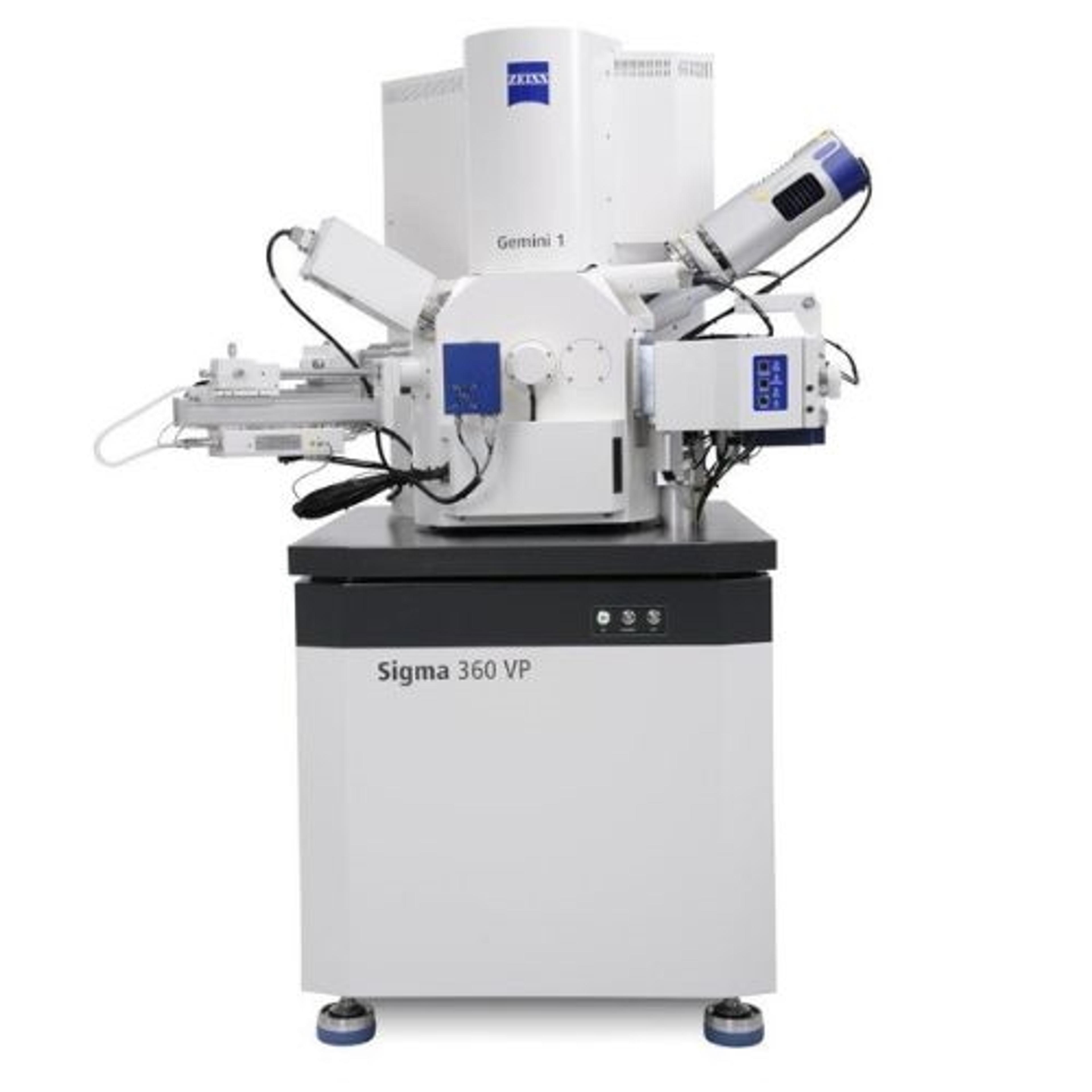
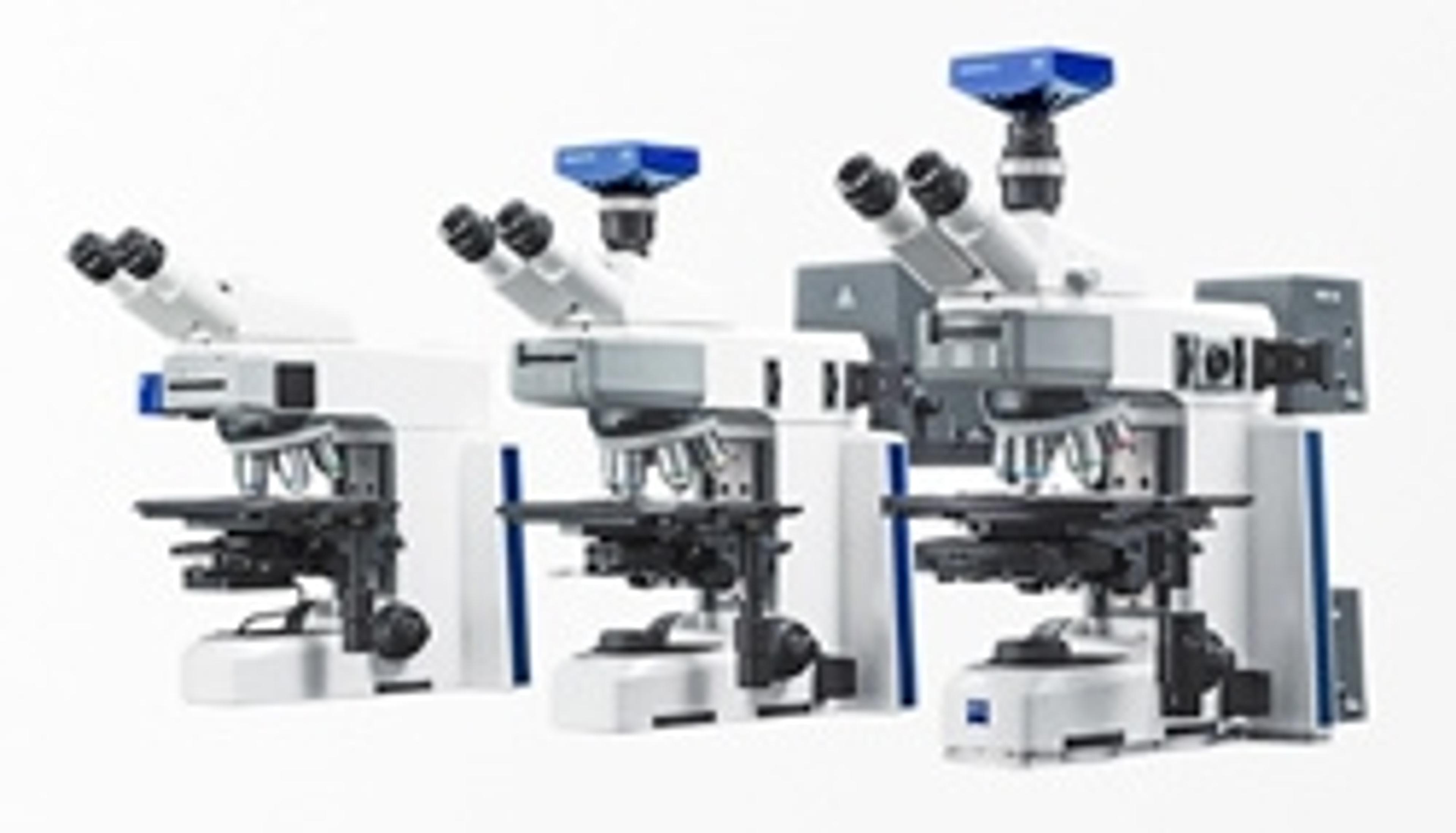
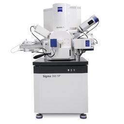
ZEISS Sigma
FE-SEM for high-quality imaging & advanced analytical microscopy


Receive your quote directly from the manufacturer.
A good choice for routine sample inspection
SEM imaging, FIB cross sectioning and lamella preparation
Reliable optics, good energy range and depth of focus, efficient detectors and add-ons. GUI complete, yet update-able
Review Date: 23 Jan 2023 | ZEISS Research Microscopy Solutions
The ZEISS Sigma family combines field emission scanning electron microscope (FE-SEM) technology with an excellent user experience. Structure your imaging and analysis routines and increase productivity. Study new materials, particles for quality inspection or biological or geological specimens. Make no compromises in high resolution imaging – go to low voltages and benefit from enhanced resolution and contrast at 1 kV or below. Execute advanced analytical microscopy using best-in-class EDS geometry and get analytical data at twice the speed and with more precision.
With the Sigma family you are entering the world of high-end nano-analysis.
- Sigma 360 is the core imaging facility’s choice — an intuitive FE-SEM for imaging and analytics.
- Sigma 560 uses best-in-class EDS geometry to deliver high throughput analytics and enable automated in situ experiments.
Microscopy with ZEN core: A unified approach to imaging and analysis
April 29, 2025 - 09:00 BST / 10:00 CEST / 16:00 CST / 17:00 JST
Join us for an informative webinar sponsored by ZEISS, where we will introduce ZEN core, a sophisticated software solution designed to enhance your microscopy workflows. This session will focus on how ZEN core provides a streamlined user interface that accommodates both light and electron microscopy, allowing users to transition seamlessly between modalities.
During the session, you will discover how ZEN core's intuitive user interface adapts to the unique needs of your laboratory allowing users of all experience levels to efficiently image, analyze, and manage data. We will explore the advanced imaging capabilities, automated workflows, and powerful AI features that make ZEN core the command center for both light and electron microscopes.
Join us for a live demonstration of ZEN core and take part in our Q&A session to address your specific inquiries. Register today to learn how ZEN core can support your microscopy research and analysis needs.
Certificate of attendance
All webinar participants can request a certificate of attendance, including a learning outcomes summary, for continuing education purposes.
If you view the on-demand webinar, you can request a certificate of attendance by emailing editor@selectscience.net.