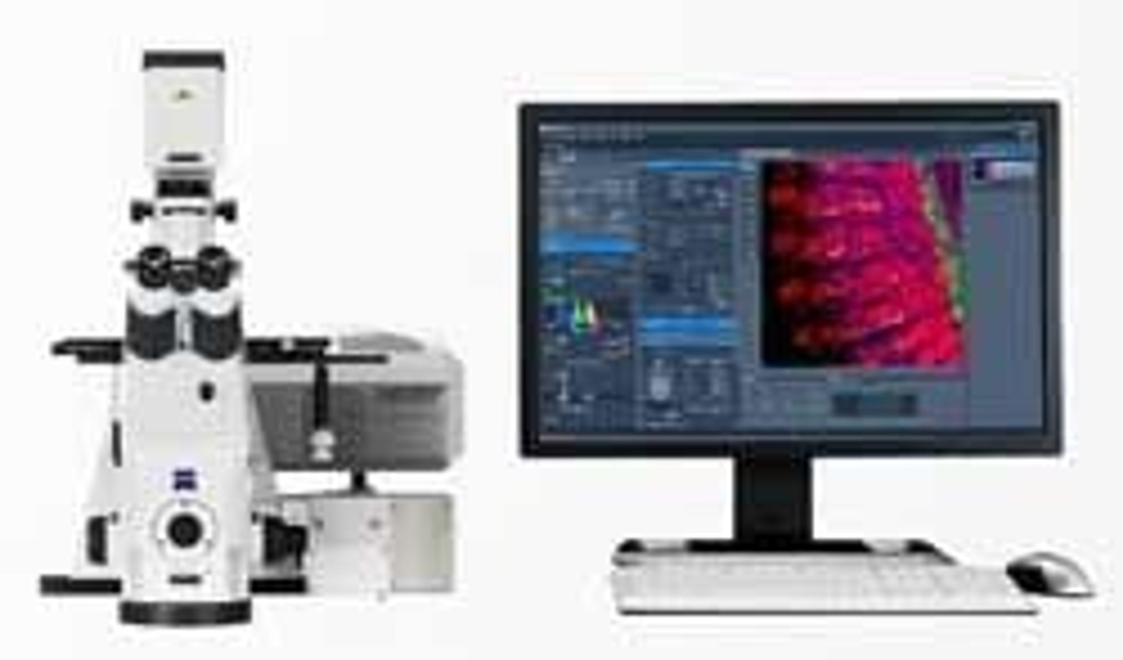
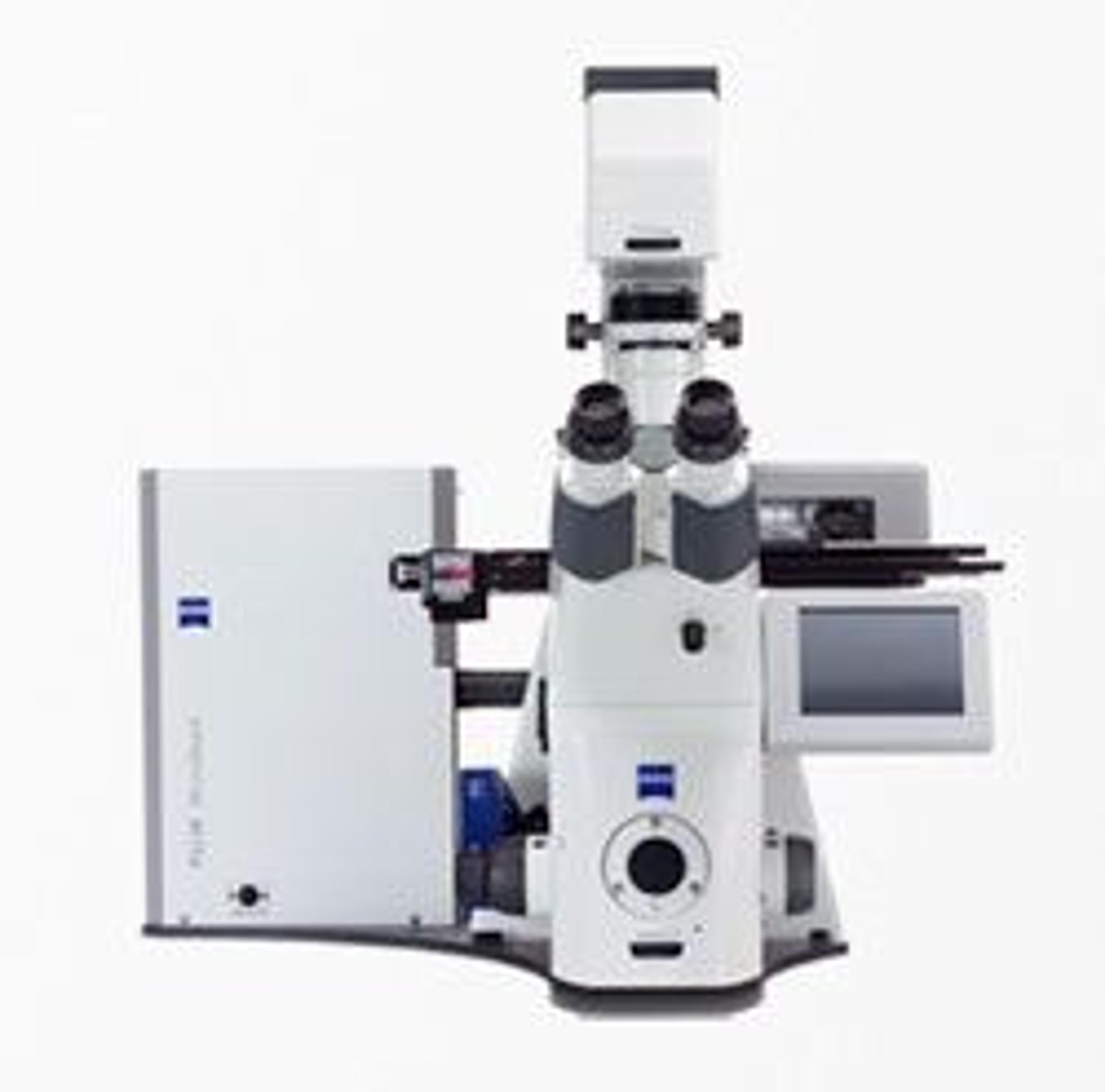
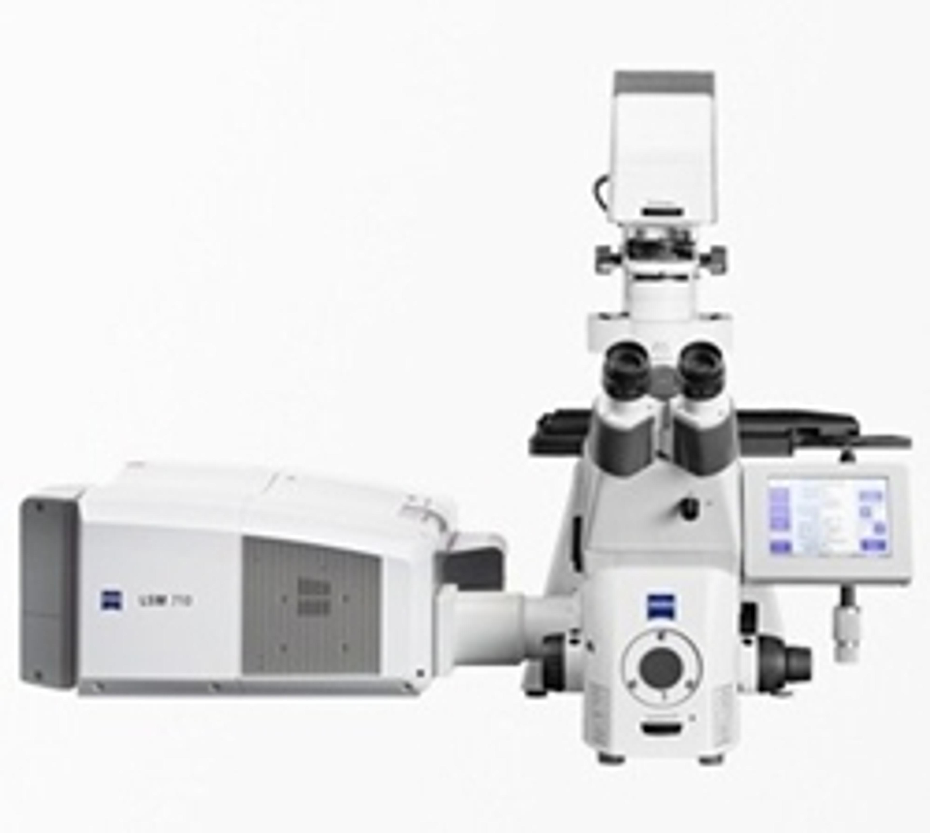
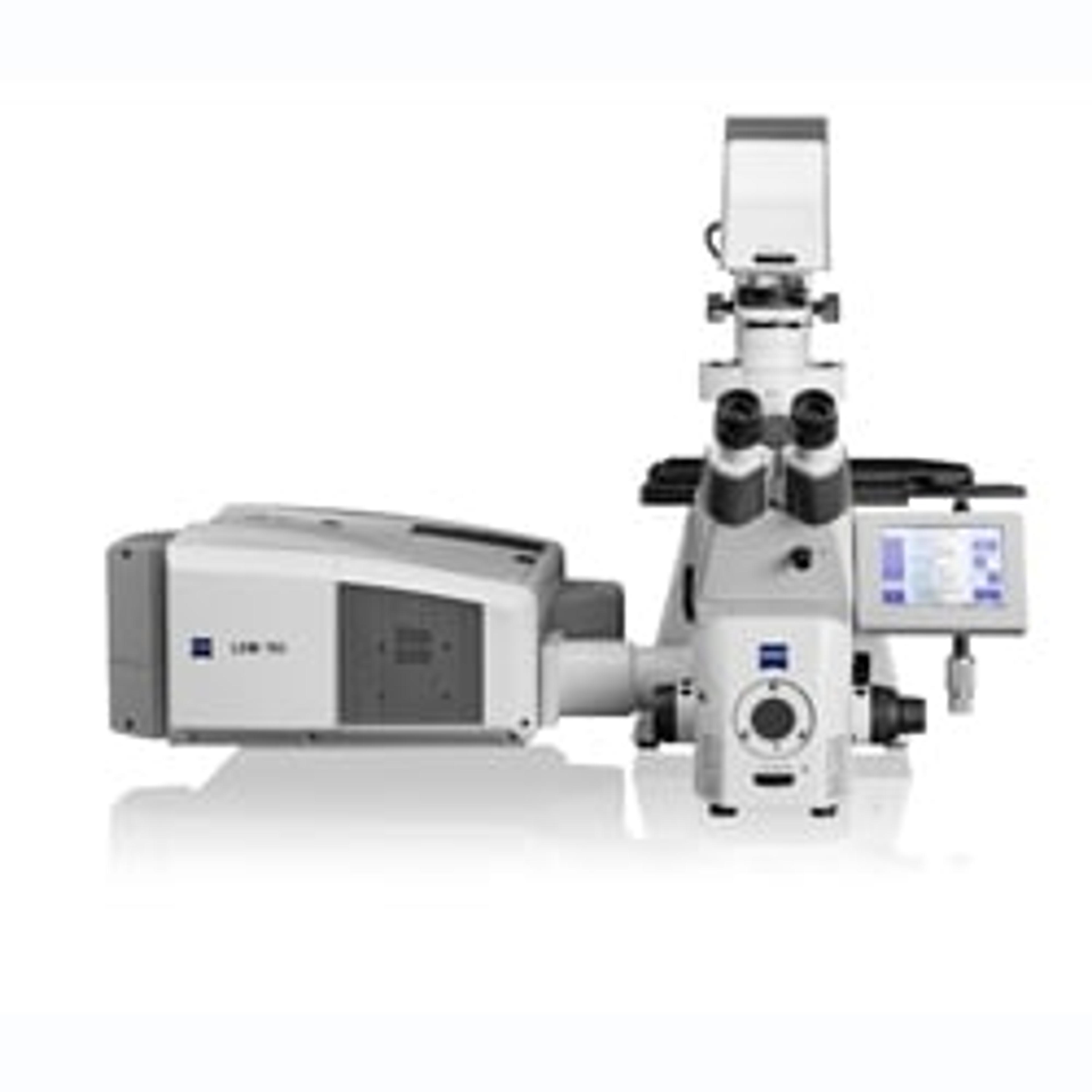
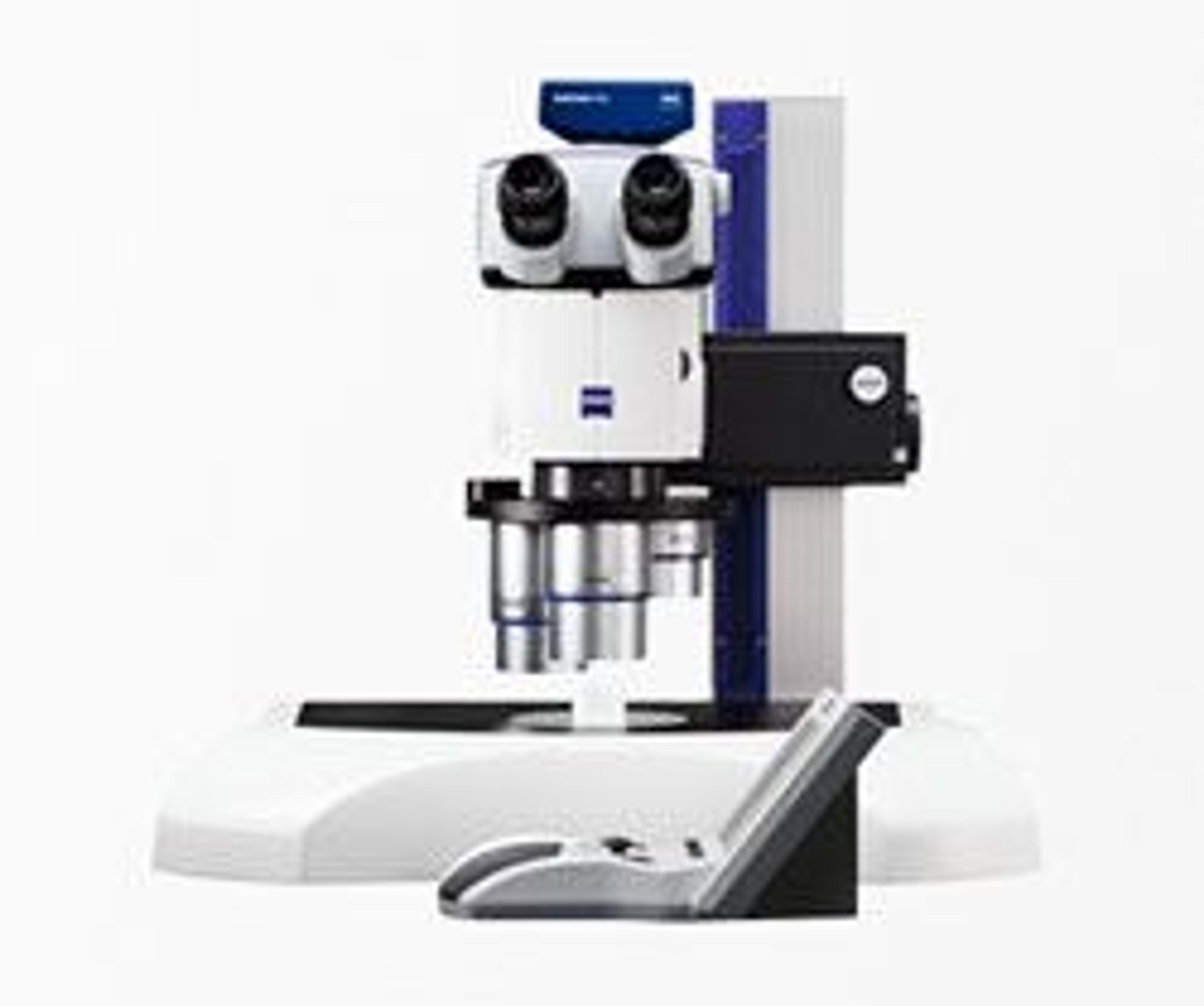
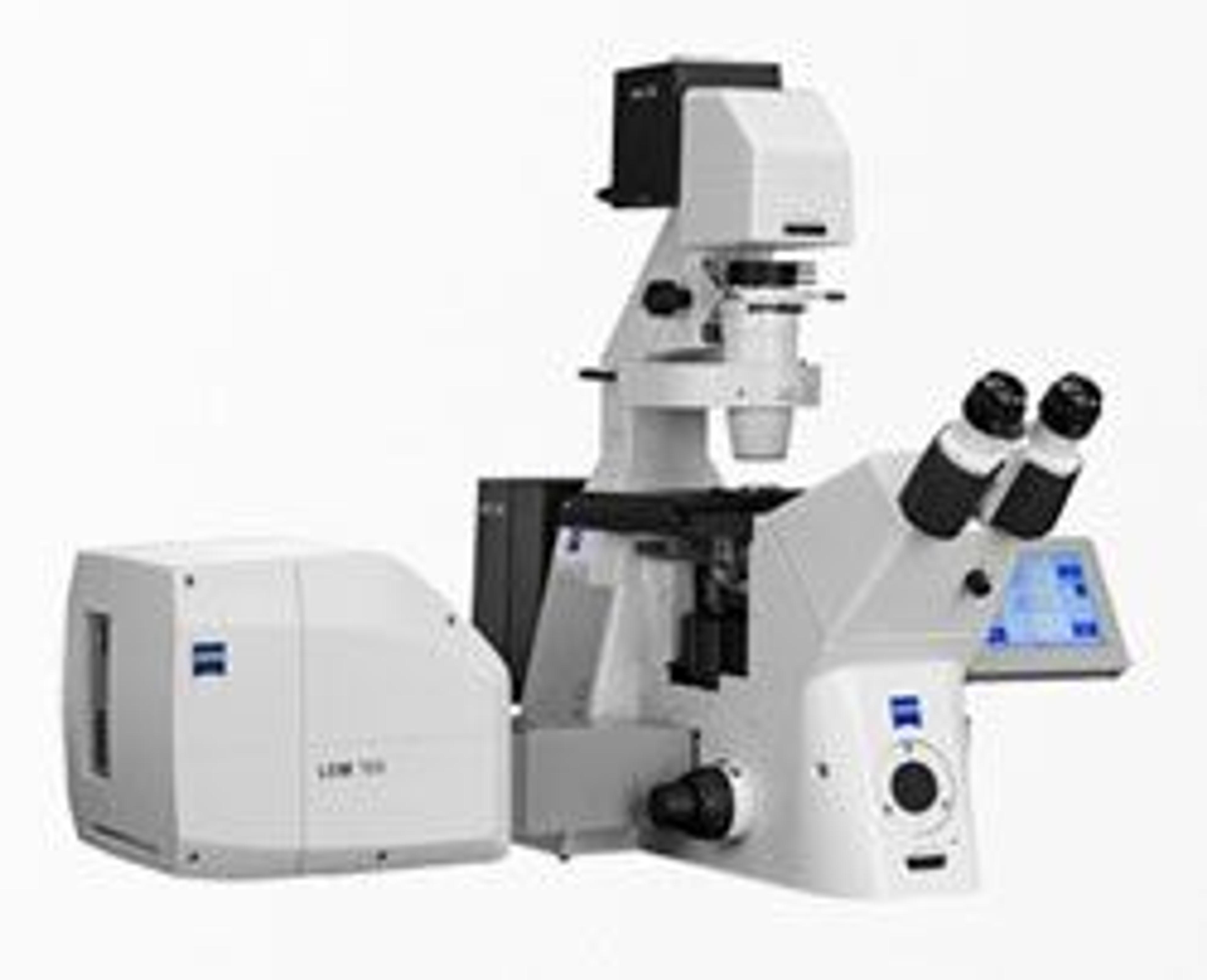
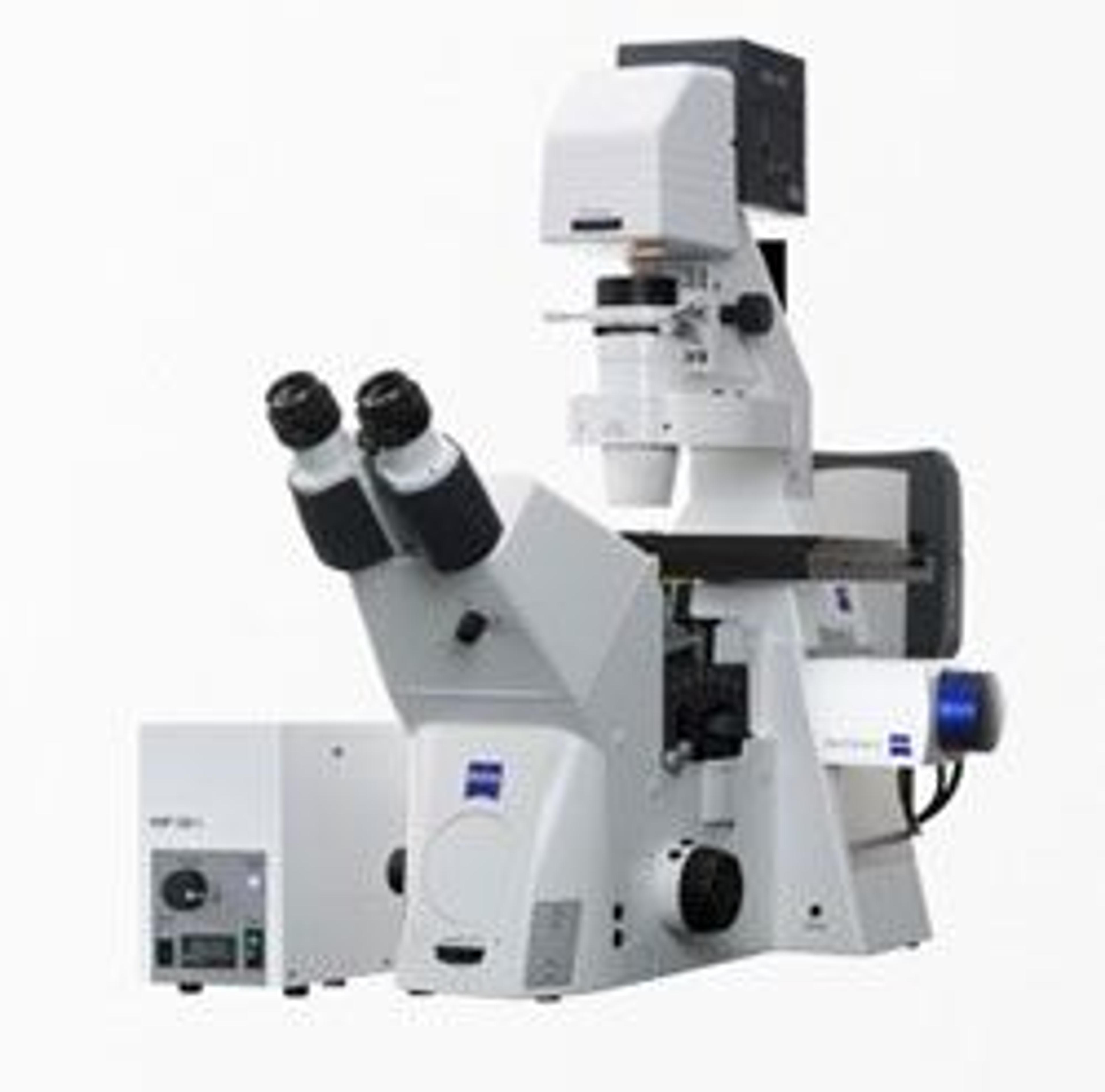
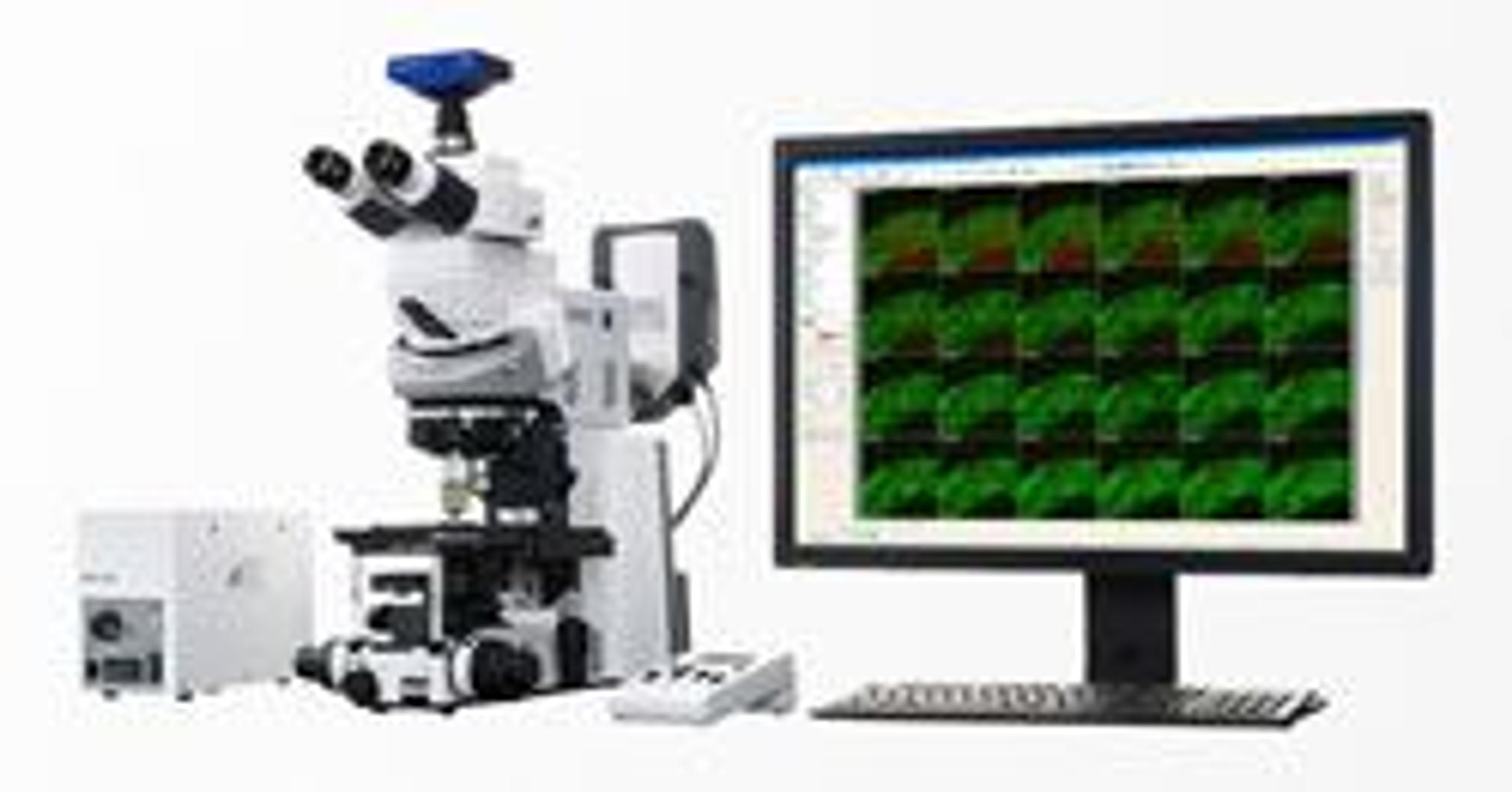
ZEISS LSM 710 NLO and LSM 780 NLO
Your flexible laser scanning microscope for multiphoton imaging Open up the world of multiphoton imaging with ZEISS LSM 710 and LSM 780 microscope systems, used in combination with the NLO add-on. On top of all-round laser scanning microscopy, you see fluorescently labeled structures down to a depth of one millimeter in living tissue. Detect spectral emission signals of your multi-labeled sample simultaneously with the 32-chan…


Receive your quote directly from the manufacturer.
Cell Biology Tissue Engineering
Relatively easy user interface high quality images possible, but takes long time to scan
Review Date: 21 Aug 2015 | ZEISS Research Microscopy Solutions
Your flexible laser scanning microscope for multiphoton imaging
Open up the world of multiphoton imaging with ZEISS LSM 710 and LSM 780 microscope systems, used in combination with the NLO add-on.
On top of all-round laser scanning microscopy, you see fluorescently labeled structures down to a depth of one millimeter in living tissue.
Detect spectral emission signals of your multi-labeled sample simultaneously with the 32-channel internal detector. Use up to 10 external non-descanned detectors. With this information, you separate signals reliably and produce highly detailed images that are rich in contrast.
And, above all, deeper in insight.

Fluorescence Polarization and Anisotropy Imaging with LSM 710/LSM 780
In this white paper, ZEISS describes a general method for the determination of instrumental correction factor G, for fluorescence polarization and anisotropy experiments utilizing an anisotropy standard.
Upgrade Your ZEISS Confocal with Revolutionary New Technology
Find out how Airyscan, the revolutionary detection concept from ZEISS, enables you to use multicolor samples with any label and get image quality. Learn how you can upgrade your 3- or 34-channel LSM 710/ LSM 780 with Airyscan and benefit from increased signal-to-noise, resolution and speed for your confocal imaging.
Correlation of Two-Photon <i>in Vivo</i> Imaging and FIB-SEM Microscopy
This white paper demonstrates that by using focused ion beam milling and crossbeam imaging it is possible to observe the same dendrites at 7 nm isotropic resolution, that were formerly imaged over weeks with a NLO-system.