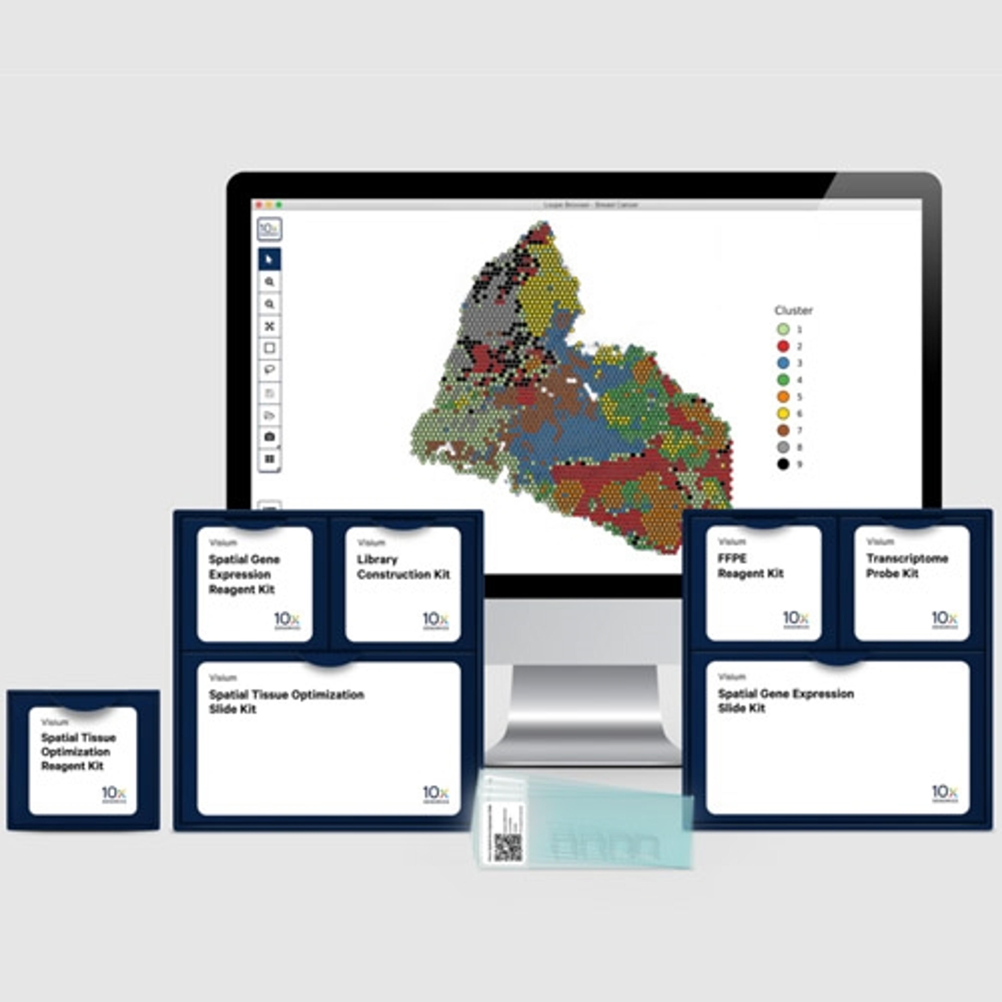
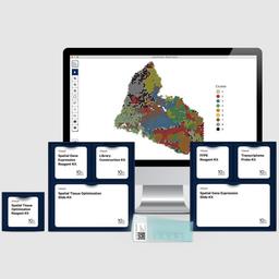
Visium Spatial Gene Expression
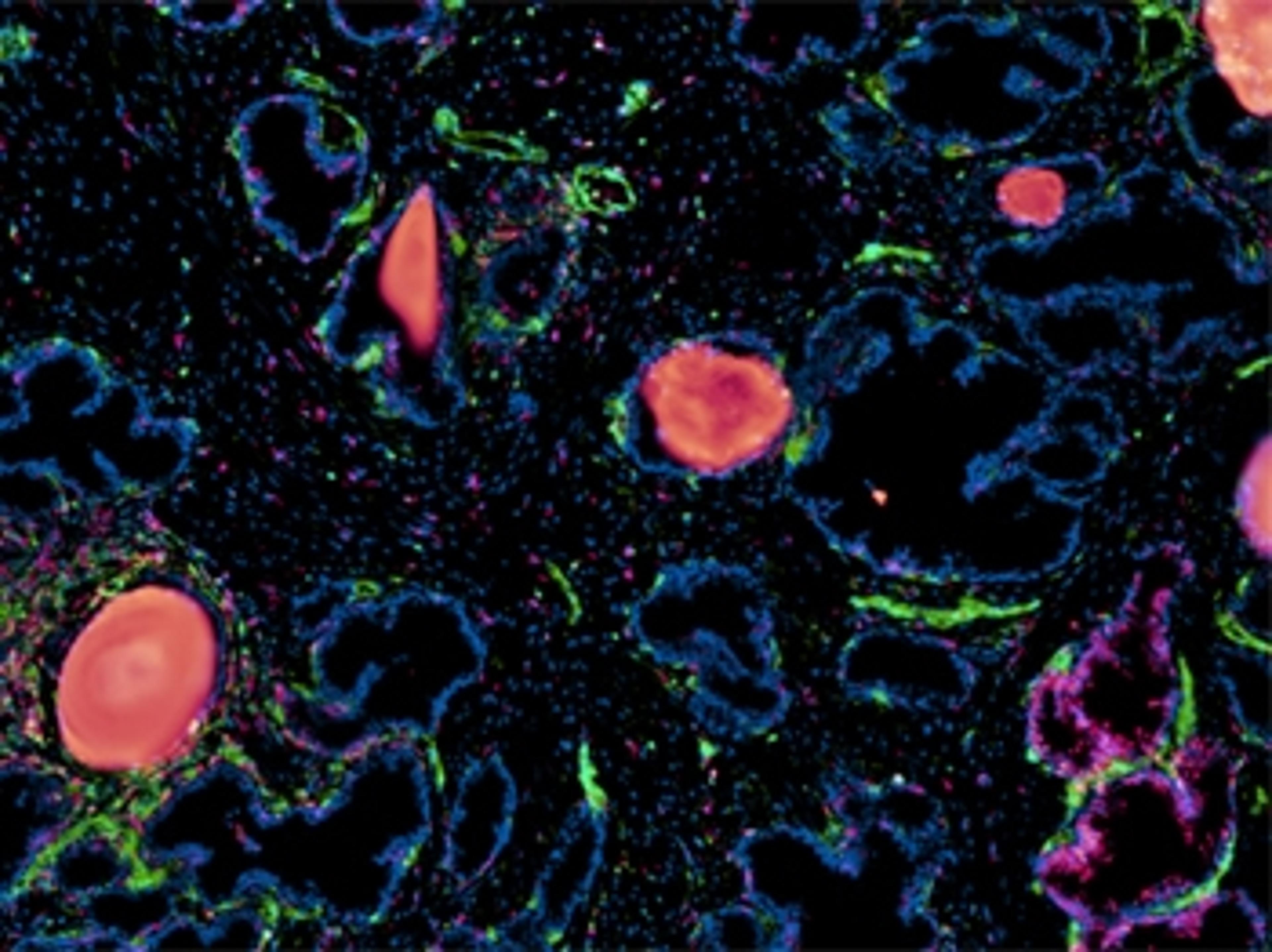
With Visium Spatial products researchers can now map the whole transcriptome for entire H&E- or immunofluorescence-stained fresh frozen, fixed frozen, or FFPE tissue sections with morphological context. Visium HD enhances the discovery power of this approach by enabling whole transcriptome spatial gene expression at single cell scale.

Visium Spatial Gene Expression

The supplier does not provide quotations for this product through SelectScience. You can search for similar products in our Product Directory.
Excellent products
reproductive multi-omics
Although a bit pricey, 10x Genomics latest line of products is fantastic for incorporating multi-omics into your research. We are using Visium now and look forward to incorporating Xenium in the future.
Review Date: 4 Apr 2023 | 10x Genomics
10 times better service
Genomics
The company offers 10 times better service than your expectations. The pioneer in spatial technology, customer service, end results, handling, ease of process, and worth purchasing.
Review Date: 5 Dec 2022 | 10x Genomics
Great. Want to apply to more samples and experiments
FFPE Visium gene expression
10x FFPE and visium is an amazing tech to investigate our cancer sample. We are able to detect copy number variation in our sample, and marker genes of clusters we are really interested. Though detachment may occur, we are able to achieve our goals. 10X has very good customer service, answering all my question in data analyses. Really appreciate their products.
Review Date: 26 Jul 2022 | 10x Genomics
This technology has helped me to have the best results in a short time
Brain Tumor
Visium Spatial Gene Expression from 10x Genomics is a novel assay that combines histology with spatially resolved whole transcriptome gene expression profiling to localize and quantify gene expression in the tissue context. Loupe Browser software (10x Genomics) makes it easy to visualize and explore spatial gene expression data on top of the morphological data from the tissue image. 10X has both Tech Support and Field Support that are readily available. The kits on sale are ready to use and no specialized instrumentation is required, consequently they can be accessible to all laboratories
Review Date: 24 Jan 2022 | 10x Genomics
- Access more sample types: Compatible with fresh frozen, fixed frozen, and FFPE tissue samples.
- Profile entire tissue sections: No need to select regions of interest - analyze the whole transcriptome from an entire section.
- High resolution: 1-10 cell resolution on average per spot depending on tissue type. With Visium HD single cell scale spatial resolution.
- Diverse sample compatibility: Demonstrated on a diverse set of organs across species (human, mouse, rat, and more).
- Protein co-detection: Combine whole transcriptome spatial analysis with immunofluorescence protein detection.
- Streamlined data analysis: Combine histological and gene expression data with easy-to-use software.
Brochures

Inside Visium spatial capture technology
In this product brochure, learn how Visium Spatial Gene Expression incorporates whole transcriptome analysis for intact tissue sections with morphological context. Discover how these complementary methods are designed to offer a previously inaccessible view of tissue biology.
Visium Spatial Gene Expression Solution
In this product brochure, learn more about the Visium Spatial Gene Expression Solution from 10x Genomics. The solution is designed for the investigation of spatially resolved whole transcriptome mRNA expression, while capturing histological information in the same tissue section.
Guide to novel approaches in spatial biology: Gain deeper insights into tissue heterogeneity
Spatial biology techniques are expanding our understanding of biological architecture with ever greater resolution in the context of tissue microenvironment. Next-generation molecular profiling solutions for the analysis of spatial gene expression look set to be game-changers, allowing us, for example, to classify tissue based on total mRNA, and enabling advances in spatially resolved gene expression that tell us where in a cell that expression is changing.
In this eBook, learn how coupling single-cell approaches with new spatial analyses of gene expression is enabling researchers to see biology in new ways through significant shifts in spatial resolution and scale, whether for cancer cell profiling or greater basic understanding in neuroscience.
Download the eBook to learn how the latest spatial biology profiling platforms from 10x Genomics can be used to resolve cancer tissue types and brain architecture in normal and diseased states, including case studies of the following tissue types:
- FFPE prostate cancer tumor samples
- Triple negative breast cancer
- Human squamous cell carcinoma
- Brain architecture
- Alzheimer’s disease markers
Spatial whole transcriptome profiling of the tumor microenvironment in prostate carcinomas
This poster describes a study using the 10x Genomics Visium Spatial Gene Expression Solution for formalin-fixed paraffin-embedded (FFPE) tissue to analyze and resolve tumorigenic profiles in sections of normal, stage III adenocarcinoma and stage IV acinar cell carcinoma human prostate samples.
Deciphering complex tumor biology with targeted multiomic spatial profiling
In this poster, Visium spatial gene expression measurements were combined with immunofluorescence allowing high-resolution intersection of transcriptomic and proteomic readouts within a whole tissue without pre-selecting regions of interest.
Introduction to Spatial Transcriptomic Data Analysis - A Case Study of Renal carcinoma
Are you interested in how spatial transcriptomics allows researchers to answer biological questions? Dr. Isaias Hernandez, postdoctoral researcher, Centre de Recherche des Cordeliers and Paris Brain Institute, will demonstrate how his team were able to unravel the differences in tertiary lymphoid structures (TLS) of responding patients to immune checkpoint inhibitors.
Key learning objectives
- Learn in practice how to analyse spatial transcriptomic data from your experiments and which pitfalls to avoid
- Discover how information from multiple Visium slides can be integrated to reveal shared and distinct features in the TLS of responding and non-responding patients
- Learn how to obtain tumoral-immune hubs that provide biologically relevant information
- Understand how to apply spatial analysis to characterize cellular heterogeneity inside and around TLS
Who should attend?
Researchers and bioinformaticians working in areas such as:
- Spatial biology
- Immuno-oncology
- Spatial transcriptomics
- Renal carcinoma
- Tertiary lymphoid structures
- Tumor microenvironment
- Cancer
Certificate of attendance
All webinar participants can request a certificate of attendance, including a learning outcomes summary, for continuing education purposes.

Advancing neurodegeneration research with single-cell and spatial multiomics
This illuminating webinar tackles the intricate pathologies of neurodegenerative diseases. By harnessing the power of single-cell and spatial technologies, we’ll explore how a multidimensional view of biology can lead to groundbreaking insights into Alzheimer’s and Parkinson’s diseases.
Key Takeaways:
- Transform your histological analysis: Learn how to enhance your understanding of plaques and tangles and their effects on the surrounding microenvironment using Visium Spatial products to integrate spatial gene expression and protein detection.
- Dissect cellular contributions: Deepen your insights into the role of disease-associated cells in pathogenesis. Find out how to identify cell-type differences, discover unique subtypes, and pinpoint novel biomarkers with Chromium Single Cell products.
- Discover cutting-edge research: Single nucleus RNA and ATAC-sequencing technologies are unraveling the molecular and cellular dynamics of neurodegeneration.
- Alzheimer’s disease focus: Recent studies utilizing multiomic approaches across millions of nuclei from hundreds of patients have revealed complex gene regulatory networks and disease-associated cell states. Learn how these insights correlate with cognitive resilience and vulnerability, shedding light on how various brain cell types—such as excitatory and inhibitory neurons and microglia—are impacted at different stages of Alzheimer’s.
- Parkinson’s disease insights: We’ll also explore findings from a study on Parkinson's that identified 13 distinct microglial subpopulations in postmortem substantia nigra tissues from PD donors. This research highlights their unique chromatin characteristics and their roles in the inflammatory processes underlying PD pathology.
This Webinar is for:
- Neuroscientists, molecular biologists, and genomic researchers seeking to deepen their understanding of neurodegenerative diseases.
- Clinicians and translational researchers focused on Alzheimer's, Parkinson's, and related dementias.
- Professionals in pharmaceutical and biotech industries exploring advanced therapeutic development.
- Academics and lab scientists interested in leveraging single-cell and spatial multiomic technologies for cutting-edge discoveries.
This session is ideal for anyone looking to enhance their research with innovative tools and insights into the cellular mechanisms driving disease progression.
All webinar participants can request a certificate of attendance, including a learning outcomes summary, for continuing education purposes.

Accelerating drug discovery and development with single-cell and spatial profiling
Watch this on-demand webinar to discover how technology from 10x Genomics can help you enhance your single-cell and spatial assays