NucView® Caspase-3 Enzyme Substrates
Fluorescent caspase-3/7 substrates for detecting apoptosis in intact cells by confocal microscopy, flow cytometry, or live cell imaging.


Place your order directly with the manufacturer.
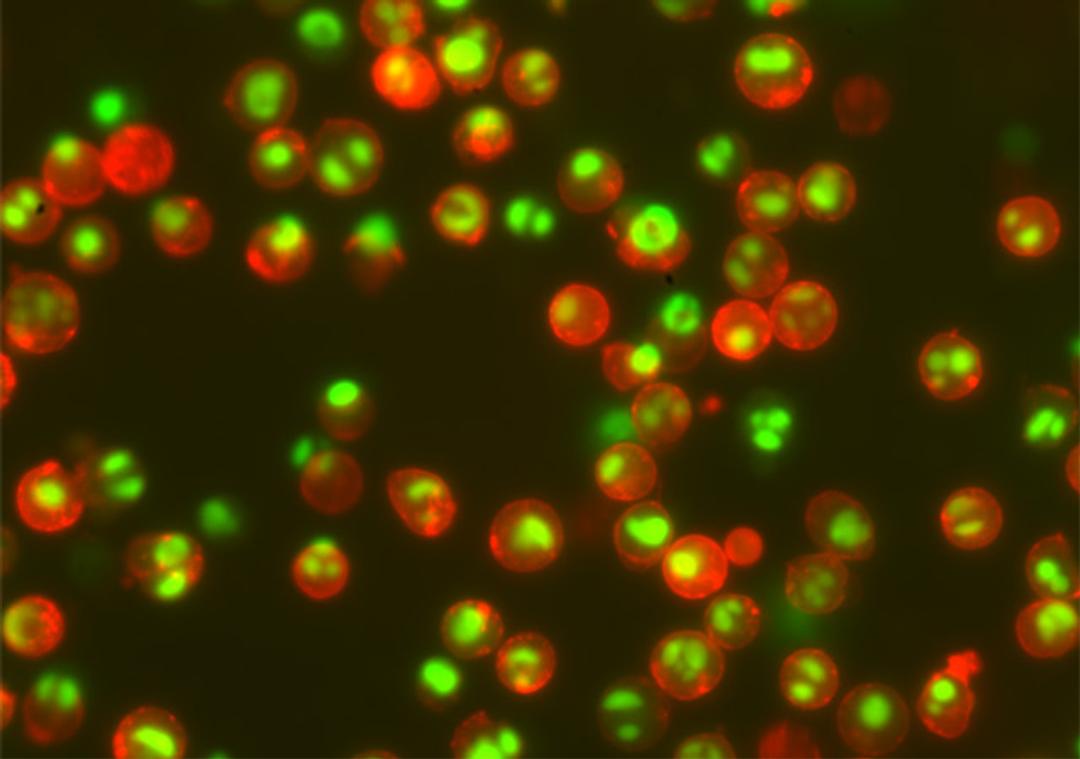
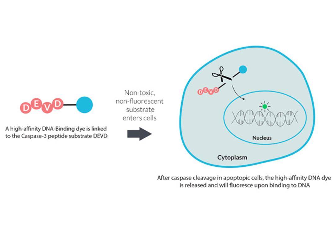
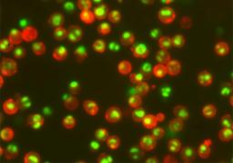
NucView® Caspase-3 Substrates are a convenient tool for detecting apoptosis in intact cells based on caspase-3/7 activity using confocal microscopy, flow cytometry, or live cell imaging.
- Rapid, no-wash, endpoint or real-time assays
- Non-toxic, allowing multi-day experiments to be performed
- Available in green, blue, or orange fluorescence
- For flow cytometry, microscopy or live cell imaging systems
- Dual detection of caspase activity and nuclear morphology
- Formaldehyde fixable
NucView® 405 Caspase-3 Substrate, 1 mM in DMSO #10405
NucView® 405 Caspase-3 Substrate, 1 mM in DMSO, Trial Size #10405-T
NucView® 405 Caspase-3 Substrate, 1 mM in 1X PBS #10407
NucView® 405 Caspase-3 Substrate, 1 mM in 1X PBS, Trial Size #10407-T
NucView® 530 Caspase-3 Substrate, 1 mM in DMSO #10406
NucView® 530 Caspase-3 Substrate, 1 mM in DMSO, Trial Size #10406-T
NucView® 530 Caspase-3 Substrate, 1 mM in 1X PBS #10408
NucView® 530 Caspase-3 Substrate, 1 mM in 1X PBS, Trial Size #10408-T
NucView® 488 Caspase-3 Substrate, 1mM in 1X PBS #10403
NucView® 488 Caspase-3 Substrate, 1mM in 1X PBS #10403-T
NucView® 488 Caspase-3 Substrate, 1mM in DMSO #10402
NucView® 488 Caspase-3 Substrate, 1mM in DMSO #10402-T
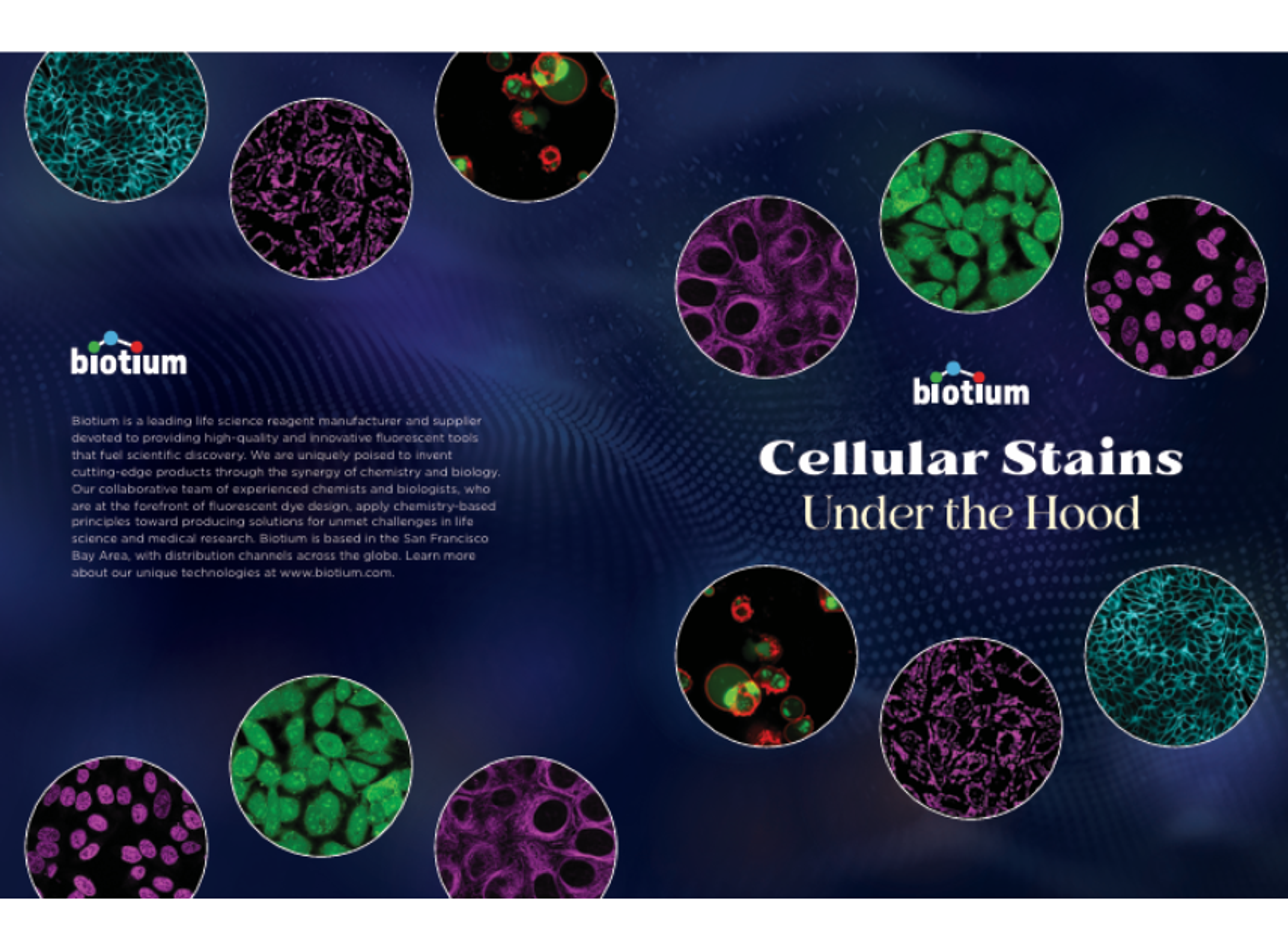
Selecting the right fluorescent cell stains for imaging and cytometry applications
This guide from Biotium provides an overview of fluorescent cell stains and their use in imaging and cytometry workflows. Discover how different probe types target specific cellular structures, including membranes, cytoplasm, nucleus, and organelles. The resource also outlines considerations for selecting appropriate dyes based on experimental needs, and highlights approaches for distinguishing live and dead cells, monitoring apoptosis, and improving signal specificity for reliable cell analysis.