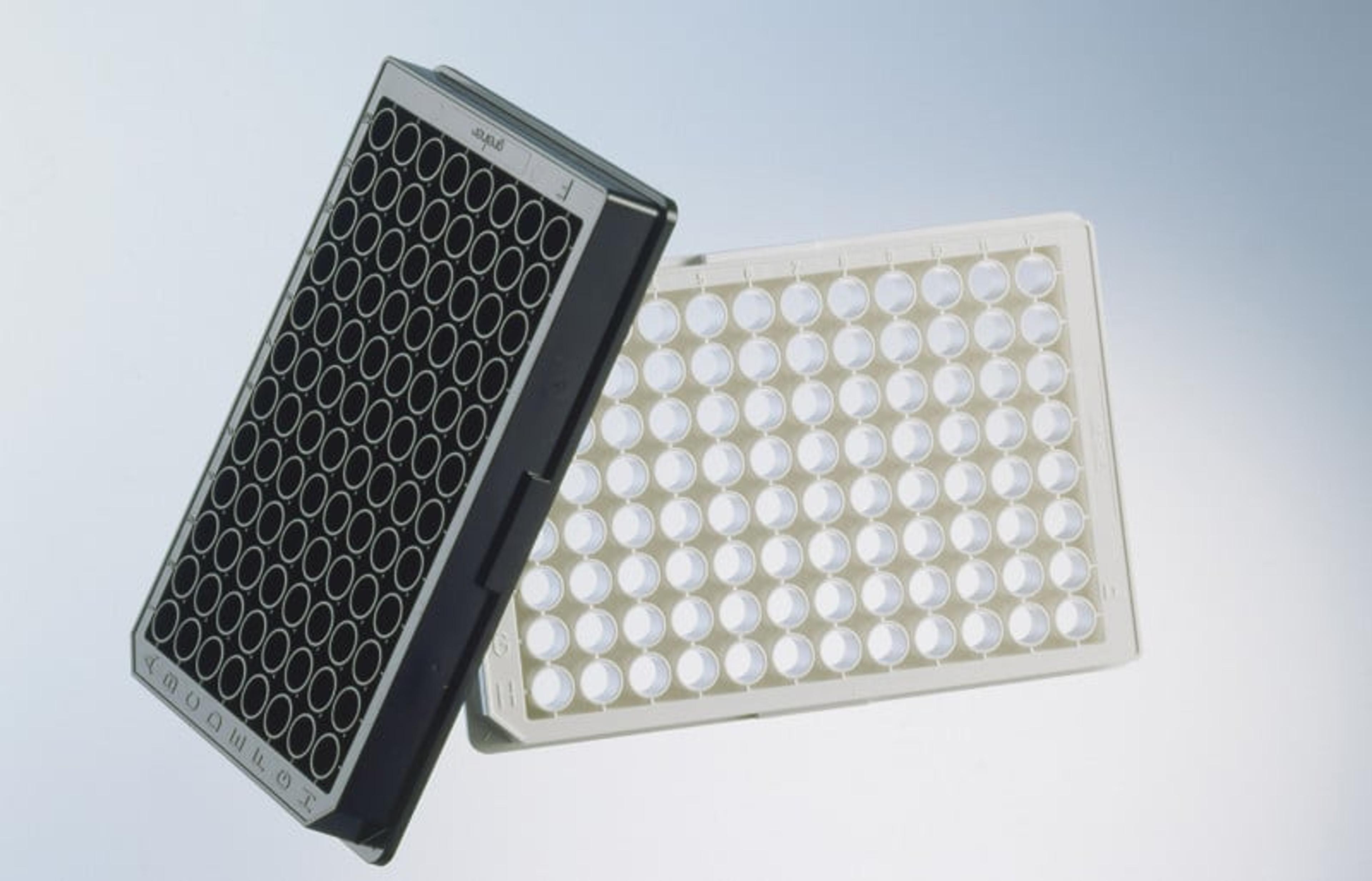
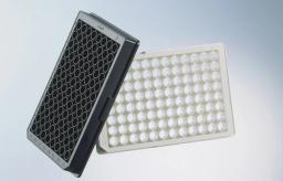
Microplates with µClear® film bottom
Black and white film bottom microplates

96 Well Microplates (µClear®bottom)
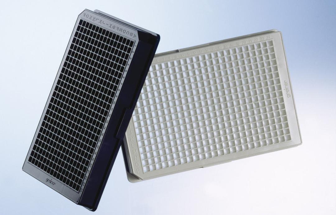
384 Well Microplates (µClear® bottom)


The supplier does not provide quotations for this product through SelectScience. You can search for similar products in our Product Directory.
Well made 96-well plates for sensitive fluorescence readouts.
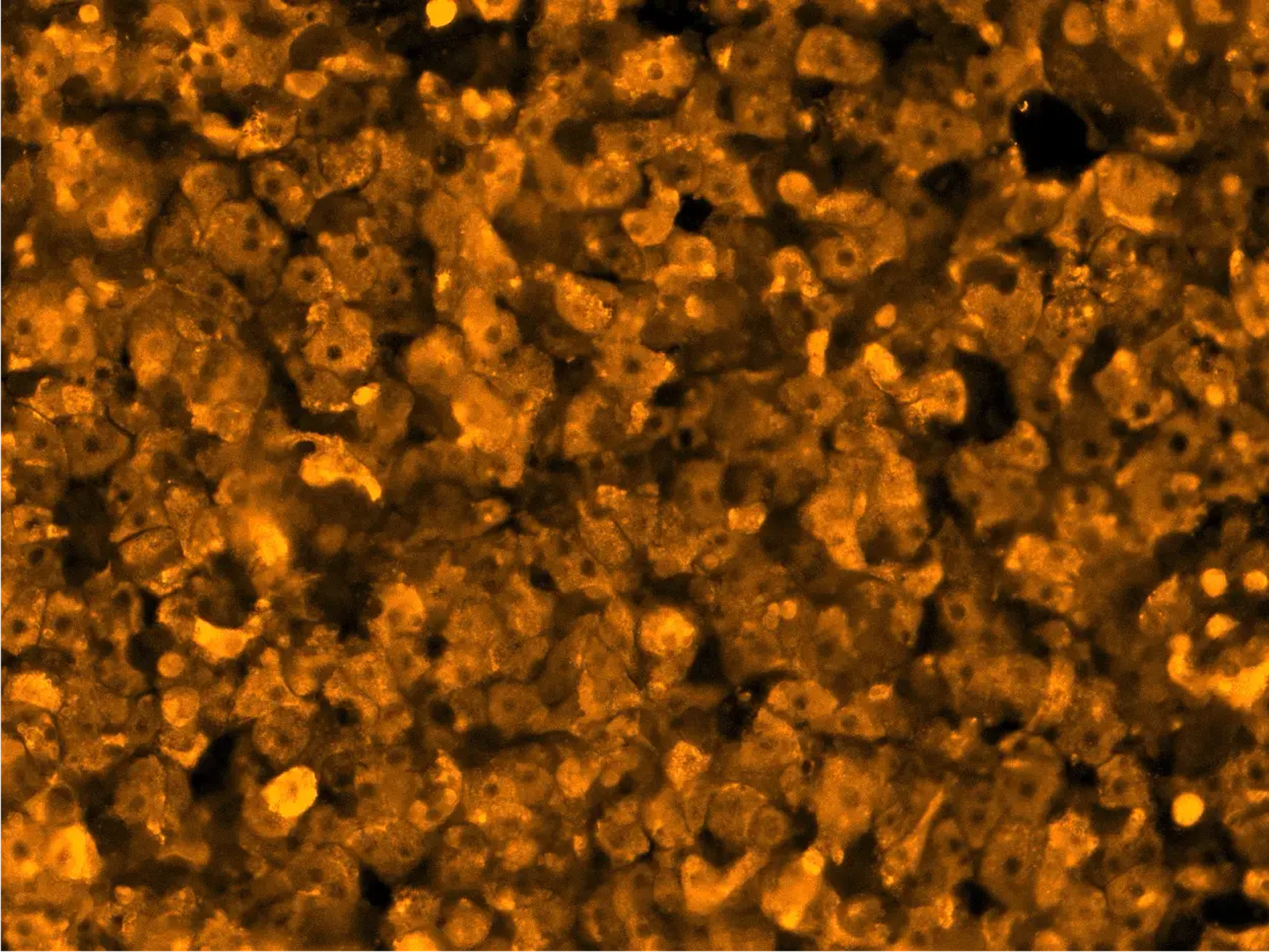
Fluorescence assays
The 96-well plates are very well made. I love the fact that it has indicators A1, A2, A3 etc. in the corner of each well so you don't need to count each time when pipetting. I use these for fluorescence assays. Great advantage is that the plates are offered in half-area, which allows me to use less sample and reagents, still getting the same sensitivity of the assay. None of the other manufacturers offer that option.
Review Date: 19 Jun 2019 | Greiner Bio-One GmbH
The development of a completely new and patented processing technique has made it possible for us to produce microplates with ultra-thin films, without the use of adhesives or solvents – the µClear® products. This special method eradicates the risk of leaking wells.
The choice of suitable films is the decisive factor, and this will influence the quality of a clear bottom microplate. Strict controls before and during production guarantee a constant quality. Polarised light is only depolarised to a slight degree and the autofluorescence of the microplates is minimized.
The 96 well µClear® microplates and 384 well µClear® microplates have a film thickness of 190 µm +/- 20 µm. In the 1536 well microplates with a transparent bottom (µClear®) the film thickness is 75 µm +/- 10 µm.
Brochures

Microplate selection guide
In this brochure, Greiner Bio-One GmbH aims to offer an overview of microplates, focusing on their applications and suitability for different research needs. Advancements in research technologies have led to various platforms used across different fields. Researchers face the task of selecting microplates tailored to their specific applications from a wide array of options. This brochure aims to provide a detailed solution to microplate selection.
Plastic labware for optimal results in modern life science microscopy using ZEISS Axio Observer and ZEISS Celldiscoverer 7
The interplay of microscope and microplate is important for achieving best results as the applicative requirements may differ significantly. In this application note, Greiner Bio-One provides an overview of the performance of two inverted microscopes – ZEISS Axio Observer (a classic stand) and ZEISS Celldiscoverer 7 (a boxed microscope system) – when working with different plastic labware products (SCREENSTAR, µClear® and CELLview) from Greiner Bio-One. The note details how ZEISS microscopes have been validated with Greiner Bio-One microplates ensuring full compatibility and highest optical performance with respect to the application requirements.
UV/VIS Spectroscopy in Microplates UV-Star, μClear, MICROLON and CELLSTAR
Ultraviolet-visible (UV/VIS) spectroscopy is a classical analytical method for determining the chemical constitution of a substance and its concentration in aqueous solution. UV/VIS spectroscopy is usually conducted in quartz glass cuvettes. However, cuvettes do not provide sufficient throughput when dealing with large amounts of samples, and microplates can be used to speed up work. In this application note from Greiner Bio-One, learn about how transparent microplates (CELLSTAR®, MICROLON) or microplates with a clear film bottom (μClear®, UV-Star®) are frequently used in UV/VIS spectroscopy. UV/VIS is a subclass of spectroscopy which uses visible light and adjacent near ultraviolet (UV) ranges for the determination of the concentrations and the characterization of dissolved substances.
Generation of Automated Label-Free Cell Migration Assays through the Incorporation of Magnetic 3D Bioprinting
Three-dimensional (3D) cell culture platforms are potential solutions as they can reconstruct tissue structure and environments in vitro. This application note presents two bioprinting procedures that were created to allow 3D wound healing and metastatic cell movement to take place in vitro. Final combined processes were tested using multiple skin and cancer cell models in 384-well format to demonstrate that the incorporation of 3D magnetic bioprinting can lead to the generation of in vivo-like cell migration data.