Multiplex immunofluorescence drives translational research
Find out why building expertise in multiplex immunofluorescence is critical in a translational clinical research setting
24 Apr 2024
Dr. Tom Lund, Scientific Lead of the Integrated Pathology Unit, Head of Digital Pathology for the Centre of Translational Immunotherapy in London
Dr. Tom Lund, Scientific Lead of the Integrated Pathology Unit (IPU) and Head of Digital Pathology for the Centre of Translational Immunotherapy in London, shares valuable insights into his team’s innovative immunotherapy research.
Lund highlights the IPU’s unique contributions to enhancing the understanding of complex cancer mechanisms, spanning from basic and translational research to supporting clinical trials, utilizing state-of-the-art tissue hybridization and digital pathology. He delves into the pivotal role of cutting-edge multiplex immunofluorescence technology in advancing clinical knowledge and enhancing patient outcomes, with implications beyond the dynamic world of immunotherapy.
The diverse landscape of translational cancer research
Introducing his team, Lund explains that the IPU is a collaborative initiative between the Institute of Cancer Research and the Royal Marsden Hospital, functioning as a vital link across clinical, translational and basic research. His team supports clinical trial research by combining emerging tissue hybridisation technologies with quantitative digital pathology to investigate the tumor microenvironment. “The IPU is a unique hybrid consisting of a core facility, a clinical lab, and an academic research facility. We apply multiplex immunofluorescence to large-scale clinical cohorts, standardizing assays as well as scientific interrogation across clinical trials over thousands of patients to generate clinically meaningful data,” elaborates Lund. “By using the cells in the tumor environment as a novel stratification criterion, we aim to evaluate predictive or prognostic patient responses to therapies ahead of time.”
Highlighting the substantial impact achieved by the IPU since its inception in 2022, he continues, “Our team has actively supported nearly 60 different clinical trials, spanning 19 different cancers, over the past 18 months.” Lund credits the IPU’s flexibility to the tissue-agnostic Akoya technology employed by his team, which requires minimal tissue-specific optimization, allowing seamless adaptation to diverse research requirements. “The Akoya Biosciences PhenoImager® HT instrument works extremely well not only for high-throughput analysis, but also bespoke analysis, offering insights specific to the clinical questions posed by a trial. Using this instrument has enabled us to quickly get to the bottom of what the clinical trial is trying to achieve, while helping eliminate concerns on whether the technology used suits a particular sample. My team has relied on Akoya instruments for a long time now, with the PhenoImager HT serving as a central resource within our laboratory.“
As a result, our lab’s analysis time has been cut by half, without compromising on accuracy, ensuring that the research output remains of a high standard.
Dr. Tom Lund, Head of Digital Pathology for the Centre of Translational Immunotherapy, on the new PhenoImager HT 2.0 upgrade
Optimizing high-throughput multiplex immunofluorescence technology
In addition to the minimal optimization required by Akoya instruments, one of the key reasons that Lund considers himself an advocate for Akoya is the prompt and robust support they provide. “The Akoya team offers support at a nearly plug-and-play level, ensuring that the instrument is operational in an incredibly short amount of time,” he says. “At the Marsden, we began to see approximately 10,000 patients a year once we launched our services. Without the luxury of a prolonged two-year optimization and validation period, we had to hit the ground running! The PhenoImager HT stood out as our natural first-choice – its robust technology and the strong chemistry of reagents backing it representing a giant leap forward compared to other available technologies in the market.”
In 2023 alone, the IPU analysed over 5,000 whole slide images, notes Lund; 8,000 whole slide images are already planned for next year. This makes the facility one of the most extensive users of high-throughput multiplex immunofluorescence technology in the world. “The Akoya workflow has allowed us to generate some of the largest multiplex immunofluorescence image databases ever assembled in record time, without requiring substantial investment on infrastructure,” he asserts.
Recently, Akoya has launched the next generation of the PhenoImager HT platform featuring new workflow additions such as on-instrument spectral unmixing to realize even higher throughput. Spectral unmixing provides the ability to distinctly isolate biomarkers of interest versus background noise, regardless of signal intensity or spectral overlap, ensuring accurate, quantitative data for insights on tumor-immune biology. The PhenoImager® HT Instrument 2.0 upgrade was officially launched in November 2023, but the IPU was an early-access user, and the new features have proved instrumental in its research, mentions Lund. “Along with spectral unmixing, this latest iteration can perform real-time image stitching during data capture from the patient sample. This presents a significant advantage as large datasets – comprising high-resolution images of up to hundreds of gigabytes in size – can be quickly saved, transferred, and analysed,” he explains. “As a result, our lab’s analysis time has been cut by half, without compromising on accuracy, ensuring that the research output remains of a high standard. Overall, this next generation of Akoya’s technology has been crucial in expediting our analysis process.”
Envisioning the future
Looking ahead, Lund emphasizes that the application of multiplex immunofluorescence is not limited to immunotherapies alone, but further holds promise across different therapeutic modalities. He identifies three key applications that appear promising for the near future. “First, in the context of cell-based therapies and cancer vaccines, where there is a need for a comprehensive understanding of specific cellular subtypes, multiplex immunofluorescence could play a pivotal role,” ventures Lund. “It has the potential to offer detailed insights into the types and activation statuses of immune cells involved in these therapies, providing a superior alternative to conventional single-color assays like standard immunohistochemistry that are used widely today.”
The second promising application of multiplex immunofluorescence is in potentially enabling a more nuanced understanding of the mechanisms underlying cytokine therapies. As an example, Lund highlights ongoing multiplex immunofluorescence-based RNA imaging research by his team, with the goal to investigate the cells producing cytokines as well as the receptors to which cytokines bind.
The third application – which Lund considers to be most important – lies in the field of combination therapies. There is a growing scientific consensus that the immune system and tumor cells are surrogates for overall tumor health, notes Lund. “As researchers enhance their understanding of immune cell behaviors in the tumor macroenvironment, their ability to predict patient responses and resistance to various therapies will also improve,” he anticipates. “This would extend to beyond immune-targeted therapies, potentially including mainstay treatments like chemotherapy, radiotherapy, complex targeted kinase inhibitors and even surgical interventions. This suggests a future where immune cells serve as biomarkers offering valuable insights for non-immune targeted therapies within combination treatments.”
“As the possibilities for the use of multiplex immunofluorescence continue to expand, it is exciting to unlock new dimensions of understanding and treating cancer that it offers, with the promise of ultimately improving patient outcomes,” concludes Lund.
Related Products
Request Quote for All Products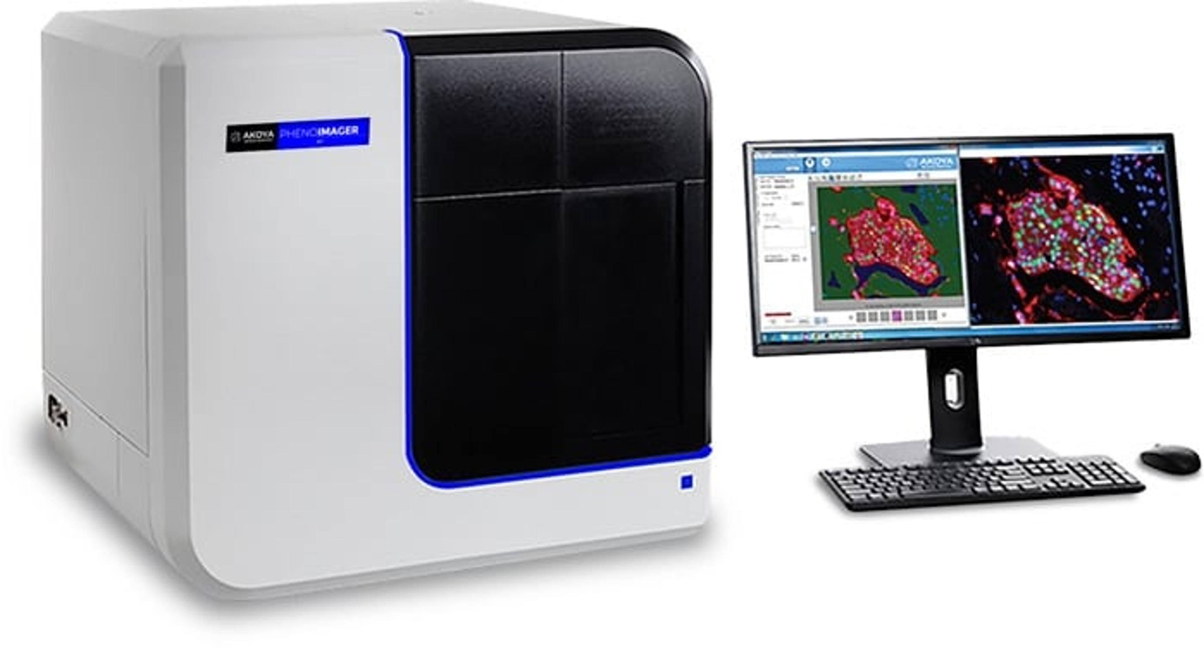
PhenoImager® HT
Akoya BiosciencesPhenoImager ® HT (formerly Vectra ® Polaris™) is the fastest and most highly cited whole-slide, single-cell resolution imaging platform for spatial phenotyping and the development of spatial signatures. Featuring Akoya’s patented Multispectral Imaging (MSI) and spectral unmixing technologies, this platform can be easily integrated into high-throughput workflows to accommodate projects regardless of your scale.