
ZEISS introduces new lattice light sheet fluorescence microscope
30 Dec 2020
With the release of ZEISS Lattice Lightsheet 7, ZEISS places a turn-key lattice light sheet instrument at the disposal of the life science research community. Based on the pioneering research and developments of Ernst H. K. Stelzer while at EMBL, Heidelberg, on light sheet technology and of Nobel laureate Eric Betzig while at the Janelia Research Campus of HHMI on structuring light sheets as optical lattices to render them thinner and longer, ZEISS Lattice Lightsheet 7 is tailored to observing cellular processes within cells and small organisms in 3D over extended time periods – all at subcellular resolution – with the minimum light dosage. Combined with full environmental control and an extremely stable optical setup, ZEISS Lattice Lightsheet 7 allows researchers to observe living specimens for hours or days.
“Lattice light sheet microscopy excels in a broad spectrum of live cell imaging applications as it provides high volume speeds with subcellular resolution and minimized light impact. Previously, however, lattice light sheet systems were reserved for experts,” says Klaus Weisshart, product manager at ZEISS. “The alignment and calibration of such systems required a physicist in the lab. Sample mounting as well as locating was not an easy task. ZEISS Lattice Lightsheet 7 overcomes these obstacles with ingenious technical implementations. I, as a biologist, can handle the system with ease, even without in-depth knowledge of the physics behind it.”
Calibration and alignment of the system fully automized
ZEISS Lattice Lightsheet 7 comes with a special structured light sheet, a so-called sinc3 beam. Calibration and alignment of the system runs fully automized in a matter of minutes. It also provides flexibility in generating light sheets of different lengths and thicknesses, all of which are available with just a click; thus, switching to the sheet that best suits the sample is very easy. Lattice light sheet generation is done by an exceptionally light-efficient beam shaping system using a spatial light modulator (SLM); only moderate laser powers are required. With its specification as a laser class 2 system and its small footprint, ZEISS Lattice Lightsheet 7 will find a space in any laboratory.
The true heart of the system is its core optics. “Since we had standard cell culture glass bottom dishes and multiwell formats in mind for use with the system, we implemented the idea of Yuichi Taniguchi from RIKEN, now Professor at the Institute for Integrated Cell-Material Sciences, Kyoto University, of an inverted configuration for our light sheet instrument,” explains Kirsten Elgass, ZEISS application engineer for 3D imaging solutions. The illumination and detection optics are arranged perpendicular like in classical light sheet set ups and at an oblique angle to the surface of the sample carrier’s cover glass. This is possible thanks to specially designed high-end optics. In this arrangement, the full numerical aperture of the detection objective lens is available. Resolutions of up to 290 nm x 290 nm x 450 nm can be achieved at an acquisition speed of up to 3 volumes per second, including standard sample preparation. The available transmitted light with its intrinsic contrast renders focusing on the experiment a simple task. With ZEISS Lattice Lightsheet 7, life science researchers can enjoy volumetric imaging at its best.
Want the latest science news straight to your inbox? Become a SelectScience member for free today>>
Related products
Request Quote for All Products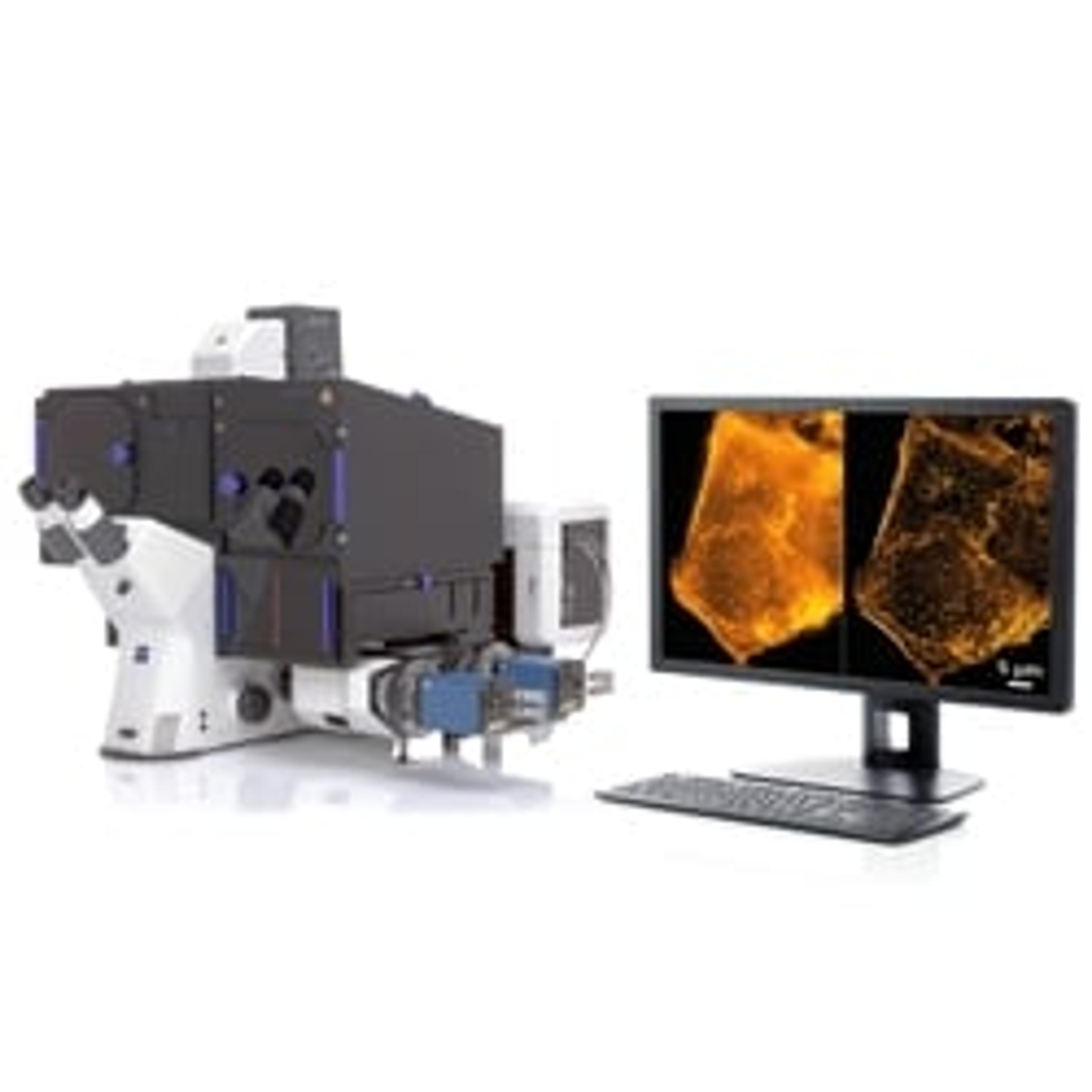
ZEISS Elyra 7 with Lattice SIM
ZEISS Research Microscopy SolutionsYour Flexible Platform for Fast and Gentle 3D Superresolution Microscopy