Workflow Applications of Continuous Live Cell Imaging
14 Sept 2016
Product news
Biomedical researchers are increasingly moving towards more advanced cellular models. These models display complex long-term dynamic biology, which places greater demands on the readouts and detection methods used to study them. Experimental methods that inform of changes in the properties of living cells over the long term are required to address added dynamic complexity. Ideally this information would be gathered throughout the life of the cells and in the context of other cells and their microenvironment.
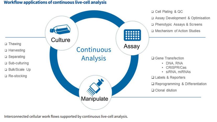
Continuous live-cell monitoring and analysis methods are beginning to address these needs and blur the lines between cell culture, cell manipulation and cell-based assay workflows to allow researchers to:
- better understand the timeline of biological processes and make informed decisions regarding their cell husbandry and analysis workflows
- increase efficiency by reducing the number of assay failures and the amount of initial assay development work that is required
- potentially streamline cell analysis workflow, save time & money and improve efficiency
Application Areas:
Cell Health & Viability
- Apoptosis
- Cell Proliferation Analysis
- Cytotoxicity
- Tumor Spheroids
Live Cell Assays
- Angiogenesis
- Cell Migration and Invasion
- Immune Cell Clustering & Proliferation
- Immune Cell Killing
- Neurite Dynamics - Label-free
- Neuronal Co-Culture - Fluorescence
- Phagocytosis
Cell Monitoring & Workflows
- Cell Culture QC
- Dilution Cloning
- Reporter Genes
- Stem Cell Monitoring & Reprogramming
- Transfection Efficiency
For more information watch this free webinar, download this whitepaper or visit the Essen BioSciences website.
Related products
Request Quote for All Products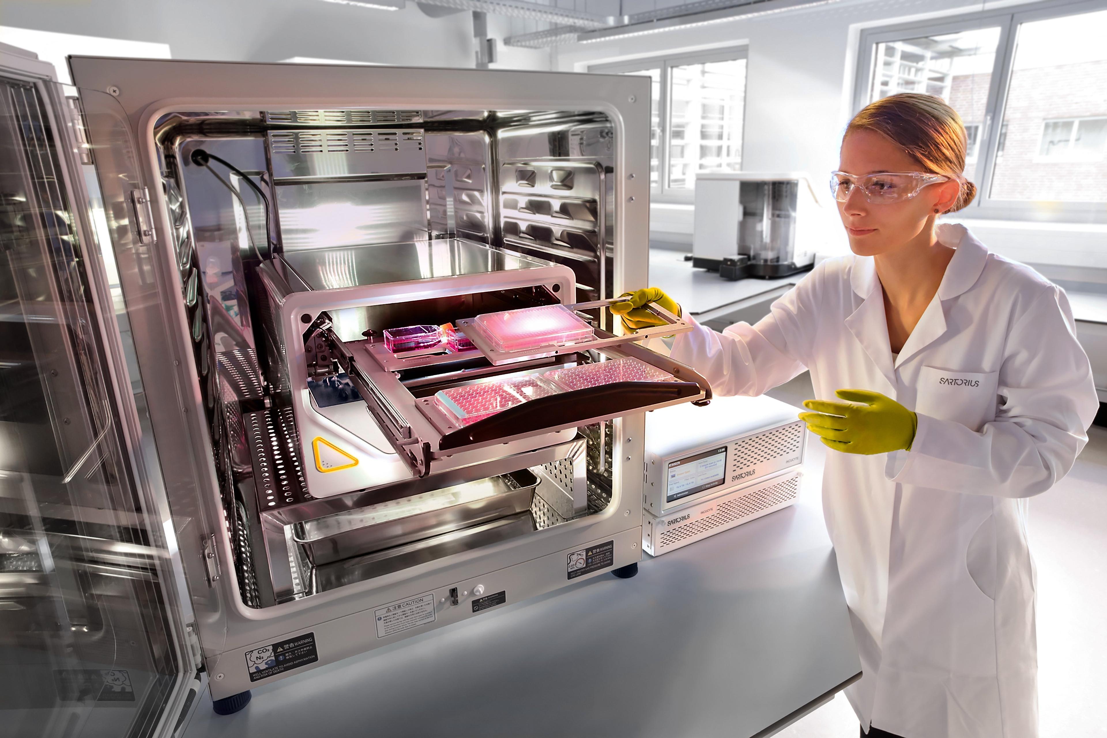
Incucyte® Live-Cell Analysis Systems
Sartorius GroupIncucyte ® , Empower Live-Cell Analysis Inside Your Incubator