
Trending in 3D cell culture: From targeting colorectal disease to tumor heterogeneity and patient-derived organoids
Discover the latest technologies and techniques that are revolutionizing 3D cell culture
26 Feb 2020
With 3D cell cultures being used increasingly for basic research and drug discovery, advancements in the field come thick and fast. From patient-derived colon cancer organoids and multidrug-resistant organisms to 3D imaging technology, keep up to date with some of the latest developments, techniques, resources and technologies in 3D cell culture in this round-up article. Plus, an expert explains the importance of the cellular microenvironment in the development of efficient drugs against complex disease.
Article: Targeting colorectal disease through patient-derived colon cancer organoids
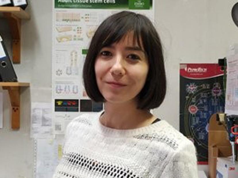
In this exclusive SelectScience interview, we talk with Dr. Asunción Fernández-Barral, Instituto de Investigaciones Biomedicas "Alberto Sols", CIBERONC, Madrid, Spain, to find more about her research into the regulatory role of vitamin D in relation to human colon stem cells and its implications in colorectal cancer. Read now>>
Article: 3D patient-derived cell models reflect tumor heterogeneity of clear cell renal cell carcinoma
Unraveling the causes and consequences of tumor cell heterogeneity is a main research goal for Dr. Hella Bolck, senior research fellow in the Department of Pathology and Molecular Pathology at the University Hospital Zurich. Studying the remarkable molecular and phenotypic variability of tumor cells is a key focus. By considering the heterogeneity of cells within individual tumors, Bolck’s research aims to help develop better strategies to treat cancer. Read now>>
eBook: Live-Cell Analysis: From Culture to Assay
In this application eBook, we look at the interconnected cellular workflows and continuous analysis cycle of cell culture to show how cutting-edge technologies from Sartorius deliver smarter cell monitoring. Download now>>
Application: 3D analysis of co-culture cancer spheroids using NoviSight™ software
In this application note, Olympus uses NoviSight 3D analysis software and the FLUOVIEW® FV3000 confocal laser scanning microscope to classify the types of cells contained in co-culture cancer spheroids and analyze cell-type-specific drug responsiveness. Download method>>
Application: Next-generation 3D patient-derived cell culture services
Reconstructed bone marrow and 3D cell culture services from zPREDICTA enable accurate testing of therapeutic agents against acute myelogenous leukemia. Download method>>
Dr. Nathalie Maubon launched HCS Pharma in 2014, inspired by an idea: in order to obtain and select more effective drugs, more relevant cellular models are needed for screening. In this exclusive interview, we speak with Maubon to find out more. Read now>>
Webinar: Multidrug-resistant organisms: Improving the time to effective treatment
In this SelectScience webinar, Dr. Jose Alexander, Director - Clinical Microbiologist at AdventHealth Orlando, discusses two key considerations when tackling the pressing issue of multidrug-resistant organisms. Watch now>>
Video: Increasing the accuracy of testing for anti-cancer therapeutics with Tecan's D300e Digital Dispenser
In this new video filmed at SLAS2020, zPREDICTA CEO and founder Julia Kirshner discusses the benefits of using Tecan's D300e Digital Dispenser for combination treatments in cell cultures to develop laboratory models for more accurate testing of anti-cancer therapeutics. Watch now>>
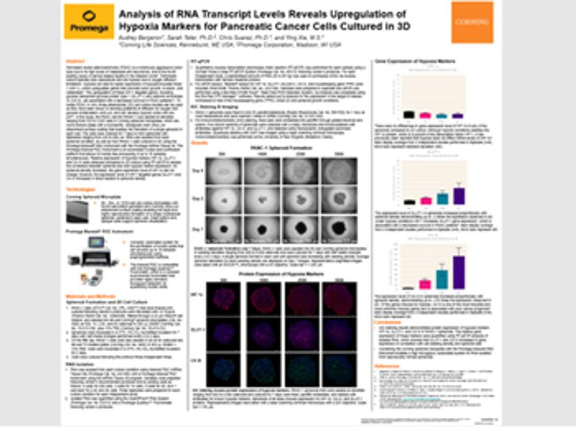
Application: Analysis of RNA transcript levels reveals upregulation of hypoxia markers for pancreatic cancer cells cultured in 3D
Pancreatic ductal adenocarcinoma (PDAC) is a notoriously aggressive tumor type due to its high levels of metastasis and recurrence and is the fourth leading cause of cancer-related deaths in the Western world. In this study, the PDAC cell line PANC-1 was seeded at densities ranging from 500 to 5,000 cells in Corning spheroid microplates and the relative expression of hypoxia markers HIF-1α, GLUT-1, and CA IX were analyzed compared to 2D culture using RT-qPCR to assess the correlation between spheroid size and hypoxia marker expression. Download method>>
Article: Cut through the haze: Can the latest 3D imaging technology help you remove out-of-focus blurring?
In this article, discover how Computational Clearing technology from Leica Microsystems quickly resolves the longstanding challenge of blurry contrast when imaging thick sections. Read now>>
Check out our 3D Cell Culture Special Feature page for more>>
Related products
Request Quote for All Products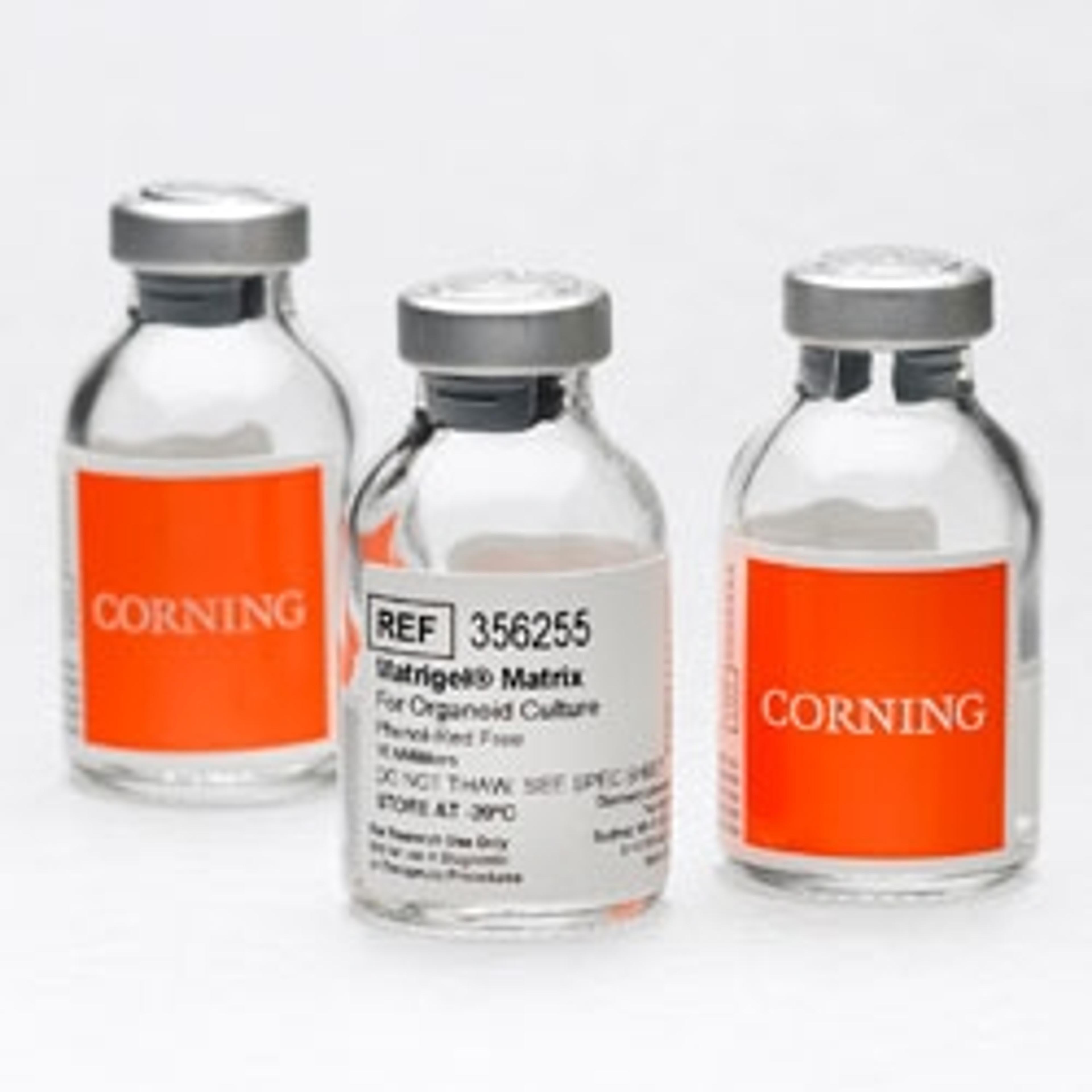
Corning Matrigel Matrix for Organoids
Corning Life SciencesAn optimized extracellular matrix that provides a convenient, ready-to-use solution, verified to support organoid growth and differentiation