The gel imaging system designed to improve workflow efficiency and fit just about anywhere
Discover an easy-to-use, compact solution promising high-resolution nucleic acid and protein gel documentation
29 Oct 2020
Editorial article

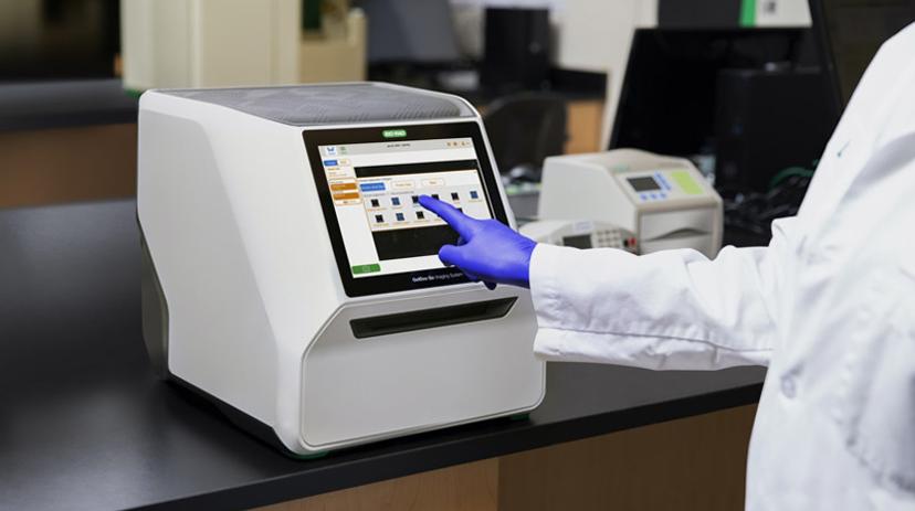
The compact design of Bio-Rad Laboratories’ GelDoc Go Imaging System makes it an ideal solution for many labs that are engaging in nucleic acid and protein research. Capable of imaging up to four mini-sized gels simultaneously, it enables researchers to capture high-resolution and publication-quality images of both nucleic acid and protein gels, all while occupying minimal space on the bench. In this SelectScience® interview, we speak with Dr. Paul Liu, product manager for Bio-Rad Laboratories’ Protein Quantification Division, to find out how this new system is set to transform the efficiency of gel imaging and analysis, and to hear his top tips on how to achieve the best imaging results.
What challenges does gel documentation present?
PL: Gel documentation and imaging has been around for a long time and the technology is pretty mature. However, there is definitely room for improvement. Researchers have become accustomed to using first film and now digital scanners to image their gels, then analyzing the image data afterward. One area for improvement is making the whole workflow, including wet lab, physical and digital aspects, more streamlined and efficient. I think we'll see a lot of companies focusing their efforts on this.
Why is gel documentation the method of choice?
PL: Gel documentation is still used because it's highly refined and able to provide the answers that scientists are looking for. In electrophoresis, for example, the aim is to separate by size and get a rough idea of quantity. Gel documentation is a very quick, inexpensive, and easy way to achieve this, whereas other technologies have some limitations. For instance, while capillary electrophoresis has the advantage of being hands-free and higher throughput, it's limited in terms of resolution, or in the case of antibody-based protein detection, not all antibodies work well in capillary systems. For now, gel documentation is still the go-to technology, and we don't see that changing for a number of years.
What are the key features of the GelDoc system and how is it addressing these challenges?
Compact size
PL: We designed the instrument to have a small footprint to conserve bench space and improve the efficiency of the physical workflow. Often labs are space constrained, especially for our academic customers, so this design allows it to fit anywhere in the lab. Customers are also able to put the instrument close to where they're running their gels. Previously, some of the larger instruments didn't fit under a bench or a shelf, meaning users would have to run their gels and then walk across the room, often spilling samples on the way, to take their images. The small design allows it to fit right next to the bench so users can do their work, take a picture, and then move on without having to move around the lab.
Data connectivity
PL: The efficiency of the digital workflow is also improved with this instrument through its easy-to-use Image Lab Touch software platform. This intuitive software is application driven, meaning the scientist can focus on being a biologist rather than a technologist. Users can take digital images and then export these either to a USB drive or to a network, which helps to smooth out the digital workflow.
Stain-Free technology
PL: The GelDoc Go, like other Bio-Rad imagers, is compatible with Stain-Free technology, which allows you to detect all proteins in a gel without a separate staining step. Bio-Rad Stain-Free gels can be run and total protein imaged directly on the GelDoc Go, reducing the amount of time and reagents needed. Ultimately, this further helps the wet workflow become more efficient.
High-resolution imaging
PL: The camera in the GelDoc Go is a high-resolution CMOS-based camera, meaning researchers can very quickly acquire an image that is publication-quality straight off the bat.
Who is the system designed for and for what applications?
PL: The system is designed for anyone performing basic research, including both academic and research-focused labs where the instrument’s small size and efficiency is an advantage, as well as pharma labs, where additional security features such as multiple user accounts and access permissions can be an advantage for regulated environments.
In terms of applications, the GelDoc supports all the common DNA and protein gel stains, from Coomassie to Bio-Rad Strain-Free and silver stains, and for nucleic acids both traditional UV stains such as ethidium bromide, as well as blue-excitable safe stains.
What do you see for the future?
PL: Like many other companies, Bio-Rad has recognized that the world is moving towards a digital workspace and we are looking to improve the integration of our instruments into that digital workflow. This includes user accounts and access, experiment data management, as well as automated data analysis. We are now positioning ourselves to jump to that next step.
Top tips for achieving success with the GelDoc Go
From Paul Liu, Bio-Rad Laboratories
- Put it right next to your gel running bench. This way you don’t have to walk across the lab and spillage is minimized.
- Keep it clean. When spillage occurs, the mat on top of the instrument is waterproof, removable, and washable. You should also remove the sample tray after imaging and clean it, as this prevents it drying out and becoming encrusted.
- Take advantage of Stain-Free technology. With this feature, you can take an image in just one minute instead of the two hours that Coomassie stains need.
- Don’t forget your password. Our Image Lab Touch software requires an admin account to be set up, so I recommend creating two separate admin accounts in case one is forgotten.
- Create individual accounts. By doing this, users can always go through their old data with ease, without going through everyone else's data.
- If your lab uses ethidium bromide, wear gloves. If accidents happen or people are messy, the outside of the instrument can accumulate ethidium bromide — wearing gloves is therefore essential.
Do you use Bio-Rad products in your lab? Write a review today for a chance to win a $400 amazon gift card >>
Related products
Request Quote for All Products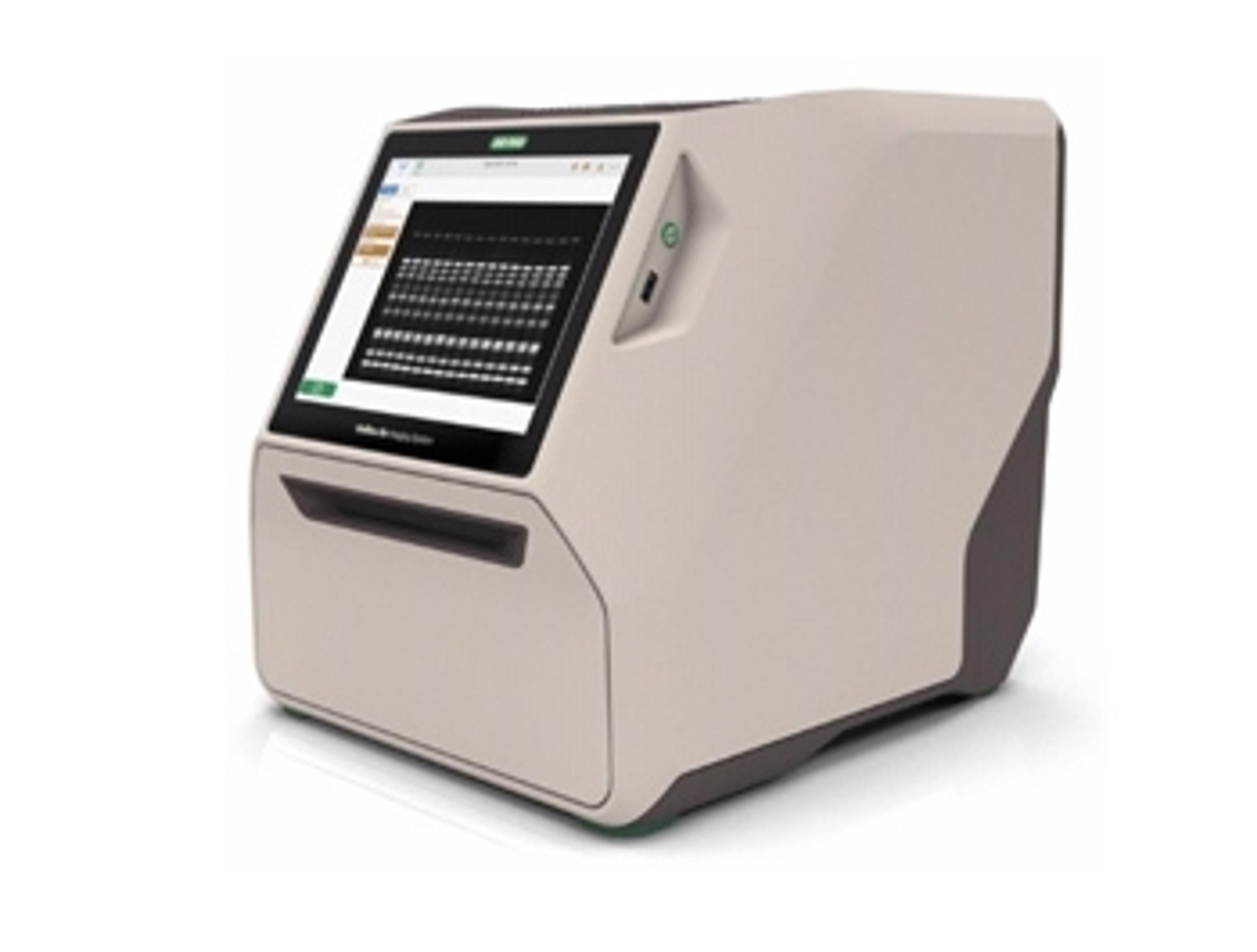
GelDoc Go Imaging System
Bio-RadThe GelDoc Go Imaging System gives you a benchtop imaging solution in a compact and evolved package.