Real-time live cell imaging revolutionizes tumor organoid research
Discover how real-time, high-throughput imaging and analysis with Countstar Spica M1 is transforming cancer research
2 Jun 2025
Editorial article
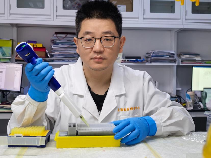
Dr. Zhenhua Li, postdoctoral researcher, Shanghai Clinical Research and Trial Center
Tumor organoids have emerged as a powerful model system, facilitating a deeper understanding of the biology of cancer microenvironments. Yet, conventional imaging methods used to analyze organoids introduce variables that compromise data quality.
As cancer rates rise globally, researchers are under pressure to find more predictive and scalable preclinical models. Unfortunately, traditional 2D and animal models often fail to reflect the complexity of human tumors truly. This has led to a surge of interest in organoid technology, miniaturized cell structures that mimic real tumors more closely than ever before. However, for these systems to materialize its potential, scientists need advanced imaging technologies that don’t interfere with the microenvironments they are studying.
A new era in cancer modeling
Dr. Zhenhua Li, a postdoctoral researcher at Shanghai Clinical Research and Trial Center, shares how the Countstar Spica M1 from ALIT LifeTech enables real-time imaging and AI-powered analysis directly within incubators, enhancing throughput, reproducibility, and experimental integrity.
Zhenhua’s research aims to enhance our understanding of thetumor micro-environment, tumor organoids, and their clinical applications. “Tumor organoids preserve the genetic background of tumor tissues in vivo and have demonstrated high specificity in drug sensitivity testing according to previous studies. Therefore, they hold great potential for future clinical applications in cancer treatment,” explains Zhenhua. “Moreover, in basic cancer research, they more accurately reflect real in-vivo conditions compared to traditional cell lines.”
The imaging challenge in organoid research
Zhenhua has encountered many challenges when analyzing organoids, particularly due to their 3D structure. “Since organoids are cultured in 3D, it is quite challenging to perform long-term tracking, imaging, and cell counting using conventional microscopy techniques,” Zhenhua explains.
Like most researchers, Zhenhua used to analyze organoids under a microscope outside an incubator and performed data analysis manually. Unfortunately, this approach introduces disturbances in the organoids and limits the throughput and reproducibility of the experiments. This is especially impactful when considering that the growth of organoids must be monitored under optimal culture conditions for extended periods.
While other high-end live cell imaging systems have been considered alternatives to traditional microscopy, they often fall short in maintaining environmental stability and are also cost-prohibitive. Subsequently, Zhenhua identified Countstar Spica M1 as an optimal solution, due to its combination of performance, affordability, and incubation stability, which are critical for long-term experiments and high-throughput settings.
By using Countstar Spica M1, Zhenhua has been able to perform non-disruptive, continuous monitoring of tumor organoids in 3D matrigel inside the incubator.
The system’s ultrafast Z-stacking technology captures clear, multi-layered images, whilst AI algorithms automatically detect, count, and analyze the organoid structure. Overall, this reduces manual user intervention, time, and potential error.
“As we can use the Spica M1 directly inside the incubator, our organoids can be cultured in a highly stable environment,” says Zhenhua. “In addition, it is equipped with various AI-powered functions, which are extremely practical.” This provides Zhenhua’s team with high-resolution, real-time data on organoid growth and morphology, which are key indicators of health, drug response, and disease progression.
The Countstar Spica M1 is designed for automated live-cell imaging and quantitative analysis of 3D cell models like tumor organoids. Its core features include:
- In-incubator imaging to preserve culture conditions
- Ultrafast Z-stacking for sharp 3D image capture
- AI-powered image processing for object recognition, labeling, and measurement
- Automated growth monitoring and real-time data visualization
Explore the features of this advanced live cell analysis system designed for both 2D cell cultures and 3D organoids.
Future organoid research for precise cancer treatment
The future of tumor organoid research is bright, with researchers in both academic and industry labs now able to incorporate high-throughput, reproducible workflows that meet clinical research standards. The addition of technologies like Countstar Spica M1 gives scientists the ability to reliably culture and analyze organoids, opening doors to precision oncology, drug screening, and longitudinal studies, which have previously been riddled with bottlenecks. “The efficiency and affordability are the most important advantages of the Spica M1,” concludes Zhenhua. “Looking to the future, it will enable more advanced cancer patients to receive and benefit from precise treatment , which will be truly exciting.”
Explore more of the latest news, reviews, and resources in our Accelerating Cancer Research Feature here.
Related products
Request Quote for All Products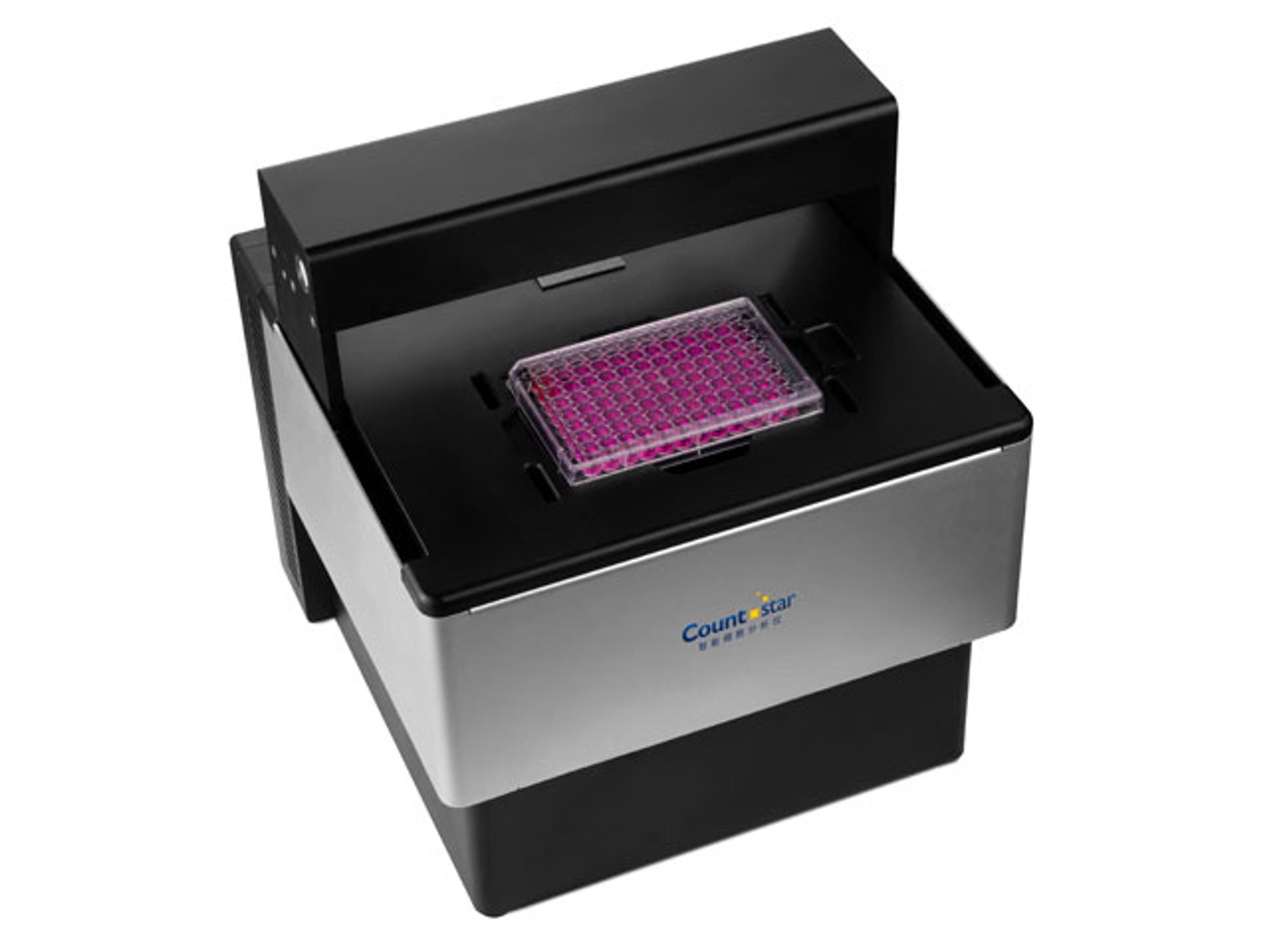
Frequently asked questions
How does the Countstar Spica M1 improve tumor organoid imaging compared to conventional microscopy?
The Countstar Spica M1 from ALIT LifeTech enables non-disruptive, real-time imaging and AI-powered analysis of tumor organoids directly inside the incubator. Unlike conventional microscopy, which requires removing organoids from optimal culture conditions and relies on manual analysis, the Spica M1 maintains a highly stable environment, supports long-term tracking in 3D matrigel, and uses ultrafast Z-stacking to capture clear, multi-layered images. Its AI algorithms automatically detect, count, and analyze organoid structures, improving throughput, reproducibility, and experimental integrity.
Why are tumor organoids important for advancing precision oncology and cancer drug screening?
Tumor organoids preserve the genetic background of tumor tissues in vivo and have demonstrated high specificity in drug sensitivity testing, making them powerful preclinical models for cancer treatment research. They more accurately reflect real in-vivo conditions than traditional 2D cell lines and many animal models, enabling researchers to study the tumor microenvironment, drug response, and disease progression with greater relevance to human tumors. This positions tumor organoids as a key technology for precision oncology, high-throughput drug screening, and longitudinal cancer studies.
What role does AI-powered image analysis play in tumor organoid research at the Shanghai Clinical Research and Trial Center?
At the Shanghai Clinical Research and Trial Center, Dr. Zhenhua Li uses the AI-powered image processing capabilities of the Countstar Spica M1 to automatically recognize, label, and measure tumor organoids in 3D cultures. The system’s AI algorithms perform automated growth monitoring and real-time data visualization, providing high-resolution data on organoid growth and morphology—critical indicators of organoid health, drug response, and disease progression. This reduces manual intervention, saves time, minimizes user error, and supports high-throughput, reproducible workflows that align with clinical research standards.