
How the race against Candida auris is advancing Candida diagnostics and susceptibility testing
Screening tools are becoming vital in the fight against Candida auris, a rapidly spreading and deadly fungal infection with growing antifungal resistance
10 Jul 2026

Dr. Nathan Ledeboer, Professor and Chief of Clinical Pathology, Medical College of Wisconsin
Candida auris (recently reclassified as Candidozyma auris) is a fungus that has been making headlines recently as the ‘super pathogen’ sweeping through hospitals that modern medicines cannot seem to stop.
The fungus readily colonizes surfaces, including human skin, and can cause severe, life-threatening infections in some patients. More concerning still, its strains are spreading rapidly and becoming increasingly resistant to many frontline antifungal treatments.
In response, researchers and clinicians are racing to implement effective screening approaches for Candida auris identification, improve antimicrobial susceptibility testing strategies, and develop novel antifungal drugs.
Among those leading the fight is Dr. Nathan Ledeboer, Professor and Chief of Clinical Pathology in the Department of Pathology and Laboratory Medicine at the Medical College of Wisconsin, and Associate Chief Medical Laboratory Officer for Froedtert Health.
The emerging threat of Candida auris
Candida species are a significant cause of human disease. While more than 15 Candida species can cause illness, more than 90% of invasive infections are caused by five: Candida albicans, Candida glabrata, Candida tropicalis, Candida parapsilosis, and Candida krusei.
Candida auris (recently reclassified as Candidozyma auris) represents one of the most significant growing threats – not because it is new, but because of how rapidly it is spreading. First identified in Japan in 2009, it has since expanded across continents and is now considered endemic in multiple countries.
Data from the U.S. paints a telling picture. Between 2013 and 2016, only five U.S. states, including Illinois, New York, New Jersey, Delaware, and Maryland, reported cases. By 2021, nearly one-third of U.S. states had identified C. auris, with many reporting between 100 and 500 cases1.
C. auris spreads so effectively because it can persist both on the human body and in the environment. It can be carried asymptomatically on the skin and other body sites, survive on surfaces for days, and even tolerate heat and some disinfectants.
Even when infection occurs, it often presents with non-specific symptoms such as fever, fatigue, and body aches, while more severe manifestations can be equally difficult to distinguish. “One of the biggest challenges that we see with Candida auris is the diversity of clinical syndromes it can cause,” explains Dr. Ledeboer. “It can affect nearly every site in the human body, including the bloodstream, skin and soft tissue, urinary tract, ear, respiratory tract, and the central nervous system.”
When invasive infection develops, outcomes can be severe, with reported mortality rates ranging from 29% to 53%2. And the impact is felt most in healthcare settings, including hospitals and long-term care facilities, where C. auris spreads readily, and critically ill patients are particularly vulnerable.
“Patients with invasive medical devices like urinary catheters are at increased risk of getting C. auris because it can colonize those catheters and then develop a biofilm, causing a clinically significant infection,” Dr. Ledeboer shares, noting that prolonged hospital stays, older age, diabetes, renal failure, and prior antibiotic use also increase the risk2,3.
Candida auris screening
With C. auris posing a growing threat in healthcare settings, early identification of infected and colonized patients is essential. As with MRSA and CRE, Dr. Ledeboer believes screening should be utilized in hospitals and other healthcare settings, particularly among high-risk patients or those suspected of colonization.
“Our team has been screening for C. auris since 2018, initially focusing on skin swabs of the palms, the webs of the fingers and toes, and perineal areas,” he explains, adding that evidence suggests these sites, as well as the nares and axilla, provide the highest likelihood of detecting the organism in a positive culture4.
There are a number of technologies available for Candida screening, and for clinical labs, the choice often comes down to a trade-off between turnaround time and cost.
“There are really good molecular techniques available that are fast – often less than 30 minutes – can be batched, and can achieve levels of sensitivity greater than 90% and specificity ranging from 95% to 99%,” says Dr. Ledeboer. “But the challenge with molecular testing is that it can be expensive.” In regions where C. auris prevalence is low, this cost can be difficult to justify for routine screening.
Traditional culture-based approaches offer a more affordable alternative but are slower and more labor-intensive. Samples must be plated and incubated at 40 °C for up to 48 hours before suspected colonies can be identified. These multiple manual steps can create bottlenecks, particularly in labs already short of staff, notes Dr. Ledeboer.
A potential middle ground is the use of chromogenic media for Candida detection, which can help streamline culture-based screening. Chromogenic media produces a distinct color change in the presence of Candida speciesand can even enable individual species identification.
“Oftentimes, chromogenic agar can provide you with a positive culture in 48 hours or less, and in many cases, 24 hours or less,” Dr. Ledeboer explains. “The performance provides high sensitivity and specificity not only for Candida auris, but for many other Candida species as well.”
Thermo Scientific™ Brilliance™ Candida 2 Agar and Spectra™ Candida Agar* from Thermo Fisher Scientific, for example, enable the identification of C. auris and differentiation of the five other clinically relevant species.
For culture-based approaches, another key consideration is whether to include a broth pre-incubation or enrichment step prior to plating. “This can improve sensitivity by 10% to 20% compared with direct plating,” says Dr. Ledeboer. However, this comes at the cost of time, potentially delaying results by a day or more and prolonging patient isolation. “You have to balance that desired optimized sensitivity against your need for a fast turnaround time,” he adds. “This will be driven by how frequently you are seeing the organism, how quickly you need results, and your infection control approach.”
Tackling antifungal resistance with susceptibility testing
Beyond its spread, what makes C. auris particularly concerning, is its growing resistance to treatment. Antifungals that once controlled infections are losing their effectiveness, with some isolates now fully resistant to azoles and showing increasing resistance to amphotericin B and echinocandins6.
“Candida auris has an incredibly diverse range of mechanisms for developing resistance,” explains Dr. Ledeboer. “This has led to a global increase in resistance, and not only to azoles. Across multiple continents, we are now seeing resistance to amphotericin ranging from 0% up to 40%, and in some countries, we're beginning to see rates of resistance to the echinocandins approaching 20%”5.
In the U.S, a study of cases in New York and New Jersey – which together account for more than half of U.S. Candida auris infections – found that between 2016 and 2020, more than 99% of isolates were resistant to fluconazole, while resistance to amphotericin B ranged from 48% to 82%6. “Fortunately, resistance to echinocandins, such as micafungin, caspofungin, and anidulafungin, remains low,” says Dr. Ledeboer. “However, even with newer antifungals like isavuconazole, we are now seeing up to 9.2% of isolates classified as non-wild type or resistant.”6
This growing antifungal resistance has led to the recommendation that any clinically significant isolate from an infected patient undergo antimicrobial susceptibility testing (AST), both to follow the epidemiology of the strain and to inform the proper treatment for that individual patient.
Laboratories have several options for AST, ranging from gold standard EUCAST and CLSI broth microdilution assays to commercial methods.
Comparative studies suggest that commercial methods generally perform well for Candida auris, particularly for echinocandins and fluconazole, with high levels of agreement with reference standards7,8,9.
For amphotericin B, if commercial methods provide an elevated minimum inhibitory concentration (MIC), it is important to consider sending that strain to a reference laboratory that uses a reference method – either from CLSI or EUCAST – to have the MIC confirmed, especially if amphotericin B is important in the management of that patient.
More on Candida testing solutions
In the U.S., England, and Wales, Candida auris is a notifiable infection, meaning healthcare facilities must report identified cases to state and local health departments. In many jurisdictions in the states, specimens are also required to be submitted to public health laboratories for strain typing and further characterization.
Accurate and reliable laboratory testing is essential for the surveillance, detection, and control of C. auris. To learn more about how laboratories can equip themselves for screening and antimicrobial susceptibility testing, explore the resources below.
- Whitepaper: Comprehensive screening of Candida auris (Candidozyma auris) to prevent healthcare-associated infections
- Whitepaper: Defending against the rising threat of drug-resistant fungal sepsis
*Thermo Scientific Brilliance Candida 2 Agar is available for purchase in Europe, while Spectra Candida Agar is available in the United States.
References
1. Lyman, M., Forsberg, K., Sexton, D. J., Chow, N. A., Lockhart, S. R., Jackson, B. R., & Chiller, T. (2023). Worsening Spread of Candida auris in the United States, 2019 to 2021. Annals of internal medicine, 176(4), 489–495. https://doi.org/10.7326/M22-3469 https://doi.org/10.7326/M22-3469
2. World Health Organization. (2022). WHO fungal priority pathogens list to guide research, development and public health action. https://www.who.int/publications/i/item/9789240060241
3. Pandya, N., Cag, Y., Pandak, N., Pekok, A. U., Poojary, A., Ayoade, F., Fasciana, T., Giammanco, A., Caskurlu, H., Rajani, D. P., Gupta, Y. K., Balkan, I. I., Khan, E. A., & Erdem, H. (2021). International Multicentre Study of Candida auris Infections. Journal of fungi (Basel, Switzerland), 7(10), 878. https://doi.org/10.3390/jof7100878
4. Proctor, D. M., Dangana, T., Sexton, D. J., Fukuda, C., Yelin, R. D., Stanley, M., Bell, P. B., Baskaran, S., Deming, C., Chen, Q., Conlan, S., Park, M., NISC Comparative Sequencing Program, Welsh, R. M., Vallabhaneni, S., Chiller, T., Forsberg, K., Black, S. R., Pacilli, M., Kong, H. H., Lin, M.Y., Schoeny, M.E., Litvintseva, A.P., Segre, J.A.,& Hayden, M. K. (2021). Integrated genomic, epidemiologic investigation of Candida auris skin colonization in a skilled nursing facility. Nature medicine, 27(8), 1401–1409. https://doi.org/10.1038/s41591-021-01383-w
5. Kean, R., & Ramage, G. (2019). Combined Antifungal Resistance and Biofilm Tolerance: the Global Threat of Candida auris. mSphere, 4(4), e00458-19. https://doi.org/10.1128/mSphere.00458-19
6. Kilburn, S., Innes, G., Quinn, M., Southwick, K., Ostrowsky, B., Greenko, J. A., Lutterloh, E., Greeley, R., Magleby, R., Chaturvedi, V., & Chaturvedi, S. (2022). Antifungal Resistance Trends of Candida auris Clinical Isolates in New York and New Jersey from 2016 to 2020. Antimicrobial agents and chemotherapy, 66(3), e0224221. https://doi.org/10.1128/aac.02242-21
7. Siopi, M., Leventaki, S., Pachoulis, I., Spruijtenburg, B., Meis, J. F., Pournaras, S., Vrioni, G., Tsakris, A., & Meletiadis, J. (2025). Evaluation of the MIC test strips for antifungal susceptibility testing of Candidozyma auris (Candida auris) using a representative international collection of isolates. Journal of clinical microbiology, 63(8), e0039925. https://doi.org/10.1128/jcm.00399-25
8. Siopi, M., Pachoulis, I., Leventaki, S., Spruijtenburg, B., Meis, J. F., Pournaras, S., Vrioni, G., Tsakris, A., & Meletiadis, J. (2024). Evaluation of the Vitek 2 system for antifungal susceptibility testing of Candida auris using a representative international panel of clinical isolates: overestimation of amphotericin B resistance and underestimation of fluconazole resistance. Journal of clinical microbiology, 62(4), e0152823. https://doi.org/10.1128/jcm.01528-23
9. Ceballos-Garzon, A., Holzapfel, M., Welsch, J., & Mercer, D. (2025). Identification and antifungal susceptibility patterns of reference yeast strains to novel and conventional agents: a comparative study using CLSI, EUCAST and Sensititre YeastOne methods. JAC-antimicrobial resistance, 7(2), dlaf040. https://doi.org/10.1093/jacamr/dlaf040
Related products
Request Quote for All Products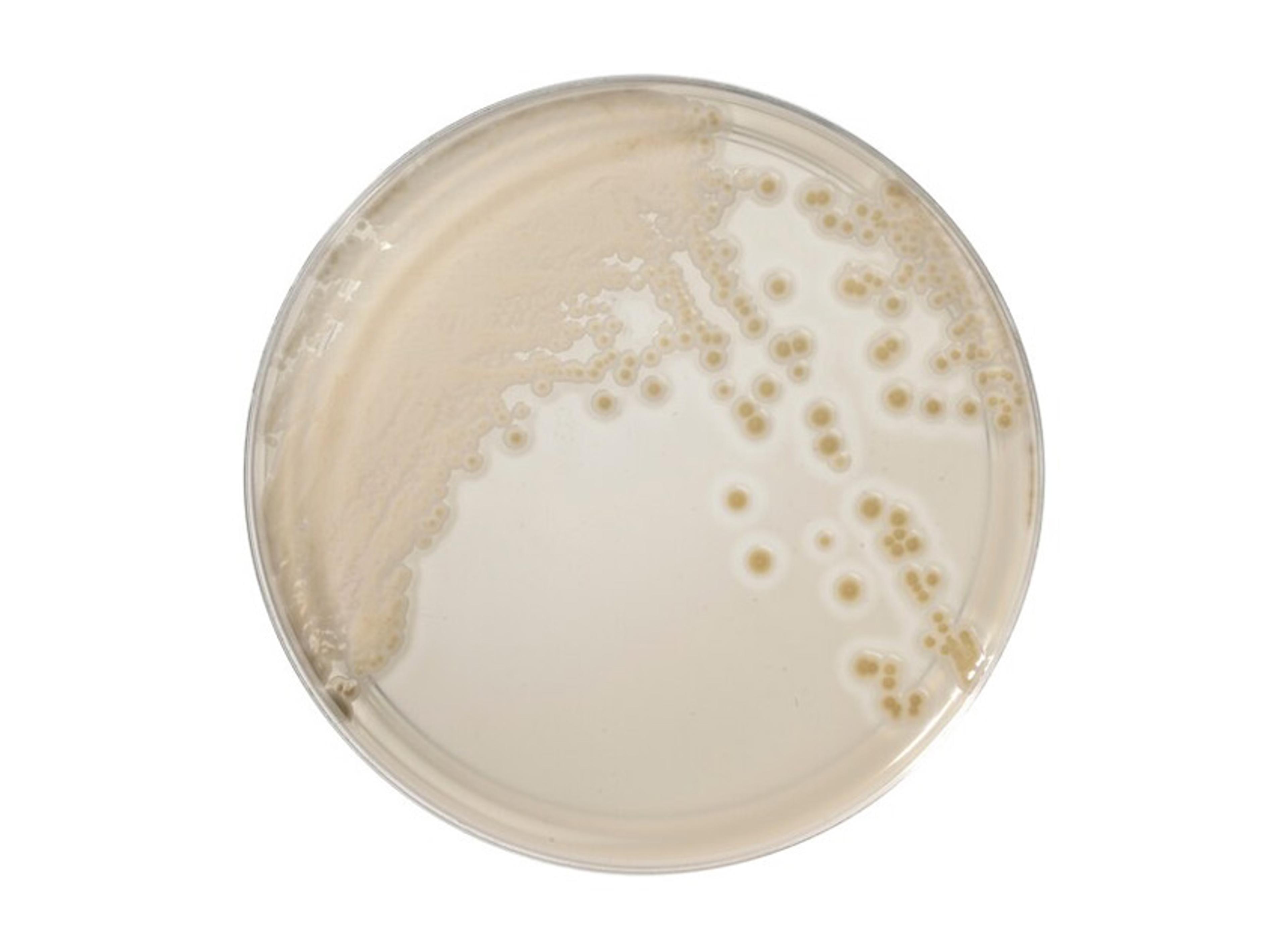
Thermo Scientific™ Brilliance™ Candida 2 Agar
Oxoid Remel MicrobiologyDiscover this innovative Candida detection and differentiation agar, designed to improve the detection and differentiation of Candida species, including screening and direct identification of C. auris and differentiation of five other clinically relevant species.
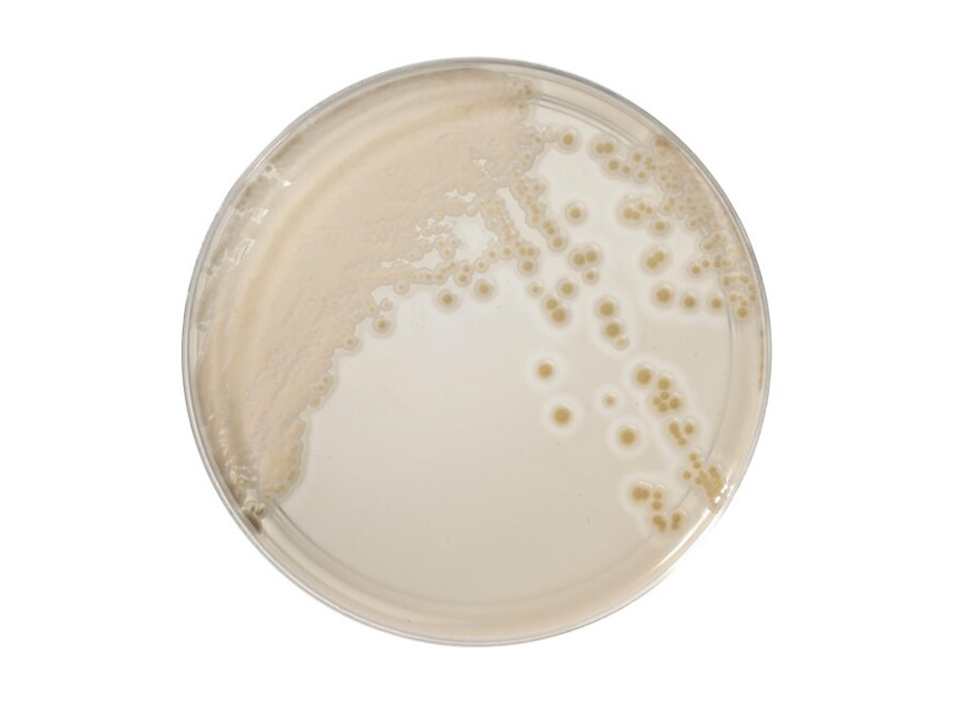
Thermo Scientific™ Spectra™ Candida Agar
Oxoid Remel MicrobiologyDiscover this innovative Candida detection and differentiation agar, designed to improve the detection and differentiation of Candida species, including screening and direct identification of C. auris and differentiation of five other clinically relevant species.
Links
Related content
Tags
Frequently asked questions
Show frequently asked questions
What is Candida auris and why is it considered a global healthcare threat?
Candida auris (recently reclassified as Candidozyma auris) is an emerging fungal pathogen that colonizes human skin and healthcare environments, causing severe invasive infections with mortality rates of 29–53%. It spreads rapidly in hospitals and long-term care facilities, persists on surfaces, and shows increasing resistance to frontline antifungals, making it a major global healthcare-associated infection threat.
How is Candida auris detected and screened in hospitals and clinical laboratories?
Hospitals screen high-risk patients for Candida auris using skin swabs from palms, finger and toe webs, perineal areas, nares, and axilla. Laboratories can use rapid molecular assays or culture-based methods, including chromogenic media such as Thermo Scientific™ Brilliance™ Candida 2 Agar and Spectra™ Candida™ Agar, which differentiate C. auris from other clinically relevant Candida species with high sensitivity and specificity.
Why is antifungal susceptibility testing critical for managing Candida auris infections?
Candida auris shows widespread resistance to fluconazole and increasing resistance to amphotericin B and echinocandins, varying across regions like New York and New Jersey. Antimicrobial susceptibility testing (AST) using EUCAST, CLSI broth microdilution, or commercial methods is recommended for all clinically significant isolates to track resistance epidemiology and guide effective, patient-specific antifungal therapy.