
Iterative Reconstruction of Tomographic Images with ZEISS OptiRecon

Using iterative algorithms, OptiRecon aims to be the latest advancement in tomographic imaging with 4X faster results

5 Apr 2018
Han Yin
Administrator / Office Personnel

Product news

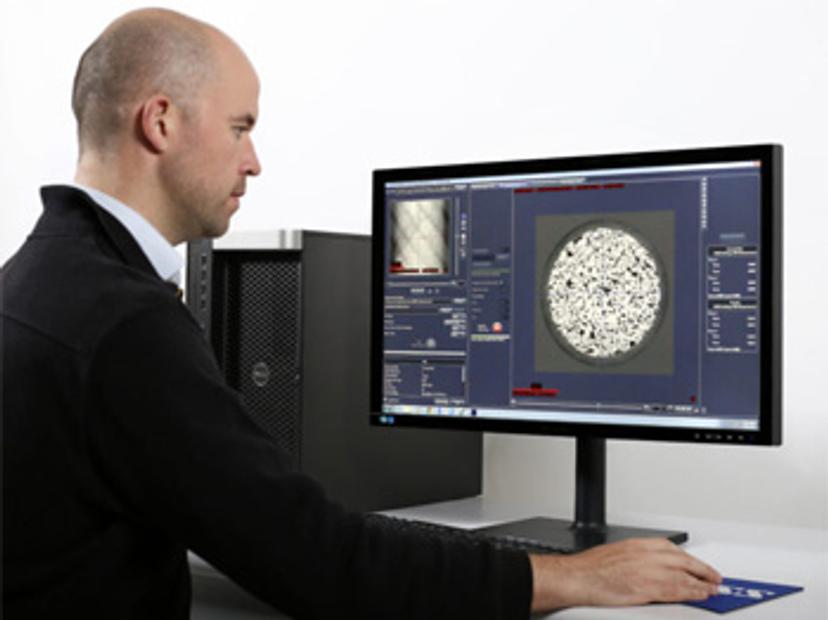
ZEISS OptiRecon is fast, efficient and user-friendly with a workflow-based user interface.

ZEISS has introduced a new module for the ZEISS Xradia Versa 500-series of 3D X-ray microscopes (XRM) that will allow users to acquire high quality images in one-quarter the time. ZEISS OptiRecon enables users to make the optimal choice for their requirements: same quality images four times faster, or superior quality in the same amount of time as standard image acquisition using an advanced new iterative reconstruction technique.

ZEISS OptiRecon is faster, more efficient and user-friendly than standard iterative reconstruction processes that are much more computationally intensive and require extensive user expertise. A combination of a workflow-based user interface that doesn’t require programming knowledge and an efficient proprietary implementation that allows reconstruction of a typical dataset deliver results in about three minutes. This technology opens the door to 3D X-ray imaging, or computed tomography, to both a wider range of industrial applications and the examination of in situ processes occurring at previously inaccessible timescales.
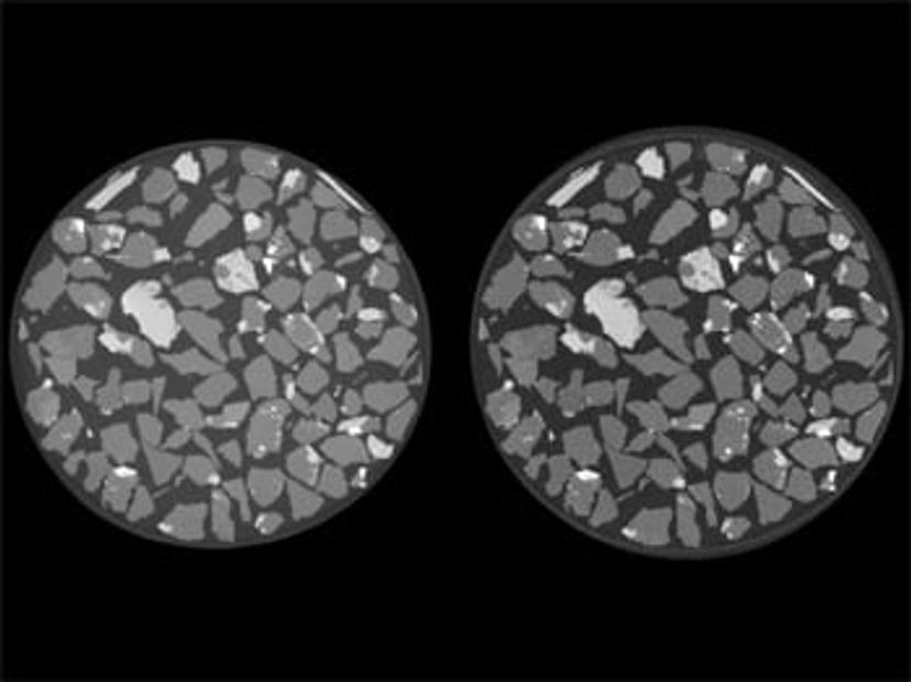
Iterative (left) and filtered back projection (right) reconstruction of a mining sample from 400 projections.

ZEISS OptiRecon has been demonstrated to be especially effective for geological or other “sparse” samples where features are large relative to voxel size. Dr. Branko Bijeljic, Senior Research Fellow, Department of Earth Science & Engineering at Imperial College London, was an early tester of the system. He says, “Iterative reconstruction is a key new technology for improving our work in high resolution X-ray microscopy. It will allow us to address in situ processes occurring at considerably shorter time scales without sacrificing image quality or data segmentability.” Dr. Bijeljic co-authored a paper on the topic that is now available from the Journal of Advances Water Resources: Optimization of image quality and acquisition time for lab-based X-ray microtomography using an iterative reconstruction algorithm.

Lourens Steger, head of X-ray microscopy at ZEISS USA, commented, “ZEISS OptiRecon exemplifies our commitment to leading the industry by bringing new advanced imaging capabilities to our award-winning X-ray microscopes. Since 2010, the ZEISS Xradia Versa family has consistently demonstrated unique capabilities for its users in electronics, materials, life sciences, and the oil and gas industry. As this latest release shows, we continue to develop based on our customers’ needs, in this case for the geological sciences in academia and industry, including oil and gas, and mining. We continue to progress this system with both hardware and software advancements to meet and exceed our users’ needs.”

Do you use the Xradia Versa series in your workspace? Write a review today for your chance to win $400 Amazon voucher.

Links

Tags

X-Ray Diffraction and SpectroscopyX-Ray diffraction & spectroscopy are used in material characterization to discern the structure and elemental composition of a sample. X-Ray diffractometers (XRD) are superior instruments in elucidating the dimensional atomic structure of crystalline materials, including powders, thin films and single crystals. For large unit cells or ordered macromolecules, consider small-angle X-ray scattering (SAXS). X-ray spectroscopic techniques include X-ray fluorescence (XRF) and X-ray photoelectron spectroscopy (XPS), both providing simple and accurate methods for determining the elemental composition of a material. Energy dispersive (EDXRF) and wavelength dispersive (WDXRF) XRF spectrometers are available, as well as handheld/portable devices. High-resolution, 3D microstructure characterization of materials can be achieved with X-ray microscopes combining sub-micron resolution imaging with 3D computed tomography. Find the best XRD and XRF spectrometers in our peer-reviewed product directory: compare products, check customer reviews and receive pricing direct from manufacturers.Robotic InstrumentsRobotic instruments can be used for high-throughput automation of many lab processes. Such processes use instruments for assays like cell based assays and ELISA, for sample preparation like shakers, centrifuges and incubators, and for analysis such as sequencing and western blot analyzers. Useful features of robotic instruments include speed, reproducibility, barcode readers, software and automation.Robotic WorkstationsRobotic workstations are automated platforms enabling the user to perform high-throughput, multi-application processes, from sample preparation tasks such as PCR set-up, DNA purification and NGS library preparation, to integrated sample prep and analysis workstations. Automation makes processes consistent, fast, precise and fully walk-away. Find the best robotic workstations in our peer-reviewed product directory: compare products, check customer reviews and receive pricing direct from manufacturers.AutomationAutomation in laboratories and manufacturing processes enhances efficiency, precision, and scalability by reducing the need for manual intervention. It plays a critical role in improving productivity, minimizing human error, and accelerating workflows in fields like diagnostics, drug development, and industrial testing. Automation technologies include robotic systems, automated liquid handlers, and process control systems that streamline complex tasks and ensure consistent, reproducible results. Explore our peer-reviewed product directory to discover the best automation solutions, compare options, read user reviews, and get prices directly from manufacturers.HD Imaging3D Imaging3D imaging technologies allow for the visualization and analysis of three-dimensional structures at high resolution. These systems are used in fields like molecular biology, material science, and medical diagnostics. 3D imaging can be applied to visualize cells, tissues, and organs, providing valuable insights into their structure and function. Browse our peer-reviewed product directory to find the best 3D imaging solutions, compare products, check reviews, and get pricing directly from manufacturers.MicroscopyMicroscopy is a technique used to observe small objects in detail, from cells to materials, using light or electron microscopes. It enables researchers to examine structures with high resolution, aiding in fields such as biology, medicine, and materials science. With advanced microscopy techniques, scientists can gain insights into cellular processes, tissue structures, and material properties. Explore the best microscopy solutions in our peer-reviewed product directory, compare products, read customer reviews, and get pricing directly from manufacturers.