HORIBA Scientific Offers Unique Solution for TERS Technique
New analytical solution brings nanoscale chemical imaging to new levels of ease and automation.
16 Sept 2015Product news
HORIBA Scientific, global leader in Raman spectroscopy solutions, has announced an innovative solution that addresses the challenges of TERS imaging.
Tip Enhanced Raman Scattering (TERS) is a technique that provides molecular information on the nanometer scale through the combination between a Raman spectrometer and a Scanning Probe Microscope. TERS has been hampered by extremely long acquisition times, measured in hours, required for collection of reasonably pixel density, and even with the last decade progress, TERS is still seen by the research community as a difficult too. In some extent, with older “TERS ready” systems offered until recently, the concerns were fairly valid.
HORIBA Scientific and AIST-NT now offer a solution that addresses all the instrumentation concerns and bring TERS as an analytical method to a completely new level today. HORIBA Scientific and AIST-NT pioneered the demonstration of TERS in out –of-the –lab conditions for the second year at number of conferences worldwide. The XploRA Nano platform in the upright and side configuration, combination with AIST-NT’s SmartSPM and HORIBA’s XploRA spectrometer, offers a very compact (complete set up stands on 900 x 760 x 55 mm optical table), fully integrated and automated solution.
At the recent SPIE conference in San Diego, the XploRA Nano equipped, with AFM-TERS tips, showed nanoscale chemical imaging of a single carbon nanotube with a spatial resolution of 8 nm, confirmed from the section analysis of the intensity of the TERS bands. The images below show an onsite demonstration of TERS mapping of a single carbon nanotube, with an optical spatial resolution down to 8 nm.
“The TERS image on the right, 100 x 100 nm scanning area, is obtained with a pixel step size of 1.3 nm, in a total acquisition time < 9 min, 100 ms per pixel, on the booth, without any active isolation table nor enclosure box, far away from ideal lab conditions,” explains Dr Marc Chaigneau, the AFM-Raman product manager, HORIBA Scientific. In this TERS map, the intensity of the D band (white and green pixels) shows the imperfection in the structure of the latter; in contrast, the areas in red correspond to the pure graphitic arrangement of the CNT through the intensity of the 2D band. Close to the local lattice defects (red circle on the map), a single step gives rise to noticeable intensity of the D peak, showing a chemical sensitivity down to 1.3 nm.”
Related products
Request Quote for All Products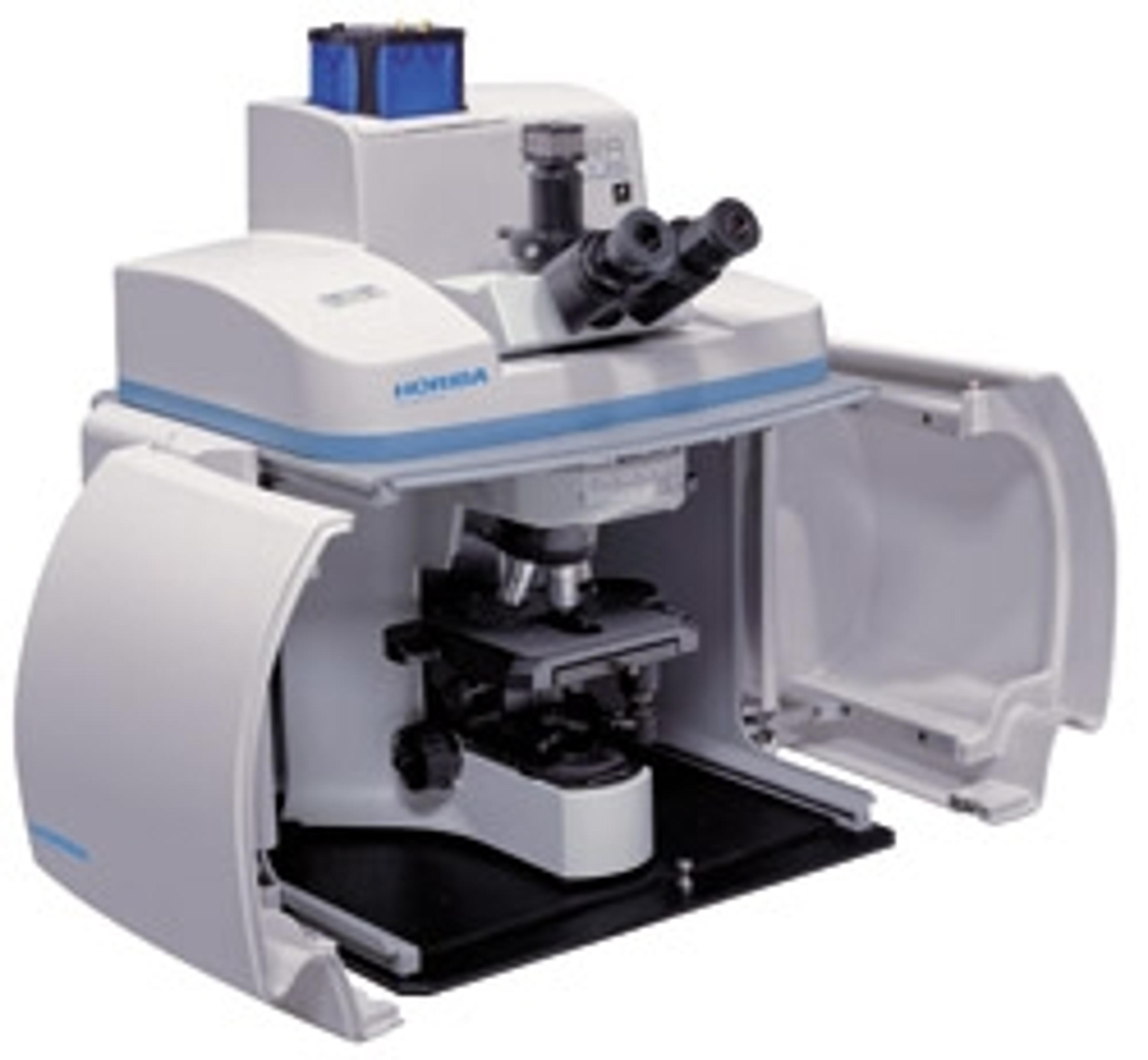
XploRA - Raman microscopy
HORIBA ScientificThe XploRA™ is a new concept in Raman microscopy, bringing Raman chemical identification directly to your microscope. The XploRA can be coupled to both upright and inverted microscopes, allowing analysis of all sample types, ranging from semiconductors and nano-materials, through to biological cells and tissues. The system provides the established performance of HORIBA Scientific Raman microscopes at a surprising price. Combining microscopy and chemical analysis the XploRA™ retains the full functionality of your microscope coupled with high performance Raman spectroscopy. The concept of the XploRA™ is straightforward – a smarter microscopy. A compact, high performance Raman spectrometer is coupled to a research grade optical microscope (upright or inverted geometry), allowing both standard optical microscopy and detailed chemical analysis on a single system. The unique design ensures full capability of the optical microscope, and complete flexibility, full automation and true research performance of the Raman analyzer.