
CRP, ESR and PV – A comparative review
This guest editorial features insights from Consultant Biomedical Scientist David Norcliffe, Advanced Clinical Practitioner David Manuel, and Bernie Benson, Founder of Benson Viscometers, on selecting inflammatory markers for clinical diagnostics
18 Sept 2026
Inflammatory markers are blood tests used by healthcare professionals to detect inflammation, which aid in the diagnosis and monitor the progression of many diseases¹. After the total and differential white cell count, which are part of the full blood count (FBC), the three most commonly used general inflammation markers to screen the wide range of organic diseases are, C- reactive protein (CRP), erythrocyte sedimentation rate (ESR) and plasma viscosity (PV).
Numerous studies have highlighted advantages of each and conversely have also demonstrated limitations. The decision to commit to a particular test can therefore be an important one for clinicians and laboratory scientists, with many factors to consider including the patient population being served. Hence, an overall review may assist in the final decision-making process.
In addition to their role as inflammatory markers, CRP and PV have also both been used as predictors of cardiovascular disease risks. PV, on the other hand, has been shown to be influential in the aid of diagnosis and monitoring progression of other disorders like diabetes, cardiovascular diseases, dementia, Alzheimer’s and COVID-19.
C-reactive protein (CRP)
Discovered in 1930, the term CRP was derived from its initial identification as a substance present in the serum of patients experiencing acute inflammation, which exhibited a reaction with the ‘c’ carbohydrate antigen found in the capsule of pneumococcus².
CRP is a calcium dependent acute phase protein of hepatic origin which binds to phosphocholine on the surface of dead or dying cells and some bacteria. It is now known to exist in at least three different forms: a monomeric (modified) form, a pentameric (natural or native) form and multimeric forms consisting of ten or more subunits³.
Other separate forms have been described such as dimers, trimers, tetramers and non-native pentameric forms⁴. Please, exercise caution when making critical decisions based on the limited information provided by the CRP.
CRP has a half-life of approximately 19 hours⁵ resulting in a continual variance in the plasma. Therefore, it’s understood to be very reactive and sensitive to changes, such as those seen in inflammation. The concentration of the reactive protein can change by approximately 5 mg/L in 6 hours and peak at 48 hours. Once inflammation subsides CRP levels reduce quickly⁶. Hence it is used by many laboratories as a general infection marker at presentation and during its progress.
There are no standard techniques for measuring and reporting CRP. It is commonly measured by immunoassay, though other techniques are sometimes used. The changes seen during acute inflammation can increase the value a hundredfold and each method must be able to encompass this, hence sensitivity is not always at the same level as some other metabolic assays. Furthermore, CRP increases with age⁷ of the patients thus affecting the baseline and therefore making interpretation challenging.
CRP is a non-glycosylated protein, but Pathak and Agarwal (2019)⁸ described different glycosylated forms in certain pathological conditions. However, the measurement of this single acute phase protein brings considerable limitations due to its isolated narrow base. Most notably, baseline levels can be influenced by patients age, gender, smoking, body weight⁹, lipid levels, blood pressure, and evidence indicates that the hormone replacement therapy (HRT) will have a profound influence on CRP which can produce higher incorrect results¹⁰.
Also, variable results in rheumatoid arthritis patients due to the heterogeneity¹¹ (the variability of the intervention effects being evaluated in different studies) and with method variation. It is also known that females have a higher CRP titer than males and a concentration hook effect can exist in particularly high levels thus introducing the possibility of erroneous quantitation¹².
Certainly, any or all of these could explain some, previous reports that CRP does not always increase in viral infections¹³. Hence CRP results obtained when differentiating between viral and bacterial infections should be interpreted with caution.
Developments have been made in CRP applications with small but significant changes being recognized as a possible cardiovascular risk factor in otherwise healthy, asymptomatic individuals. Hence high sensitive assays (hs-CRP) have since been developed using immunonephelometry or immunoturbidity which mostly focus on concentrations at or below the stated reference range but are both more expensive than standard CRP assays and less commonly used. Furthermore, hs-CRP assays should not be used in the presence of other physiological inflammation, limiting the reliable use of hs-CRP especially when diagnosing cardiovascular risks.
Point-of-care testing (POCT) CRP assays can be used in emergency care for possible detection of inflammation. This can be a useful guide and is also suitable for outreach or satellite laboratories. It has also been reported that a CRP to albumin ratio of less than 32 has a negative predictive value of up to 89% for ruling out sepsis¹⁴ but the numerous influences on the baseline must be considered in its interpretation.
Normal reference ranges for CRP vary with the technique used and other factors but typically are usually approximately 3–10 mg/L¹⁵. Health care professionals and clinicians are still not in an agreement for an international numerical standard for a definitive normal range and abnormal range of CRP results. Perhaps this is due to the wide range of results produced by the many different manufacturers of CRP analyzers and the varying analytical testing methods.
Erythrocyte sedimentation rate (ESR)
The ESR is almost certainly the oldest laboratory blood test still in use; being first described over 100 years ago¹⁶. It is based on the principle of determining the rate of red cell (hematocrit) sedimentation falling through plasma under the influence of gravity, in a diluted stationary sample measured over one- hour¹⁷. An increase above the reference range may be an indicator of a disease process. It is influenced by the albumin/globulin (AG) ratio, particularly fibrinogen.
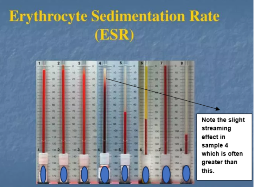
Fig 1: Illustration of ESR tests reflecting the tendency of red blood cells to settle more rapidly in the face of some disease states⁽²⁰⁾
Although variations of the original technique had been used since 1921 it was standardized by Dr. A Westergren, who was a founding member of the expert panel of International Committee for Standardization in Hematology (ICSH); being published in 1973.¹⁸
The ESR test process mechanism is triphasic, producing a sigmoid curve, with each phase varying in length between patients. The three phases are: aggregation, sedimentation and packing of the red blood cells (alternatively this process is known as lag, log and syneresis). The ESR result is analyzed and recorded after one hour on the assumption that the third phase is complete.
The traditional Westergren test requires an EDTA anticoagulated sample, less than 4 hours old¹⁹. An aliquot of this sample is then diluted with trisodium citrate to reduce the zeta potential of the red cells, using a ratio of 4 parts blood to 1 part sodium citrate. This is gently mixed and transferred into a plugged, graduated tube measuring 200 mm length and 2.5 mm internal diameter. Then the tube is left to stand vertically away from direct sunlight, vibration, and from any form of movement, which are known as serious interfering factors in the process.
The result is where the demarcation zone between plasma and red cells occurs and is reported in millimeter (mm) in 1 hour. However, due to variations in the length of each phase between patients, it is not uncommon for ‘streaming’ to occur. This is where the third phase is incomplete and a hazy, and unclear line of demarcation is seen, often occupying up to 20 mm of the graduated tube, thus making an accurate and clear results which is subjective and difficult to achieve. In the author’s experience this occurs in approximately 10% of ESR tests.
There is a plethora of information about the numerous limitations of the ESR test, one of which is the dependence on the hematocrit, which gives rise to the different reference ranges for males, pre- and post-menopausal females. Drawbacks with ESR test includes the unphysiological nature of the test, the age of the sample prior to testing, the patients age, and use of medication (particularly aspirin and steroids which can often distort a high result towards normal).
Some external interfering factors could affect the test results including, laboratory temperature, stable vibration free test environment. Most importantly the ESR test does not have realistic quality control and quality assurance materials to validate the test carried out.
The author has found that the accuracy of the dilution with trisodium citrate was of particular importance. Automated ESR analyzers, which considerably shorten the duration of testing, will only provide an approximation of the results obtained by a traditional Westergren test.
The International Council for Standardization in Hematology (ICSH) indicated in a report²¹ that while the Westergren method remains the gold standard, the results produced by new instruments may vary from those obtained using the Westergren method by as much as 142%. Furthermore, the ICSH report highlighted that various non-Westergren methods exhibited discrepancies among themselves of up to 42%.
Plasma viscosity (PV)
Plasma viscosity (PV) is renowned as a quick, non-specific test, which is accurate, reliable, repeatable, is very useful for screening and eminently suitable for condition monitoring. The viscosity (a fluid’s resistance to flow) of plasma is determined by size of the protein, its concentration and importantly, molecular symmetry.
Fibrinogen has a very significant effect on the viscosity of plasma. This is due to fibrinogen having a length to diameter ratio of approximately 18:1²². So, elevation in the total levels of acute-phase proteins and fibrinogen, contributes to an increase in plasma viscosity.
Therefore, the viscosity of plasma is significantly influenced by changes in the size and the levels of protein and importantly the volumes of fibrinogen molecules, which occur as a consequence of infection, inflammation, and physical trauma.
PV measurement in clinical practice has been used since 1946, following studies referencing the mechanism of the ESR, John Harkness published what was to be a seminal paper²³.
In 1963 he designed a semi-automated capillary viscometer for PV measurement which was later developed, manufactured and marketed as the Coulter Harkness Plasma Viscometer. This instrument eventually (1972) then became the gold standard for clinical plasma viscosity measurement.
The analyzer test temperature was chosen to be 25°C, room temperature. It was considered easier and more economical to manufacture the viscometer testing and reporting at 25°C rather than the more obvious and physiological human body temperature of 37°C. Reporting at 25°C is still widely used today, though modern automated clinical viscometers will test precisely at 37°C but can report at a choice of 37°C or 25°C depending on the user’s preference.
The PV measurement is most accurately achieved using a capillary viscometer. The theory is quite simple. Thin, low viscosity fluids will travel faster through a capillary than thick high viscosity fluids. The time taken for an aliquot of plasma (under the influence of air pressure) to travel through a calibrated capillary is detected by two measured infra-red optic points and recorded. The recorded viscometer ‘run time’, is used by an algorithm which will determine the viscosity of the fluid.
A modern automated clinical viscometer aspirates only 50 µL of sample therefore, adult and pediatric repeat tests are feasible. The PV test can be carried out using the residue from the full blood count tube, eliminating the need for additional blood samples.
Other methods that have been used historically to measure plasma viscosity are the cone and plate viscometer which measures the resistance (drag) in an aliquot of plasma when a cone or plate is rotated in the fluid. However, this method is far less sensitive than the capillary method. The cone and plate instrument required a much larger sample volume, a greater assay time and manual pipetting.
Another early viscosity test method once used for determining PV was the falling ball viscometer. This viscometer is often used in industry, and it determines the viscosity by measuring time for a uniform ball to fall through a vertical column of liquid. Both these methods are now generally considered inappropriate in a modern clinical laboratory environment.
The viscosity of plasma with the human body will automatically increase as part of the normal response to infection, inflammation, and due to the effects of traumatic physical injury. A PV will give an accurate reflection of the intensity of the patient’s condition at the time of test.
The PV test is cost-effective, quick and available in many hematological laboratories. The test can be carried out using the plasma sample from the residue of the full blood count tube, eliminating the requirement for additional blood sample.
The PV test has the capability to diagnose and monitor many clinical conditions. It has been exclusively used in the diagnosis and detection of Waldenström’s macroglobulinemia and hyperviscosity syndrome. It exhibits significantly greater versatility in behavior across various other disease states.
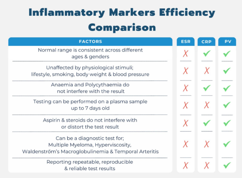
Hence plasma viscosity is considered to have a dramatic diagnostic impact leading to the most appropriate treatment for conditions such as, multiple myeloma, polymyalgia rheumatica, rheumatoid arthritis, temporal arteritis, cardiovascular disease, COVID-19, Waldenström’s macroglobulinemia, hyperviscosity syndrome, diabetes, sepsis, meningitis, vascular dementia and Alzheimer’s.
Most notably in a 2021 paper, by Gleghorn et al²⁴ who reported an increase in viscosity and a predictive value of greater than 90% of patients with SARS COVID-19 developing complications and requiring hospitalization.
Further interesting, published papers indicate that PV can be used to diagnose and differentiate between vascular dementia and Alzheimer’s disease.²⁵ It has been long established that PV can be used to great advantage as a predictor of cardiovascular diseases.²⁶
The PV normal reference range for individuals aged 3 years and above is considered to be, a result within 1.50–1.72 mPa.s⁽²⁷⁾ reported at 25°C (room temperature) and 1.12–1.27 mPa.s reported at 37°C. Importantly minor variation in a patients PV of 0.05 mPa.s is deemed to have clinical significance.
Discussion and conclusions
Over the last 20 years it has been common for hematology and biochemistry labs to combine to form Blood Science departments. The traditional soft barriers have been breached, though dedicated specialist staff remain in their particular disciplines to maintain service quality and expertise. These changes have seen the emergence of the Managed Service Plan (MSP) for lease and maintenance of all major equipment and reagents, often over both disciplines, using a single combined contract.
Many of the major biochemistry analyzer manufacturers incorporate CRP testing within their devices, subsidizing the relatively expensive assay from the cheaper and more frequently requested tests such as urea and electrolytes thus increasing its attraction to the customer when operating within a tight financial budget and considering an MSP.
Some laboratories have retained their historical traditional discipline specific inflammatory markers, offering two inflammatory tests, whilst others have made the difficult decision to provide just one. Hence it is not uncommon for some blood sciences labs to offer ESR or PV from hematology and CRP from biochemistry, whilst some labs only rely on a single marker.
Whilst an accurate direct conversion factor between any two or all three test methods cannot be produced due to variations and specificity, it can be seen from the evidence in this review that CRP appears to have some virtues, particularly its use as a predictor of cardiovascular disease and its point of care platform. However, it does have several considerable limitations.
The numerous influences make the baseline figure less than certain which could impact on clinical decision making. Furthermore, there is a significant unclear (gray) area in results and interpretation.
Whilst the ESR has earned its place in history, the numerous limitations and interfering factors appear to make it an anachronism in the modern laboratory. Whilst a grossly raised ESR may be a good indicator of disease, particularly at the diagnostic stage, the tendency for the ESR to incorrectly report as ‘normal’ puts in to question the true value of the ESR test.
When looked at objectively the ESR is unphysiological and does not reflect any part of the in-vivo circulation. Additionally, there are no realistic quality control or quality assurance schemes which accurately reflect the test conditions.
Furthermore, in cases of IgG myeloma, the ESR result is dramatically clear but paradoxically in IgM myeloma or Waldenström's macroglobulinemia the result is commonly reporting in the normal range due to the plasma being too viscous for the hematocrit cells to fall through under the effect of gravity, even despite the dilution with trisodium citrate.
The PV test is sensitive and is a true reflection of the ‘in vivo’ conditions. Additionally, it is meticulously regulated through well-established quality control and quality assurance schemes, notably at the UK, National External Quality Assessment Service, (UK NEQAS).
PV has a very narrow, specific reference range and it measures the overall collective effect of protein changes rather than a specific protein and offers several worthwhile advantages. These include that testing can be performed on a plasma sample up to 7 days old, and the result is unaffected by factors such as anemia, patient age, gender or smoking habit, while showing stronger correlation with disease activity.
Also, PV results are not distorted or affected by medication such as salicylates (aspirin) or steroids, and test is independent of the hematocrit²⁸,²⁹,³⁰,³¹. Moreover, the combination of PV and serum viscosity ratios has been used as a predictor of chronic arthritis in patients with early rheumatic disorders³².
Where laboratories offer two different inflammation markers for best clinical practice, then the evidence above would indicate CRP and PV as the prime choice due to the many notorious limitations of the ESR.
All three markers are well known to increase in bacterial, viral infections, sepsis, and temporal arteritis. However, caution must be exercised in the interpretation of CRP results where it conflicts with that of PV due to the limitations above. PV has fewer limitations, the test is quick, sensitive, reliable, and inexpensive. Both CRP and PV have been used as predictive indicators of cardiovascular disease in addition to their role as inflammation markers.
The changes which occur in the viscosity of the plasma in multiple disease states have often been described and compared. Notably, the UK government recently announced 10-year NHS plan places a large emphasis on further development of medical analytical technology.³³
Where only a single inflammation marker is provided by the laboratory then evidence would indicate that PV is clearly likely to be the best choice due to its indisputable baseline and narrow range. PV is a sensitive test and a change as small as 0.05 mPa.s considered to be diagnostically significant. There are fewer influencing factors than CRP and its versatility appears to be at least as wide, as supported by a plethora of publications in numerous disease states.
Additionally, its use as a cardiovascular risk predictor does not require a separate assay technique and can be performed from the same sample or residue of a routine full blood count. In summary, while the on-board CRP assay presents some attractions, the PV appears to excel in terms of versatility, performance, and reliability compared to the other two inflammatory tests.
The PV test has the capability to diagnose and monitor any condition that the ESR test is utilized for including screening or monitoring. However, it is crucial to acknowledge that a PV test cannot always be substituted by using the CRP or ESR tests.
PV test results are reliable, repeatable, and reproducible with little or no ambiguous and equivocal areas thus making interpretation easier than the ESR or CRP. The benefits and rewards of PV testing are substantial for the patient’s clinical outcome.
PV can add useful information in the diagnosis and treatment of many common and also rare disorders. Therefore, it is recommended that plasma viscosity tests should be exploited and utilized more frequently in clinical medicine.
References
1. Watson J. Inflammatory markers explained. (National Institute for Health and Care Research, 2022) https://arc-w.nihr.ac.uk/ news/inflammatory-markers-explained/
2. Nehring SM, Goyal A, Patel BC. C Reactive Protein. In: StatPearls. Treasure Island (FL): StatPearls Publishing; July 10, 2023.
3. Boncler M, Wu Y, Watala C. The Multiple Faces of C-Reactive Protein-Physiological and Pathophysiological Implications in Cardiovascular Disease. Molecules. 2019;24(11):2062. doi:10.3390/molecules24112062
4. Wang J, Tang B, Liu X, et al. Increased monomeric CRP levels in acute myocardial infarction: a possible new and specific biomarker for diagnosis and severity assessment of disease. Atherosclerosis. 2015;239(2):343-349. doi:10.1016/j.atherosclerosis.2015.01.024
5. Markanday A. Acute Phase Reactants in Infections: Evidence-Based Review and a Guide for Clinicians. Open Forum Infect Dis. 2015;2(3):ofv098. doi:10.1093/ofid/ofv098
6. Bray C, Bell LN, Liang H, et al. Erythrocyte Sedimentation Rate and C-reactive Protein Measurements and Their Relevance in Clinical Medicine. WMJ. 2016;115(6):317-321.
7. Thomas L, editor. Labor und Diagnose: Indikation und Bewertung von Laborbefunden für die medizinische Diagnostik. Frankfurt: TH-Books-VerlagGes. 2008
8. Pathak A, Agrawal A. Evolution of C-Reactive Protein. Front Immunol. 2019;10:943. doi:10.3389/fimmu.2019.00943
9. Buchbinder S, Kratzsch J, Fiedler GM, et al. Body weight and oral contraceptives are the most important modulators of serum CRP levels. Scand J Clin Lab Invest. 2008;68(2):140-144. doi:10.1080/00365510701487727
10. Sproston NR, Ashworth JJ. Role of C-Reactive Protein at Sites of Inflammation and Infection. Front Immunol. 2018;9:754. doi:10.3389/fimmu.2018.00754
11. Orr CK, Najm A, Young F, et al. The Utility and Limitations of CRP, ESR and DAS28-CRP in Appraising Disease Activity in Rheumatoid Arthritis. Front Med (Lausanne). 2018;5:185. doi:10.3389/fmed.2018.00185
12. Roberts B. C-reactive protein (serum, plasma). (Association for Laboratory Medicine, 2012) https://labmed.org.uk/ our-resources/science-knowledge-hub/analyte-monographs.html
13. Henderson R. Podcast: EP 86 - Inflammatory marker testing. (GPnotebook, 2023) https://gpnotebook. com/en-GB/podcasts/immunology/ep-86-inflammatory-marker-testing
14. Sisto UG, Di Bella S, Porta E, et al. Predicting sepsis at emergency department triage: Implementing clinical and laboratory markers within the first nursing assessment to enhance diagnostic accuracy. J Nurs Scholarsh. 2024;56(6):757-766. doi:10.1111/jnu.13002
15. Tidy C. Blood tests to detect inflammation. (Patient, 2024) https://patient.info/ treatment-medication/blood-tests/bloodtests-to-detect-inflammation
16. Grzybowski A, Sak JJ. Who discovered the erythrocyte sedimentation rate?. J Rheumatol. 2011;38(7):1521-1523. doi:10.3899/jrheum.101312
17. GPnotebook. Erythrocyte sedimentation rate. (GPnotebook, 2018) https:// gpnotebook.com/en-GB/pages/haematology/erythrocyte-sedimentationrate
18. Jou JM, Lewis SM, Briggs C, et al. ICSH review of the measurement of the erythocyte sedimentation rate. Int J Lab Hematol. 2011;33(2):125-132. doi:10.1111/j.1751-553X.2011.01302.x
19. Hu QL, Li ZJ, Lin L, et al. Effect of storage temperature and time on erythrocyte sedimentation rate. Eur J Med Res. 2022;27(1):76. doi:10.1186/s40001-022-00701-3
20. Leblanc P. Erythrocyte Sedimentation Rate (ESR). (Slideserve, 2014) https:// www.slideserve.com/patience-leblanc/erythrocyte-sedimentation-rate-esr
21. Kratz A, Plebani M, Peng M, et al. ICSH recommendations for modified and alternate methods measuring the erythrocyte sedimentation rate. Int J Lab Hematol. 2017;39(5):448-457. doi:10.1111/ijlh.12693
22. Lowe GD. Blood rheology in vitro and in vivo. Baillieres Clin Haematol. 1987;1(3):597-636. doi:10.1016/s0950-3536(87)80018-5
23. Harkness J, Houston J, Whittington RB. Plasma Viscosity: A Clinical Test. Br Med J. 1946;1(4442):268-270.
24. Gleghorn DM, Olayiwola O, Omer A, Lynn R, Norcliffe D, Robinson M. Diagnostic value of plasma viscosity testing for patients with COVID-19. Int J Lab Hematol. 2021;43(4):O227-O229. doi:10.1111/ijlh.13532
25. Aras S, Tek I, Varli M, et al. Plasma viscosity: is a biomarker for the differential diagnosis of Alzheimer’s disease and vascular dementia?. Am J Alzheimers Dis Other Demen. 2013;28(1):62-68. doi:10.1177/1533317512467682
26. Koenig W, Sund M, Filipiak B, Döring A, Löwel H, Ernst E. Plasma viscosity and the risk of coronary heart disease: results from the MONICA-Augsburg Cohort Study, 1984 to 1992. Arterioscler Thromb Vasc Biol. 1998;18(5):768-772. doi:10.1161/01.atv.18.5.768
27. Hutchinson RM, Eastham RD. A comparison of the erythrocyte sedimentation rate and plasma viscosity in detecting changes in plasma proteins. J Clin Pathol. 1977;30(4):345-349. doi:10.1136/jcp.30.4.345
28. Kenny MW, Worthington DJ, Stuart J, et al. Efficiency of haematological screening tests for detecting disease. Clin Lab Haematol. 1981;3(4):299-305.
29. Pickup ME, Dixon JS, Hallett C, Bird HA, Wright V. Plasma viscosity – a new appraisal of its use as an index of disease activity in rheumatoid arthritis. Ann Rheum Dis. 1981;40(3):272-275. doi:10.1136/ard.40.3.272
30. Ballas SK. The erythrocyte sedimentation rate, rouleaux formation and hyperviscosity syndrome. Theory and fact. Am J Clin Pathol. 1975;63(1):45-48. doi:10.1093/ajcp/63.3.45
31. Alexander GJ, Blake DR, Holman RL, Bacon PA. Predictive value of paired plasma and serum viscosity in early rheumatic conditions. Br Med J (Clin Res Ed). 1981;282(6271):1198. doi:10.1136/bmj.282.6271.1198
32. GOV.UK. 10 Years Health Plan working groups. (GOV.UK, 2024) https://www.gov. uk/government/groups/10-year-healthplan-working-groups
Links
Tags
Frequently asked questions
Show frequently asked questions
What are the three most commonly used inflammatory markers in blood tests?
After the full blood count’s total and differential white cell count, the three most common general inflammatory markers are C-reactive protein (CRP), erythrocyte sedimentation rate (ESR), and plasma viscosity (PV). Healthcare professionals use these blood tests to detect inflammation, support diagnosis, and monitor disease progression.
How do CRP, ESR, and plasma viscosity compare as inflammation tests?
CRP responds quickly to inflammatory changes but has a baseline influenced by factors including age, gender, smoking, body weight, and medication. ESR has numerous interfering factors and lacks realistic quality-control materials. Plasma viscosity is described as quick, sensitive, reliable, repeatable, independent of hematocrit, and less affected by patient characteristics or medication.
Why may plasma viscosity be the best single inflammatory marker for clinical laboratories?
The article identifies plasma viscosity as the preferred single marker because it has a narrow reference range, fewer influencing factors, and reflects in-vivo conditions. A change of 0.05 mPa.s may be clinically significant. PV can support diagnosis and monitoring across conditions including cardiovascular disease, COVID-19, diabetes, sepsis, rheumatoid arthritis, Alzheimer’s disease, and hyperviscosity syndrome.