
Ask the Expert: High-Content Cell Analysis in Drug Discovery Applications
AstraZeneca scientist Dr. David Baker discusses live-cell imaging techniques for drug discovery applications, and how to optimize your assay development
16 Oct 2018

Characterizing cell models and optimizing assays are key components of the drug discovery pipeline. SelectScience was recently joined by Dr. David Baker, senior research scientist in AstraZeneca’s cell assay development team, for an information-packed webinar and live Q&A session. Webinar attendees heard Baker discuss using longitudinal live-cell imaging techniques in assay development and analysis. Key examples, such as measuring proliferation rate, transfection efficiency and cytotoxic compound screening, were explored.
The live Q&A session gave attendees the opportunity to ask their own questions, catch the session highlights below, and register to watch the full webinar on demand here >>
Are there any effects on the imaging signal when you change the growth media?
DB: Not so far in my experience. I’ve used a lot of the different media standards, mostly with tumor cell lines; this has been both with and without phenyl red and I haven’t seen any effects in terms of the quality of cell imaging. Of course, if this is a worry, then test out a few options.
How do you activate CD8+ T cells? Is this by anti-CD3 and anti-CD28 stimulation, and how different is this model from tumor-educated T cells in co-culture assays?
DB: Wow, I hadn’t thought of this – I’m certainly thinking of testing tumor-educated T cells now! The activation I’ve done so far is in anti-CD3 and anti-CD28 stimulation. Whilst we haven’t tested the tumor educated T cells, if I was to guess, I would say that the anti-CD3 and anti-CD28 stimulation would be the strongest.
I would like to understand more on TCA multiplex assays. Could you share a general protocol?
DB: The protocol that I follow directly is made by Intellicyt. You can get the assay protocol from them if you contact them directly.
What are the advantages of the IncuCyte over just looking at your cells?
DB: Yes, you could quite simply take them out of the incubator and just look at them every day. But there are several advantages to the IncuCyte. First of all, it’s objective analysis: you’re removing any bias that one might introduce into your cells by simply looking at them. You can also set it up at any time of the day – so you can have your result ready for when you get back to work in the morning. Finally, multiple plates can be analyzed at the same time, and the software analyzes images as they’re being taken, in real time. This means you can have your results graph as soon as you turn on the software. In my experiments, I am able to generate results from six different plates immediately.
Are measurements skewed by heterogenous cell populations, since cells are at different stages of division and protein expression levels, at different times?
DB: It does average the whole cell population of the well, including confluence and transfection efficiency. That’s the advantage of flow, you can see every single event and divide them into different populations.
Is it possible to analyze cells in suspension on the IncuCyte?
DB: This is more tricky because it depends upon where your cells will be lying in the well and whether it can autofocus. I mainly use it for adherent cells but also for analyzing cells in co-culture experiments – where it’s much easier to analyze adherent cells. Perhaps contact IncuCyte to find out more.
Do you ever use the IncuCyte for drug screens?
DB: Not really, if we did, it would be further down the cascade with a smaller number of compounds. As I showed in the presentation we generally use it to characterize the cell model and look at the start and end point of cell assays. If we have a lot of compounds to put through an assay, however, we would use a simpler plate reader-based assay to interrogate large numbers, then use the IncuCyte once certain compounds have been triaged out. The strength of the system is in small-scale screens not high-throughput screens.
On the IncuCyte, have you seen any issues with cross-contamination?
DB: No, we haven’t, we make sure that all the different cleaning procedures are followed using fresh buffers. You can set up washes in between samples or treatments to negate cross-contamination but we haven’t really seen any issues so far.
How long does it take to run a 384-well plate on the IncuCyte?
DB: In the high-throughput screening group they’ve managed to read a 384 plate in around 11 minutes, but it completely depends on what you want from your well. If you want to interrogate, say, a population of cells that comprises 5% of your well population then you will want to interrogate each well for much longer. With the IncuCyte, you can test to see how stable your plates are for assay duration, and that can be part of the optimization process.
How does cytokine detection on the iQue screener compare to other cytokine detection methods?
DB: We’ve done comparisons between the iQue, and other cytokine detection platforms or methods, such as Luminex and MSD, and the trends for all of them seemed to appear the same. Absolute quantification varies between technologies but in screening we look for percentage increase instead, so we’re confident with the iQue results. The method for the iQue is easier, you don’t have to perform any washes. You just pour the supernatant off, add to beads, incubate and read. It’s also just generally more amenable to our workflows than the other two methods.
Which type of multichannel pipette do you use, and how do you deal with bubbles?
DB: We use the Thermo Fisher Scientific 16-channel pipette and I pre-wet tips with blank media, as well as making sure I have a bit of reserve left. For example, if I need to pipette 20µl, then I’ll have 25µl ready to pipette — that usually deals with bubbles.
Watch the full webinar on demand here>>
Related products
Request Quote for All Products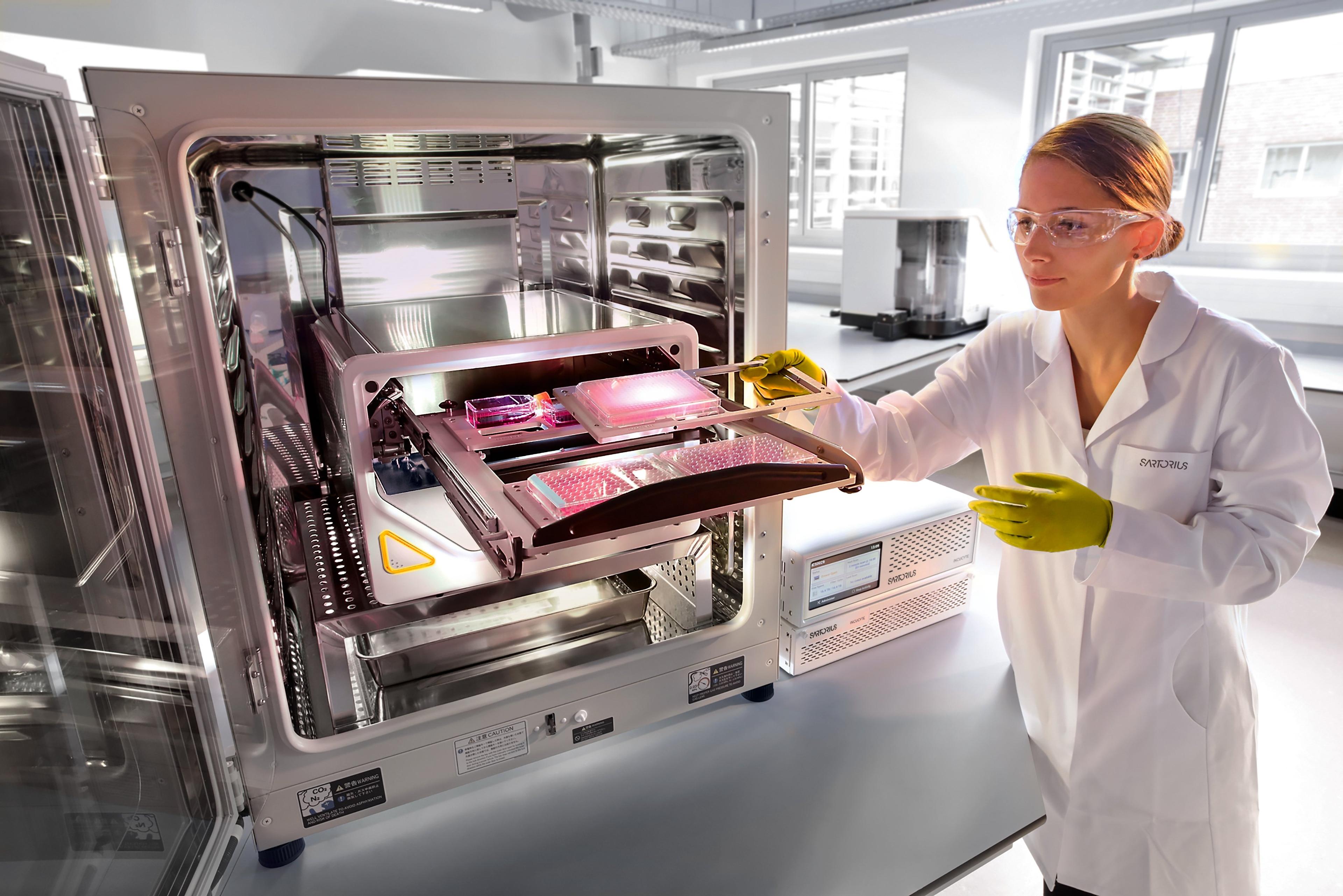
Incucyte® Live-Cell Analysis Systems
Sartorius GroupIncucyte ® , Empower Live-Cell Analysis Inside Your Incubator