Transform your discovery research with spatial phenotyping
20 Oct 2021Spatial phenotyping of whole tissue sections enables the study of different cell phenotypes and how they interact in situ to impact disease pathology and progression. In areas such as oncology, immunology, and neurodegenerative disease, this is rapidly increasing our understanding of underlying cell-level biology and holds the potential to drive a new wave of therapeutic discovery.
In this new eBook, we provide a user guide to spatial phenotyping and highlight a range of its benefits and applications, including how it can enable researchers to:
- Unlock single-cell discoveries in FFPE tissues
- Explore tumor heterogeneity
- Visualize tumor-infiltrating lymphocytes
- Perform highly multiplexed hypothesis-free biomarker discovery
- Conduct hypothesis-driven biomarker discovery and high-throughput translational research
Related Products
Request Quote for All Products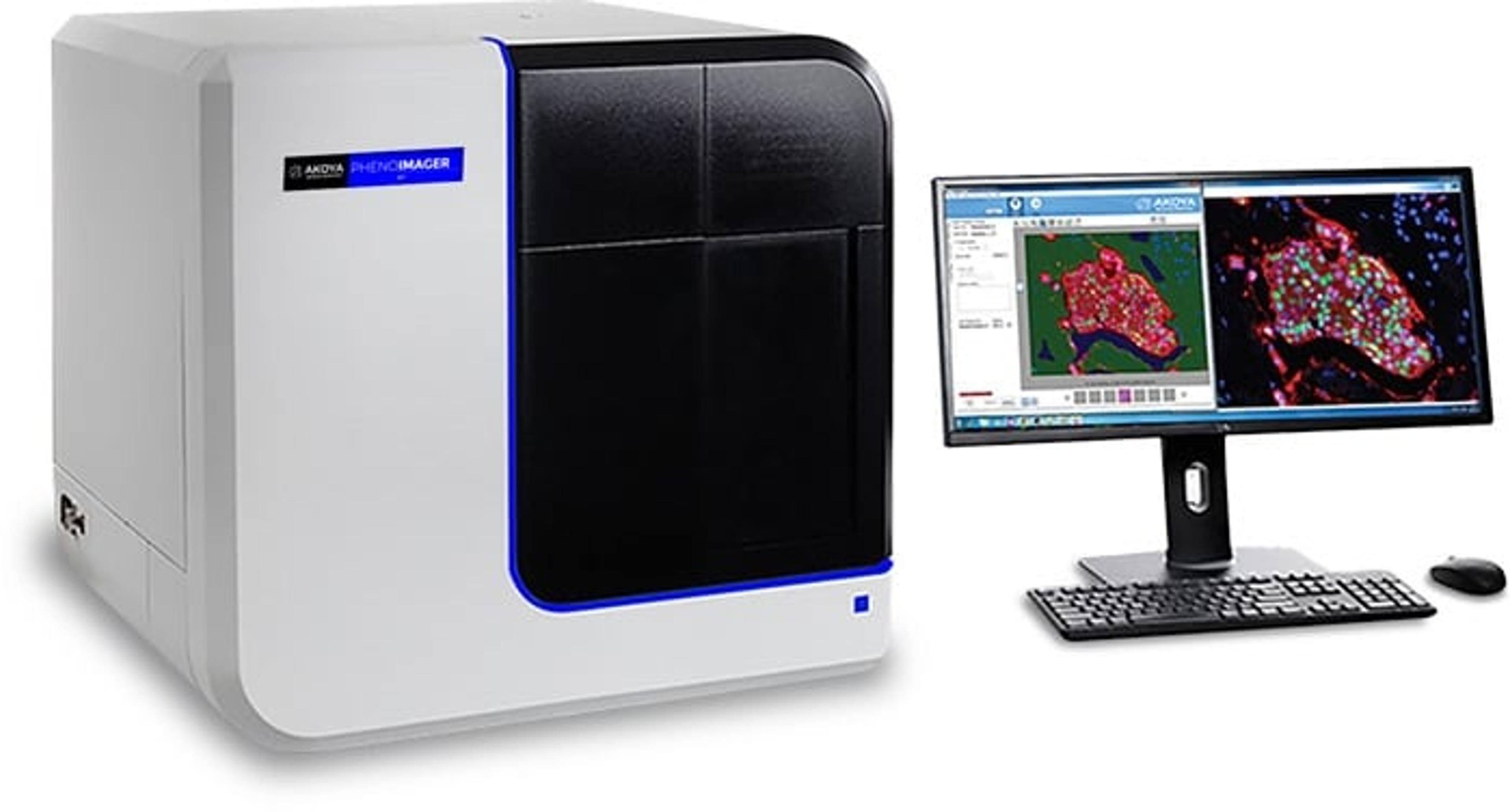
PhenoImager® HT
Akoya BiosciencesPhenoImager ® HT (formerly Vectra ® Polaris™) is the fastest and most highly cited whole-slide, single-cell resolution imaging platform for spatial phenotyping and the development of spatial signatures. Featuring Akoya’s patented Multispectral Imaging (MSI) and spectral unmixing technologies, this platform can be easily integrated into high-throughput workflows to accommodate projects regardless of your scale.
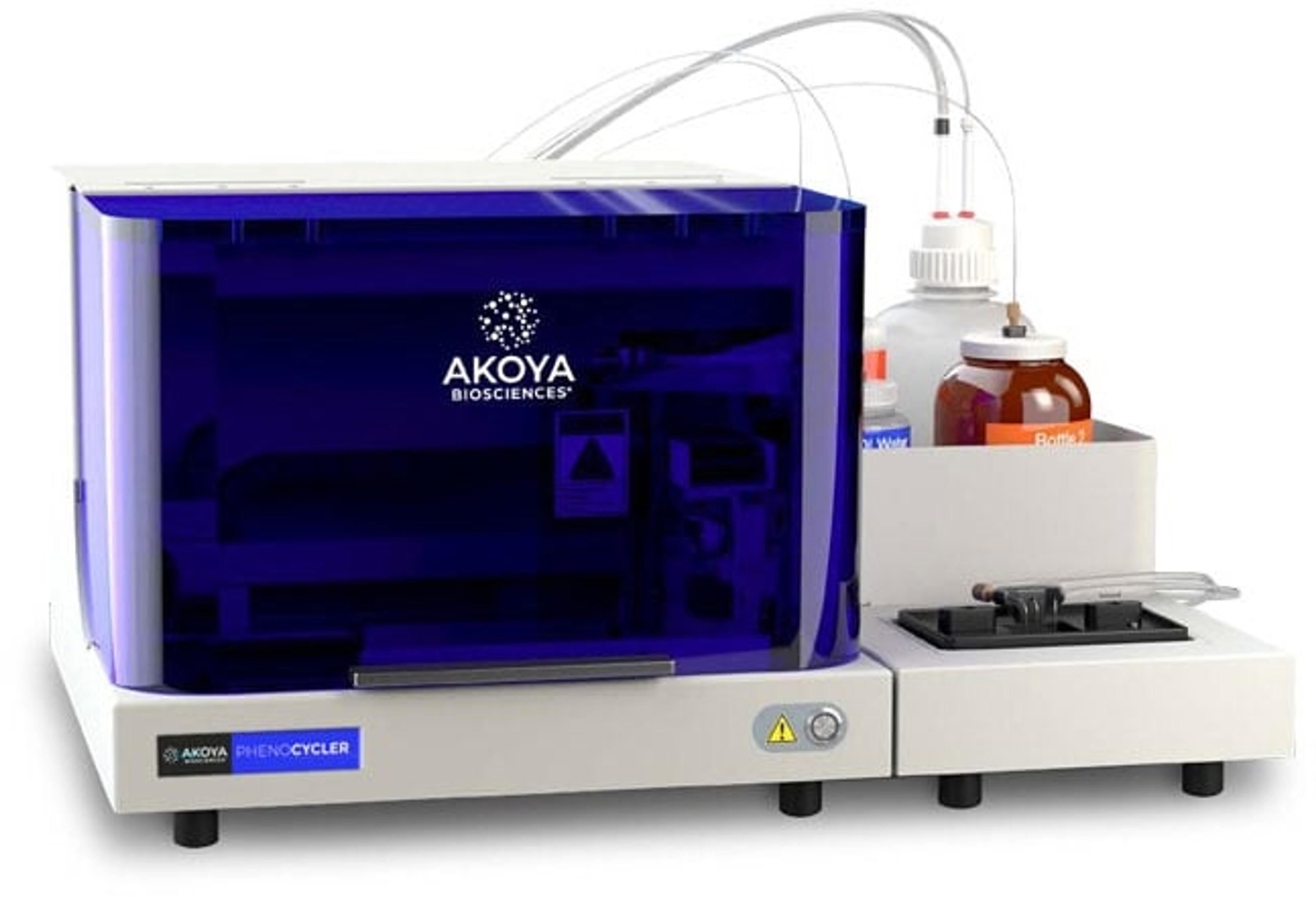
PhenoCycler®
Akoya BiosciencesThe PhenoCycler ® is a cutting-edge fluidics control unit that integrates with the PhenoImager Fusion imaging platform creating the fastest spatial biology solution with unparalleled throughput.