Nanoscopy meets Lifetime: Introducing τ-STED
19 Sept 2021Fluorescence lifetime provides a new perspective for STED nanoscopy. With the unique τ-STED functionality from Leica Microsystems, your scientific research will benefit from super-resolution STED offering excellent performance with a much lower light dose and getting rid of undesired background signal. τ-STED combines the optical signals from STED and the photophysical information from the fluorescence lifetime at unprecedented speeds. This new approach to STED uses phasor analysis in a novel way. τ-STED enables an increased STED resolution and elimination of uncorrelated background noise, even at low excitation and STED powers.
In this application note, find out how you can use τ-STED to perform:
- STED imaging at cutting-edge resolution with dramatically lower excitation and STED light dose
- Gentle live-cell STED imaging for extended time-lapse experiments
- Multicolor applications with the best STED probes
Related products
Request Quote for All Products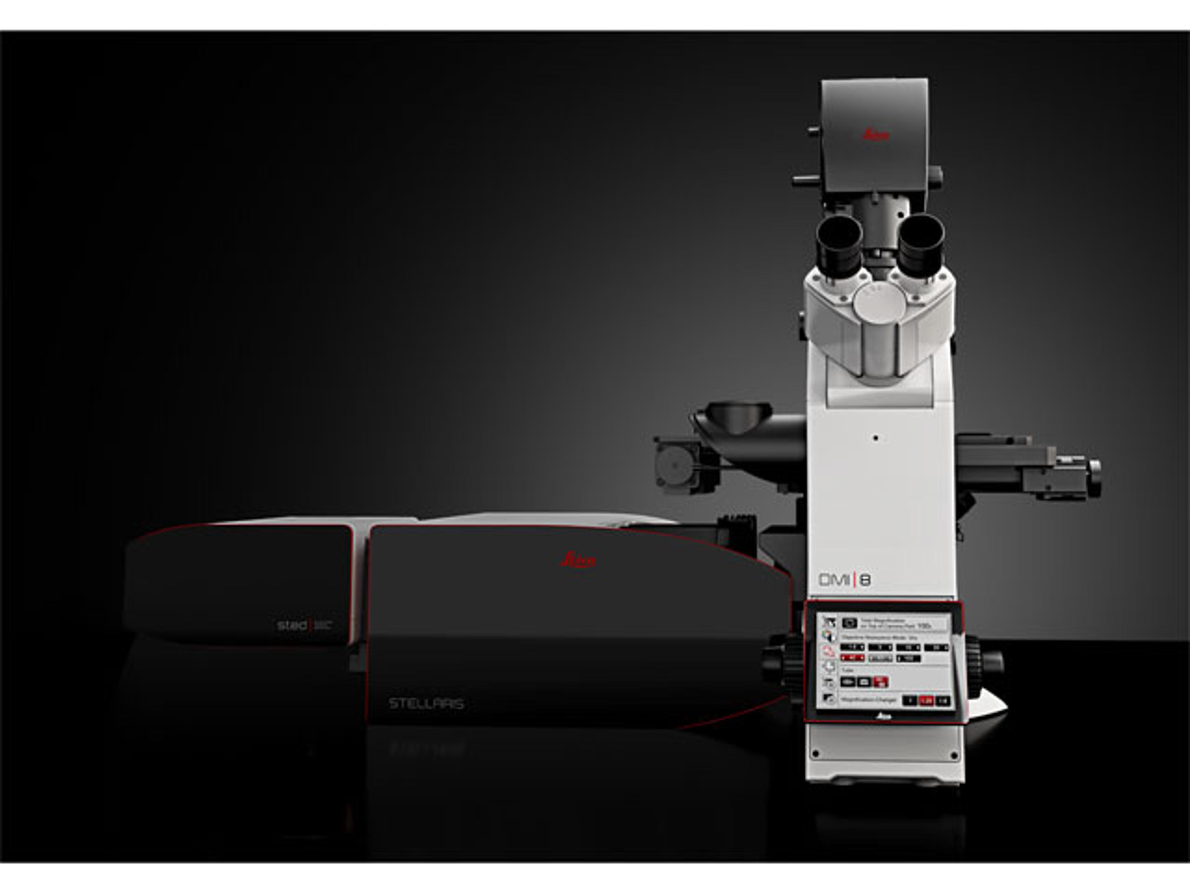
STELLARIS STED Microscopes
Leica Microsystems EuropeOur STED (stimulated emission depletion) technology joins the STELLARIS platform to provide you the fastest way of imaging beyond the diffraction limit. Obtain cutting-edge nanoscopy results in no time with astounding image quality and resolution, while protecting your sample. STED super-resolution allows you to study multiple dynamic events simultaneously, so you can investigate molecular relationships and mechanisms within the cellular context.