SelectScience InterviewsLife Sciences
How to ensure physiological relevance in neonatal models with live-cell imaging
2021-sartorius-sandraleibel-V_2.txt
2 Jun 2021
Dr. Sandra Leibel, assistant professor at University of California, discusses her work investigating surfactant metabolism in neonatal patients and explains the challenges that arise when attempting to model fetal lungs. Dr. Leibel highlights how the Incucyte Live-Cell Analysis System helps streamline modeling workflows by enabling the close monitoring and imaging of developing lung cells within a petri dish.
Related Products
Request Quote for All Products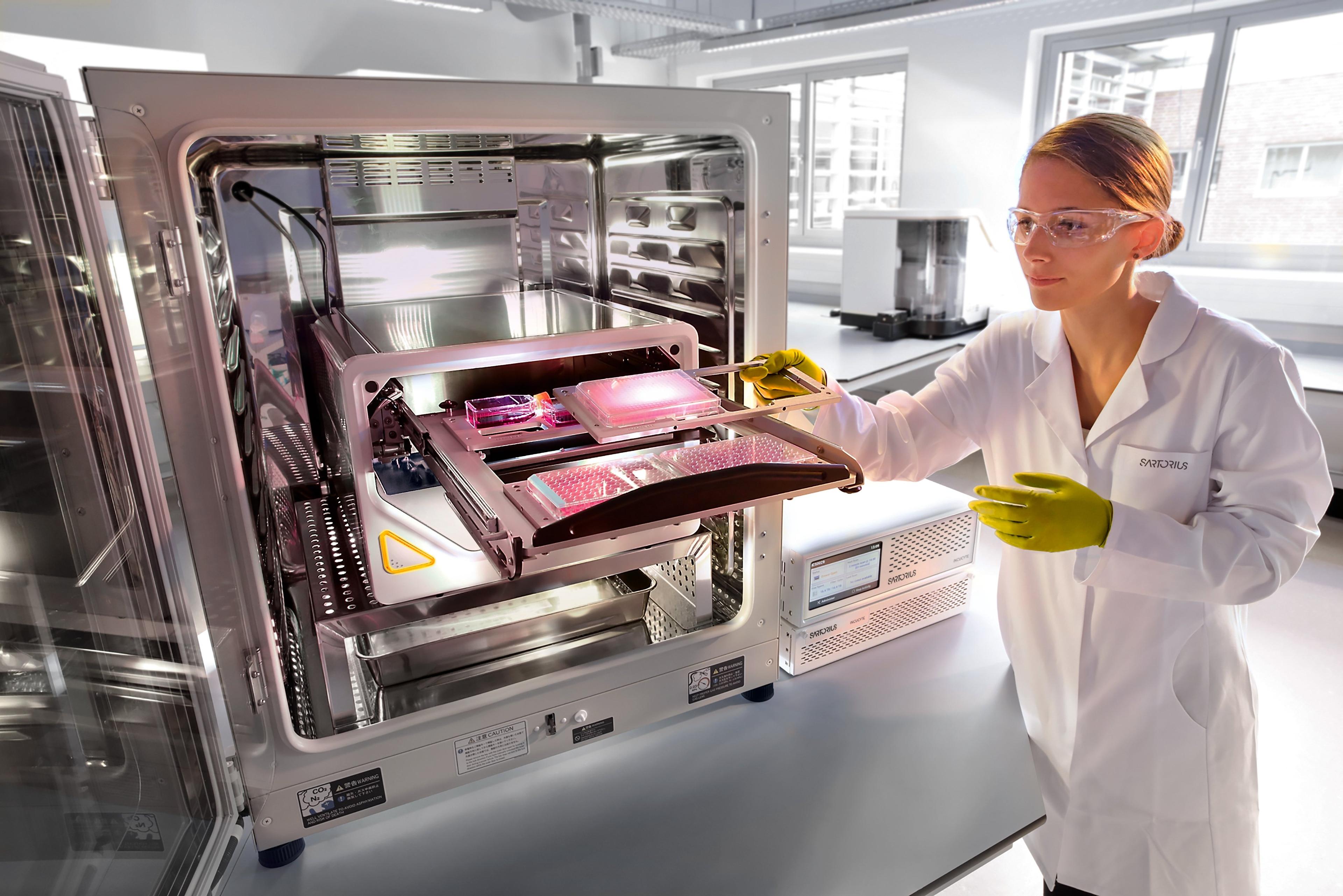
Incucyte® S3 Live-Cell Analysis System
Sartorius GroupOur flagship, supports the workflow and workload of larger laboratories.