
TechTalk: Personalized risk stratification for prostate cancer and the potential of whole-slide IR imaging
Friday, October 30 at 17:00 GMT | 18:00 CET | 13:00 EDT | 10:00 PDT
The molecular and spatial heterogeneity of prostate cancer complicates risk assessment and treatment planning. A key challenge is to map molecular tissue characteristics without losing the histopathological context, while meeting the throughput and reproducibility requirements of clinical research.
In this TechTalk, we will explore how label-free, stain-free whole-slide IR imaging can add a new dimension to tissue analysis. Using prostate cancer as a key application, we will show how spectral pathology combines chemical imaging with spatial information to characterize tissue and investigate its potential for more personalized risk stratification.
Certificate of attendance
If you attend the live TechTalk, you will automatically receive a certificate of attendance, including a learning outcomes summary, for continuing education purposes. If you view the on-demand TechTalk, you can request a certificate of attendance by emailing editor@selectscience.net.
TechTalk details
- Cost: Free to attend
- Location: Online
- Duration: 20 minutes
Registration is required to secure your place. If you register but can’t attend live, you will receive a link to the on-demand recording once it becomes available.
Speakers

Who should attend?
- Pathologists and clinicians
- Digital pathology specialists
- Bioinformaticians working with tissue imaging
- Researchers developing new diagnostic and personalized medicine approaches
Featured products
Request Quote for All Products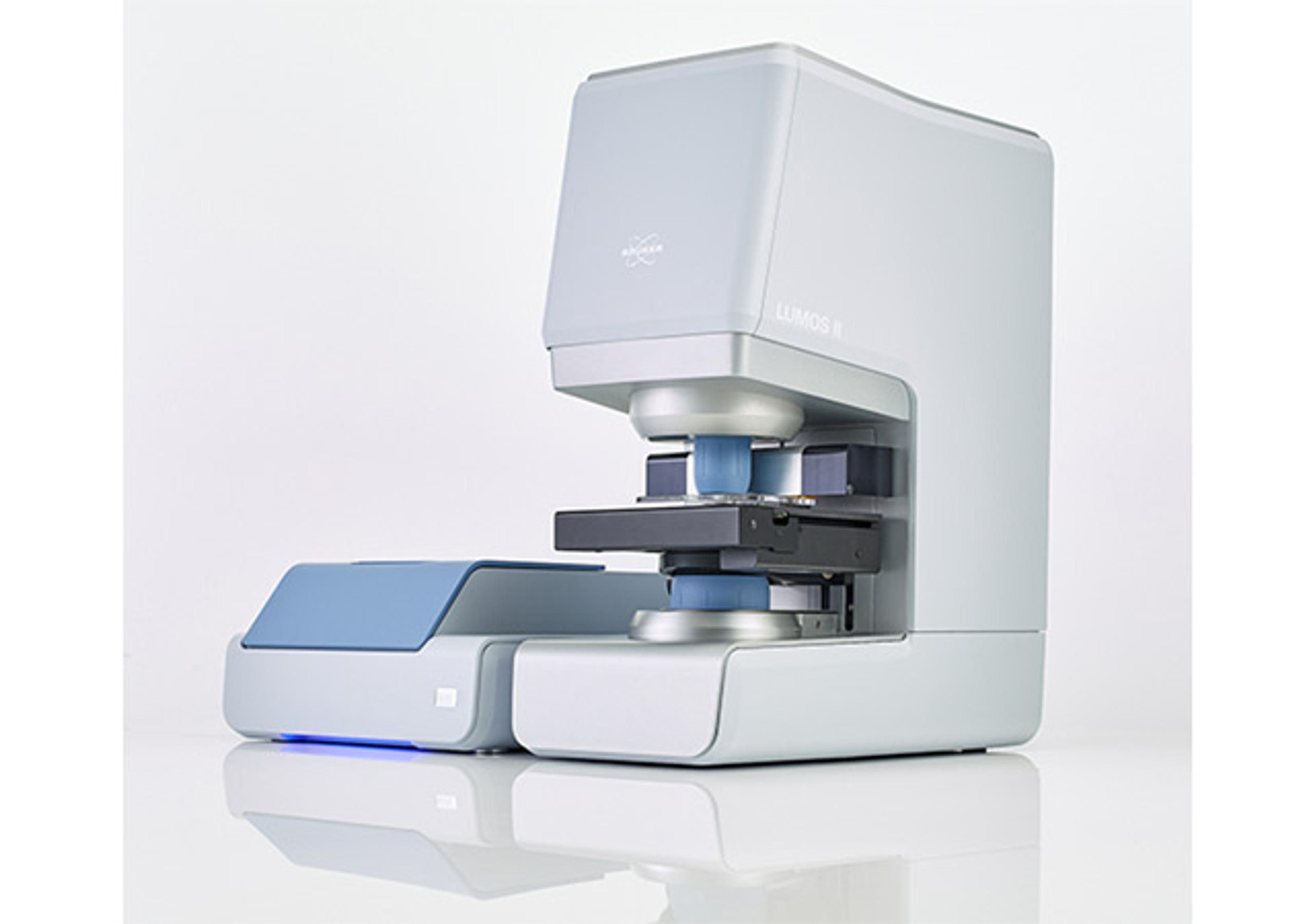
LUMOS II ILIM
Bruker OpticsLUMOS II ILIM redefines infrared imaging speed and precision. With patented Infrared Laser Imaging technology, it delivers hyperspectral data at a pace that transforms workflows: what used to take months can now be done in days. It’s not just faster—it’s smarter, combining automation, AI-driven analysis, and multimodal imaging for research and routine applications alike.
What will this webinar cover?
- How spectral pathology can complement conventional morphological tissue assessment
- How spectral information could contribute to more objective risk stratification and support treatment decisions
- How whole-slide IR imaging reveals label-free chemical information across tissue sections