The Importance of a Stable Fluorescence Light Source in FRET Measurements
26 Apr 2011This application note reviews a few of the most relevant sources of error in FRET-based measurements and point out how having a stable and precisely controllable fluorescent light source, like that offered by the X-Cite® exacte, can help to reduce these complications by providing stable fluorescence illumination. The FRET ratios in HEK cells using two light sources with differing output stability are compared.
Related products
Request Quote for All Products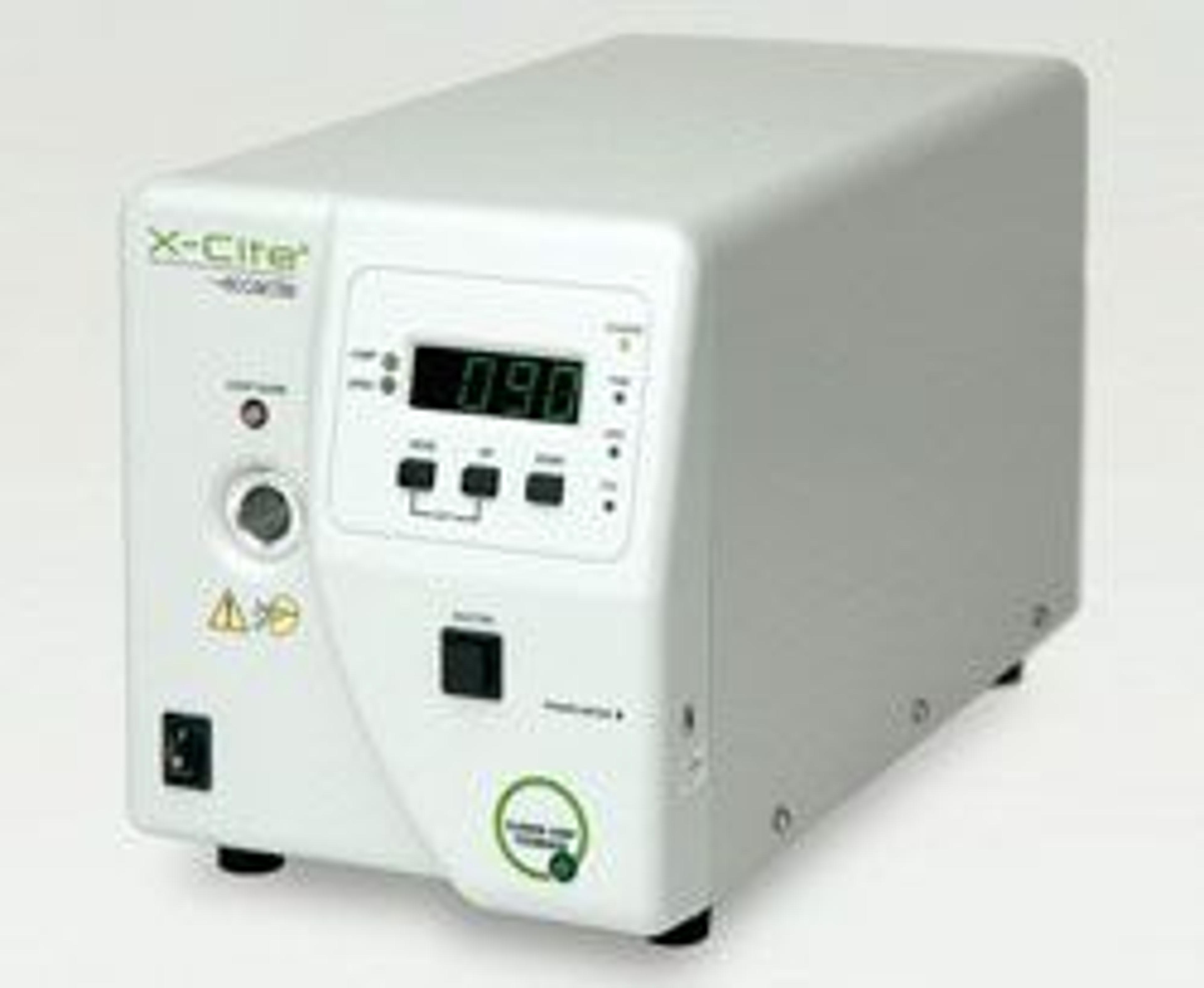
X-Cite Exacte
Lumen DynamicsThe X-Cite® exacte employs a DC-powered mercury lamp coupled with patented Closed-Loop Feedback technology to provide ultra-stable illumination, for imaging protocols lasting from milliseconds to days. With the X-Cite® Optical Power Measurement System, light output can also be calibrated in absolute power units (watts), ensuring truly repeatable experiments. Features of the X-Cite® exacte: Lamp: 200W, Pre-aligned, Intelli-Lamp® technology, 2000 hour guaranteed life. Includes: Closed-Loop Feedback technology, Adjustable iris in 1% increments, Computer Control & GUI, Internal high speed shutter. Compatible with Carl Zeiss, Leica, Nikon, Olympus and Motic microscopes. Additional Specifications of the X-Cite® exacte: Certifications: CE Marked, Certified to IEC, Canadian and US Standards