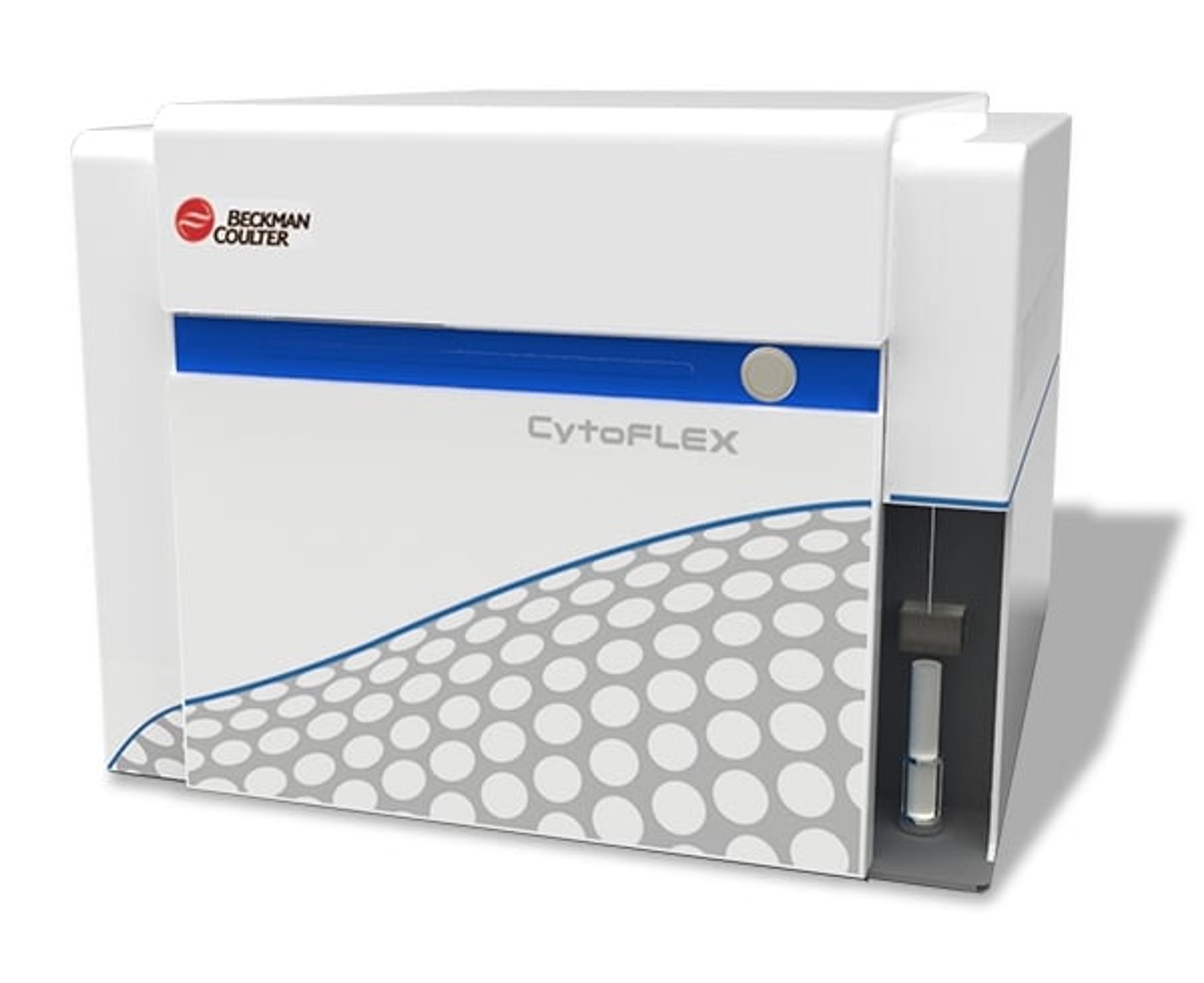
Set-Up of the CytoFLEX for Extracellular Vesicle Measurement

Set-Up of the CytoFLEX for Extracellular Vesicle Measurement

16 Aug 2015

This application note describes how to set-up and standardize the CytoFLEX for particle measurement and discuss some pitfalls which should be avoided to get the best information from EVs detection. Extracellular vesicles are a heterogeneous cell-derived particle population in a size range between 50nm to 1,000nm. There is a growing interest not only from academic research groups to determine EV in several fluids such as cell culture supernatant, in plasma samples or in whole blood but also in clinical research since it has been shown that the measurement of microparticles (MPs) might be of clinical relevance. Compared with other methods of detection, flow cytometry has the big advantage that EV can be detected as rare events, in high numbers and by antigens on the surface, which characterize their cellular origin.

CytoFLEX Flow Cytometer

Beckman Coulter Life Sciences

Providing quality and performance at any configuration, the CytoFLEX system provides powerful sensitivity and resolution for the simple to the most challenging applications. CytoFLEX delivers and surpasses capabilities expected in top tier analyzers, with excellent performance and nanoparticle resolution.

(23)

Links

Tags

AntibodiesAntibodies are used in techniques such as confocal and fluorescence microscopy, flow cytometry, ELISA, ELISPOT, immunohistochemistry, western blotting and immunopreciptation. Select specific antigen reactivity, high specific affinity, low non-specific binding, monoclonal or polyclonal, primary or secondary antibodies and associated conjugates such as an enzyme or dye for visualization.Flow Cytometry / Cell CountingFlow cytometers are used to count, sort and examine multiple characteristics of cells. Other cell analysis equipment includes image cytometers, cell counters, fluorescence-activated cell sorters (FACS), magnetic-activated cell sorters (MACS), and a range of flow cytometry assay kits. Flow cytometers can reveal information on cell viability, cell proliferation, apoptosis and cell cycle progression, as well as identify cell populations and intracellular or cell-surface molecules. Additionally, some flow cytometers, known as FACS, have an additional sorting function after analysis. Cell counters and image cytometers count live and dead cell populations and can also conduct cell proliferation assays. Find the best flow cytometers, cell counters and cell sorters in our peer-reviewed product directory: compare products, check customer reviews and receive pricing direct from manufacturers.BiomarkersBiomarkers are biological markers which can be measured and evaluated to indicate a biological state. The use of biomarkers in research and diagnosis can indicate a normal or disease state or drug response of cells / tissues. Biomarkers include genetic markers, cell surface markers such as antigens, antibodies or receptors and secreted molecules such as cytokines. An assay system is required for identification of biomarkers. :CytometryCytometry is a broad term for the quantitative analysis of cells and cell systems. The most popular methods to study cytometry are flow cytometry and image cytometry.Human PlasmaHuman plasma is the liquid component of blood, which carries blood components - nutrients, hormones, proteins, and waste - throughout the body. Plasma-based studies provide insights into biomarkers, immune responses, and disease mechanisms. Compare plasma handling tools and systems in our peer-reviewed product directory.