ResourceLife Sciences
Monitoring confluency of adherent cells in multi well plates using the CELENA X High Content Imaging System
28 May 2026This application note demonstrates how the CELENA® X High Content Imaging System from Logos Biosystems can be used to monitor and quantify cell confluency in multi-well plates. Using automated brightfield imaging and image analysis pipelines, the system enables accurate measurement of cell coverage over time, supporting reproducible and objective assessment of cell growth. Discover how the workflow allows high-throughput analysis and reduces reliance on subjective visual estimation in cell culture experiments.
Related products
Request Quote for All Products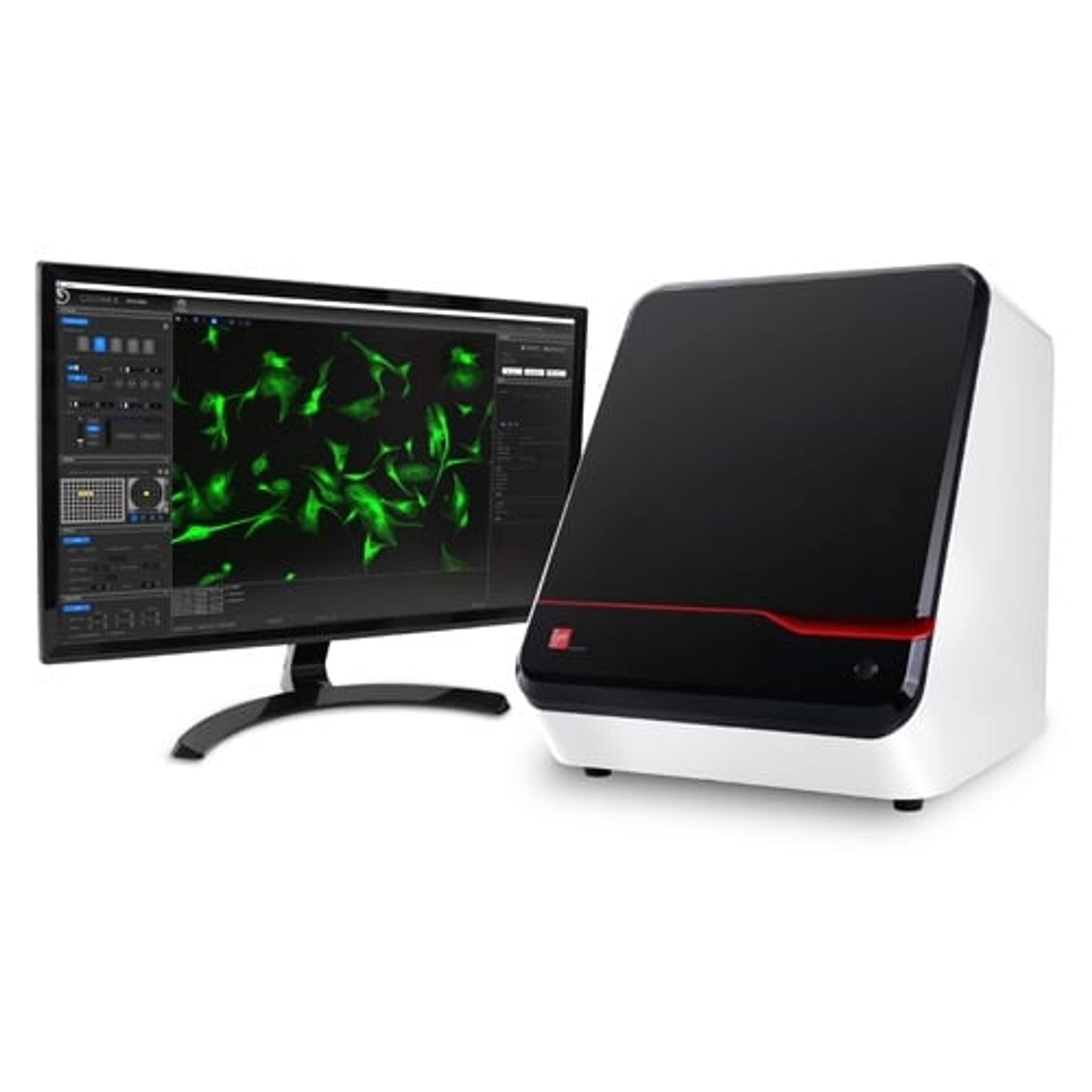
CELENA® X High Content Imaging System
Logos BiosystemsThe CELENA ® X High Content Imaging System is an integrated imaging system designed for rapid, high content image acquisition and analysis.
Links
Tags
Cell ImagingCell imaging can be achieved using a number of techniques including confocal microscopy, transmission electron microscopy, atomic force microscopy, and light sheet microscopy.High Throughput ScreeningImage AnalysisImage analysis involves the extraction of meaningful information from images, often using software to quantify and interpret visual data. It is widely used in cell biology, material science, and diagnostics. Increasingly, AI is being used to streamline image analysis. Explore image analysis tools in our peer-reviewed product directory; compare products, check reviews, and get pricing directly from manufacturers.Cell CultureCell culture involves growing cells under controlled conditions, typically outside of their natural environment, to study cellular behavior, drug response, and disease models. Cell culture is crucial in biotechnology and biomedical research. Explore cell culture products in our peer-reviewed product directory; compare products, check reviews, and get pricing directly from manufacturers.Bright Field MicroscopyHigh Content ImagingHigh content imaging is a method combining two or more fluorescent microscopy experiments to identify substances that alter a cell’s phenotype in a desired manner. The process is adapted to multi-well plates and both the image acquisition and analysis are automated.