High-content imaging for morphological profiling of 3D breast acini
10 Apr 2023With the development of quantitative tools for image analysis and classification, diagnostic accuracy of breast cancer is significantly increasing. High-content imaging (HCI) represents a powerful image-based paradigm to extract high-content data from biological images. This imaging system consists of acquisition, processing and analysis steps used to dramatically increase imaging throughput and quantification; each step is highly computer-driven, allowing users to accelerate repetitive operations and reduce human intervention and bias. In this application note, CrestOptics presents a method for automated imaging and analysis of breast cancer in three-dimensional (3D) epithelial breast cell cultures. In particular, it developed an image analysis framework for the automated imaging of in vitro 3D breast epithelial acini through the characterization of acinar structure morphology.
Related products
Request Quote for All Products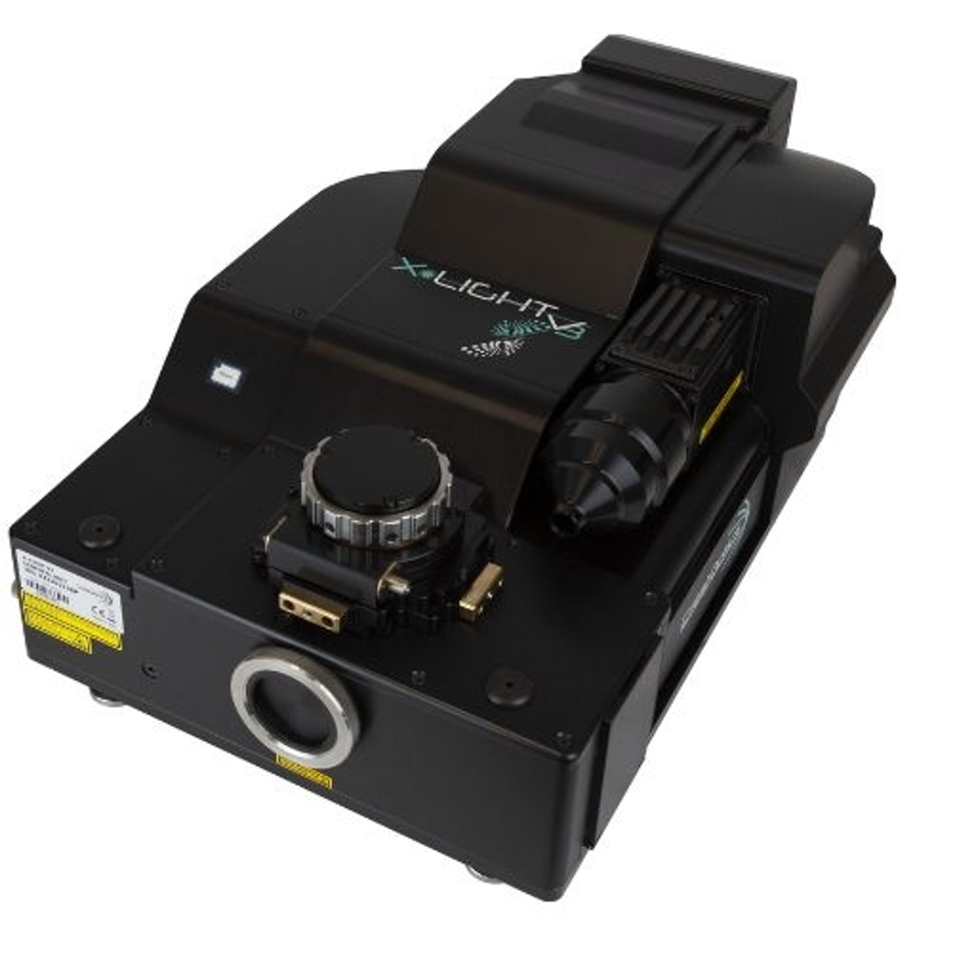
X-Light V3
CrestOptics S.p.A"SEE BRIGHTER, SEE FASTER, SEE MORE". X-Light V3 is the next generation of spinning disks, and it relies on cutting-edge technology, advanced optical design, and engineering solutions developed by CrestOptics to meet the very high-end specifications required by modern fluorescence microscopy applications.