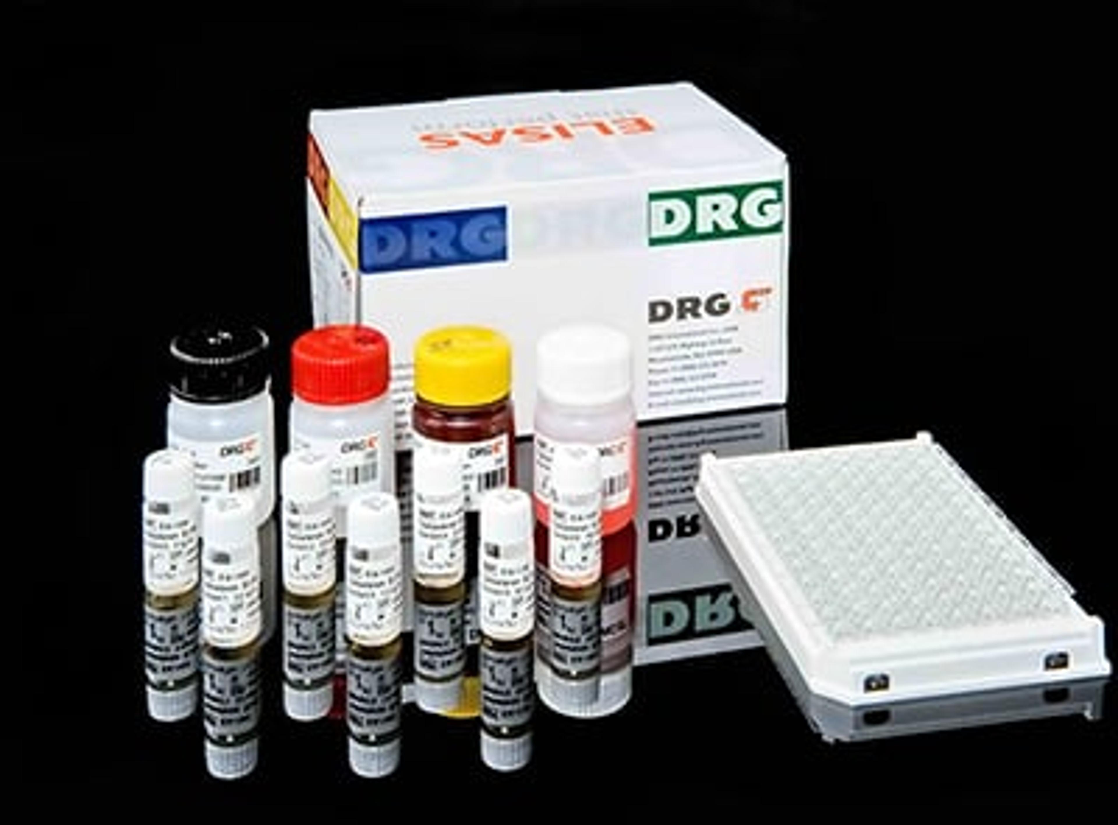
Ureaplasma urealyticum IgG ELISA
DRG International Inc.High Quality Assays with Reproducible and Reliable Results
High Quality Assays with Reproducible and Reliable Results
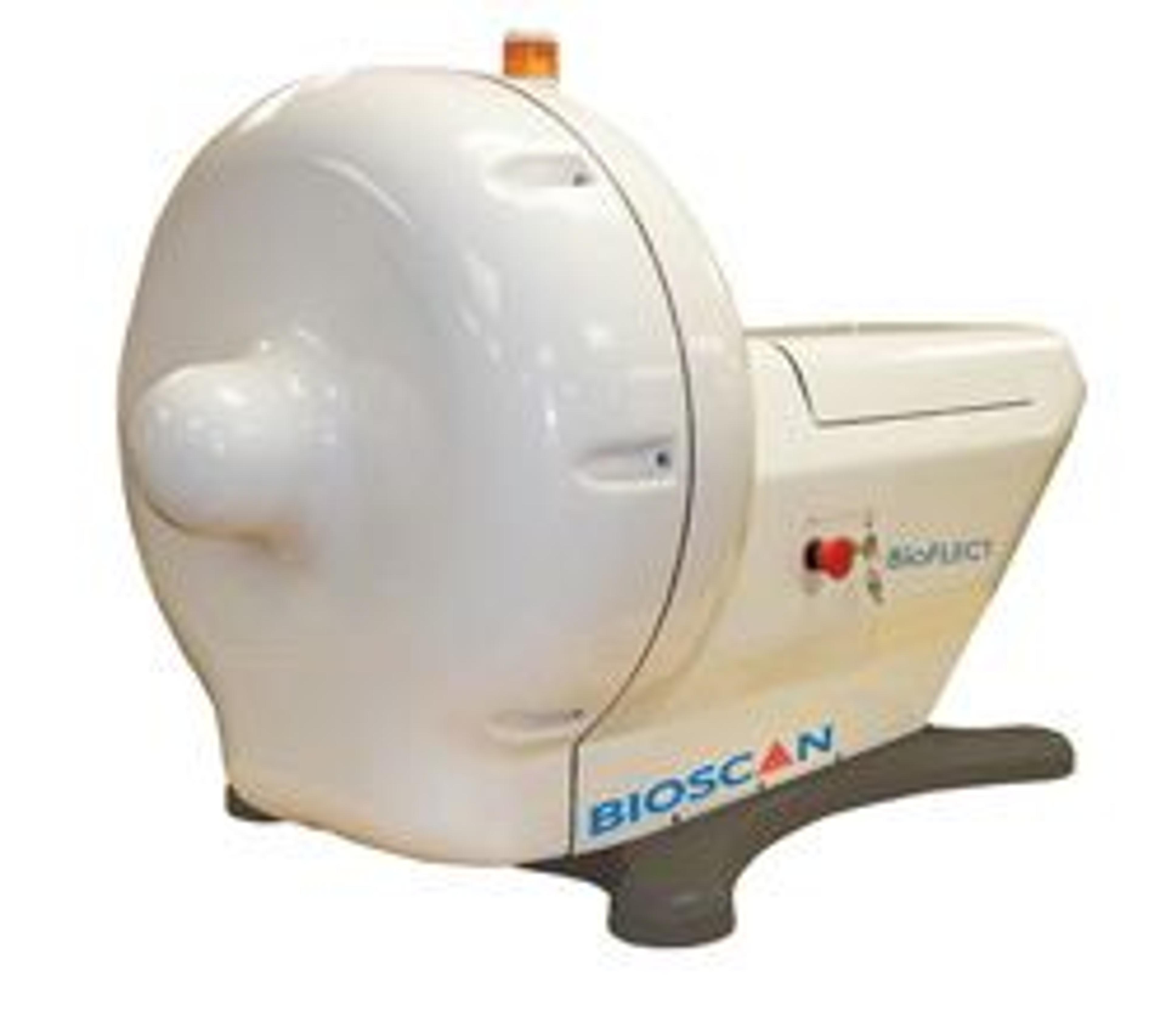
BioFLECT™ In Vivo Preclinical Imager - The Only True 360° Tomographic Optical Imager With the launch of the BioFLECT, Bioscan will enable researchers to realize the full potential of in vivo optical imaging for the first time. With BioFLECT’s true 360° tomographic system, researchers will no longer have to choose between imaging only near the surface of the subject or partial tomography solutions that distort the subject or c…
High Quality Assays with Reproducible and Reliable Results
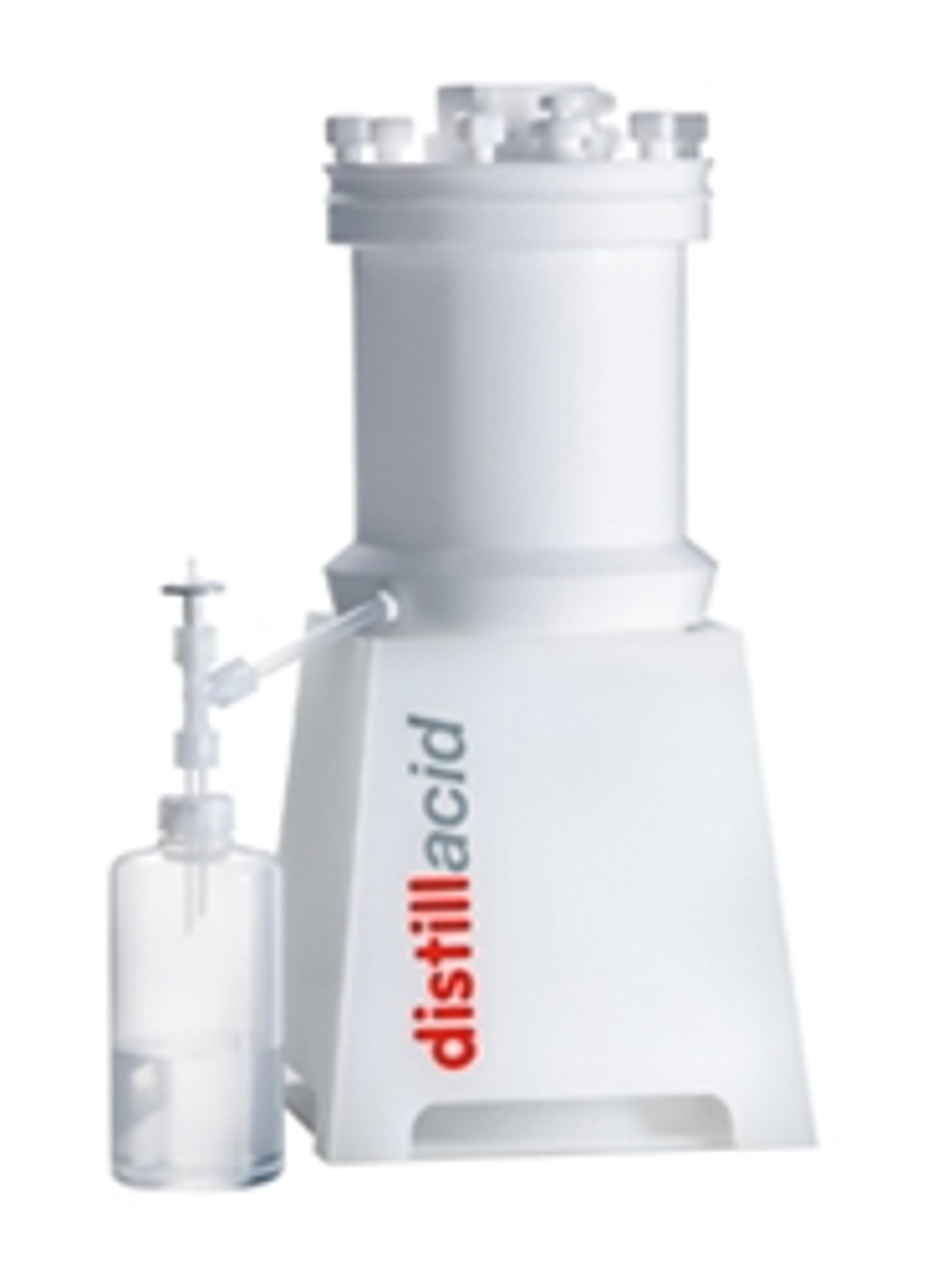
High-Purity Acids, Always fresh to hand thanks to Subboiling. As rule, chemical prices increase exponentially as the chemical’s purity increases. Therefore, even where annual consumption is apparently minimal, high-purity acids can quickly become a significant cost factor for trace analysis laboratories. These costs can be avoided by the employment of a subboiling apparatus to produce high-purity acids from more economical…
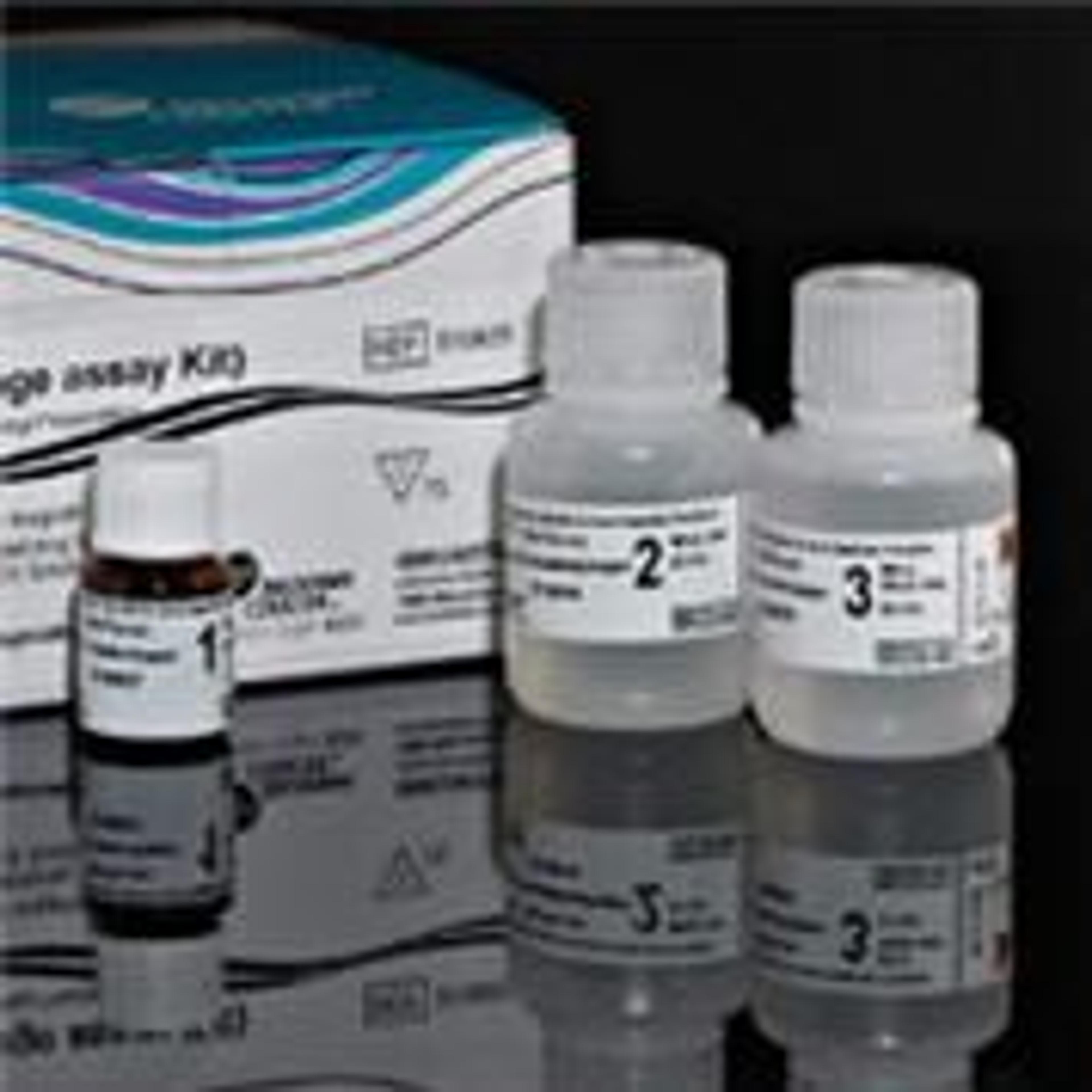
The PerFix EXPOSE Kit (Phospho-Epitopes Exposure kit) is a Fast & Easy Procedure for Cell Signaling.
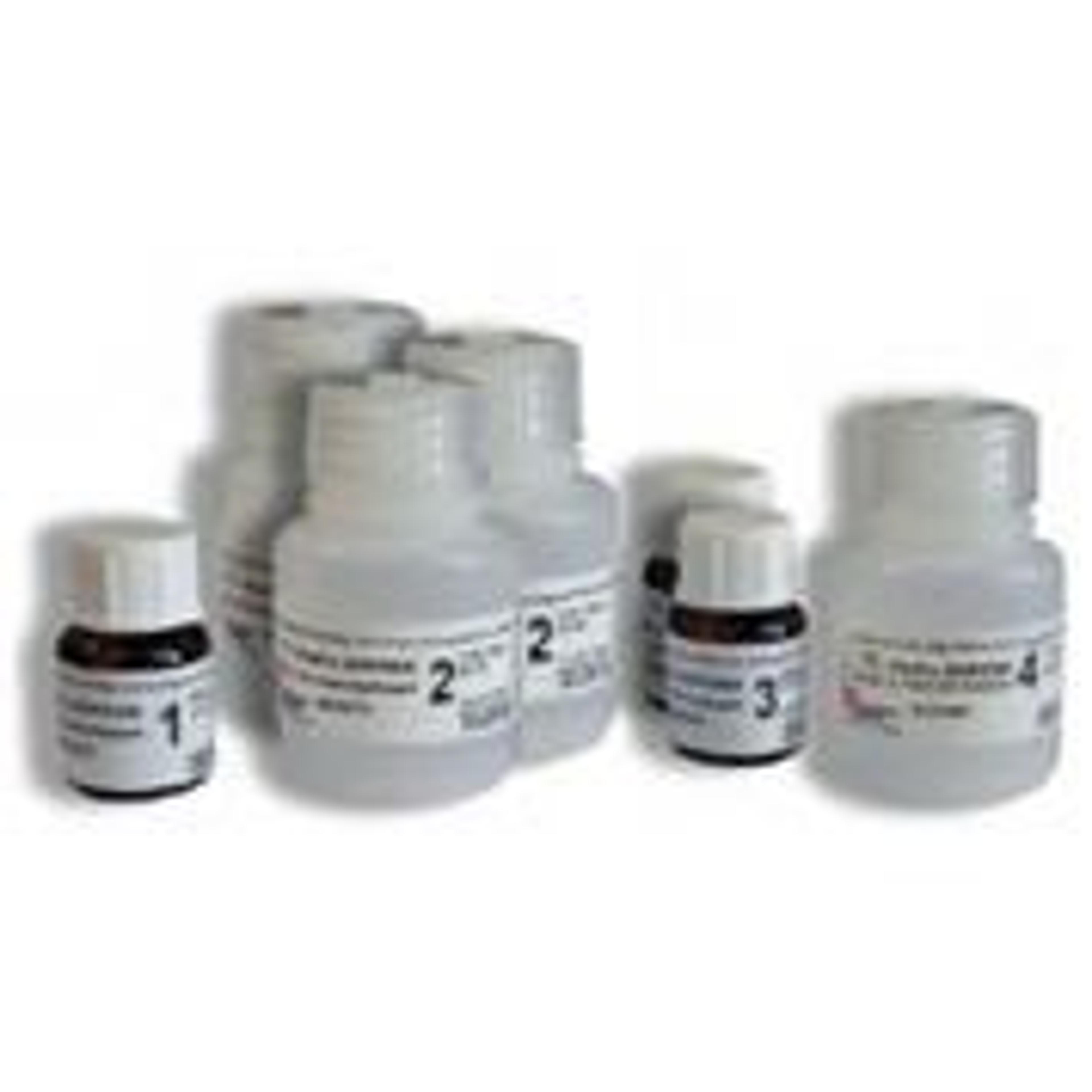
The PerFix-nc Kit (no centrifuge assay Kit) has been developed to enhance the signal-to-noise ratio of intracellular staining and simplify the workload necessary for the sample preparation.
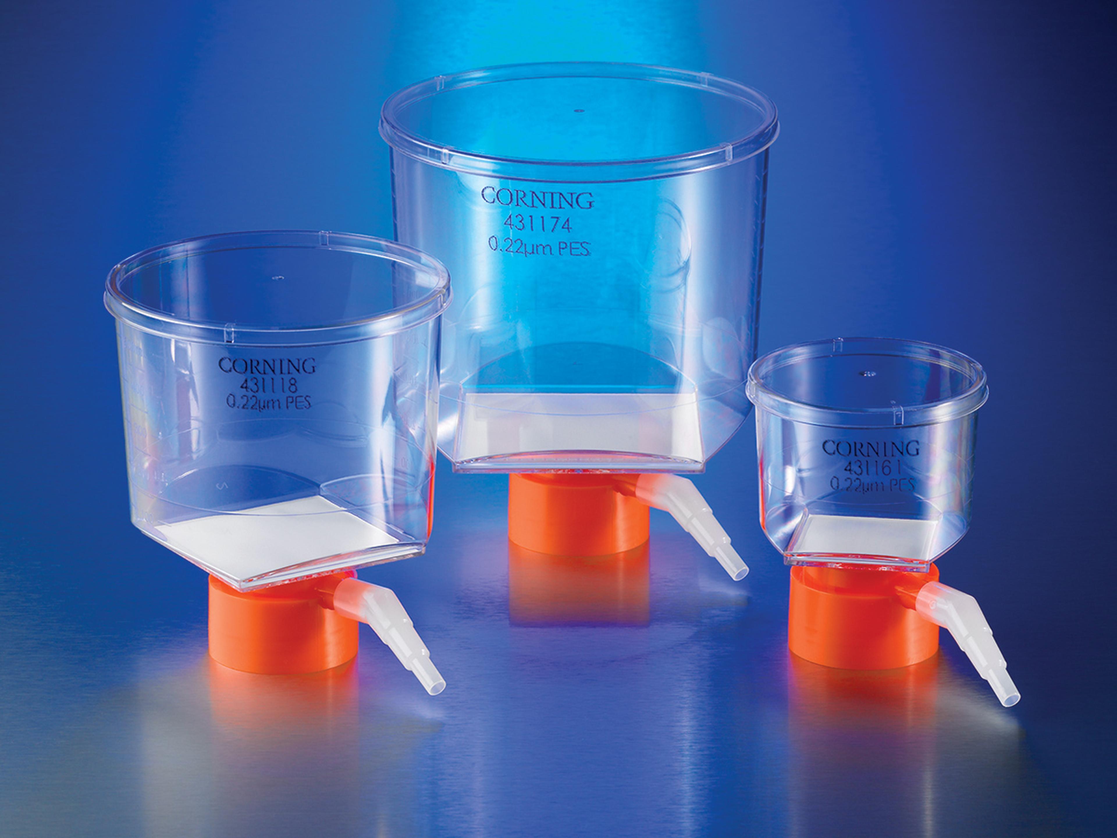
Fits plastic or glass media bottles with 33mm neck sizes. Cellulose acetate (CA) membranes provide fast flow rates and low protein binding and are good for filtering cell culture media. Angled hose connector simplifies vacuum line attachment. Individually packaged, sterile and certified nonpyrogenic. Each system has the membrane material and pore size printed on the unit and is color-coded by membrane type for easy produ…
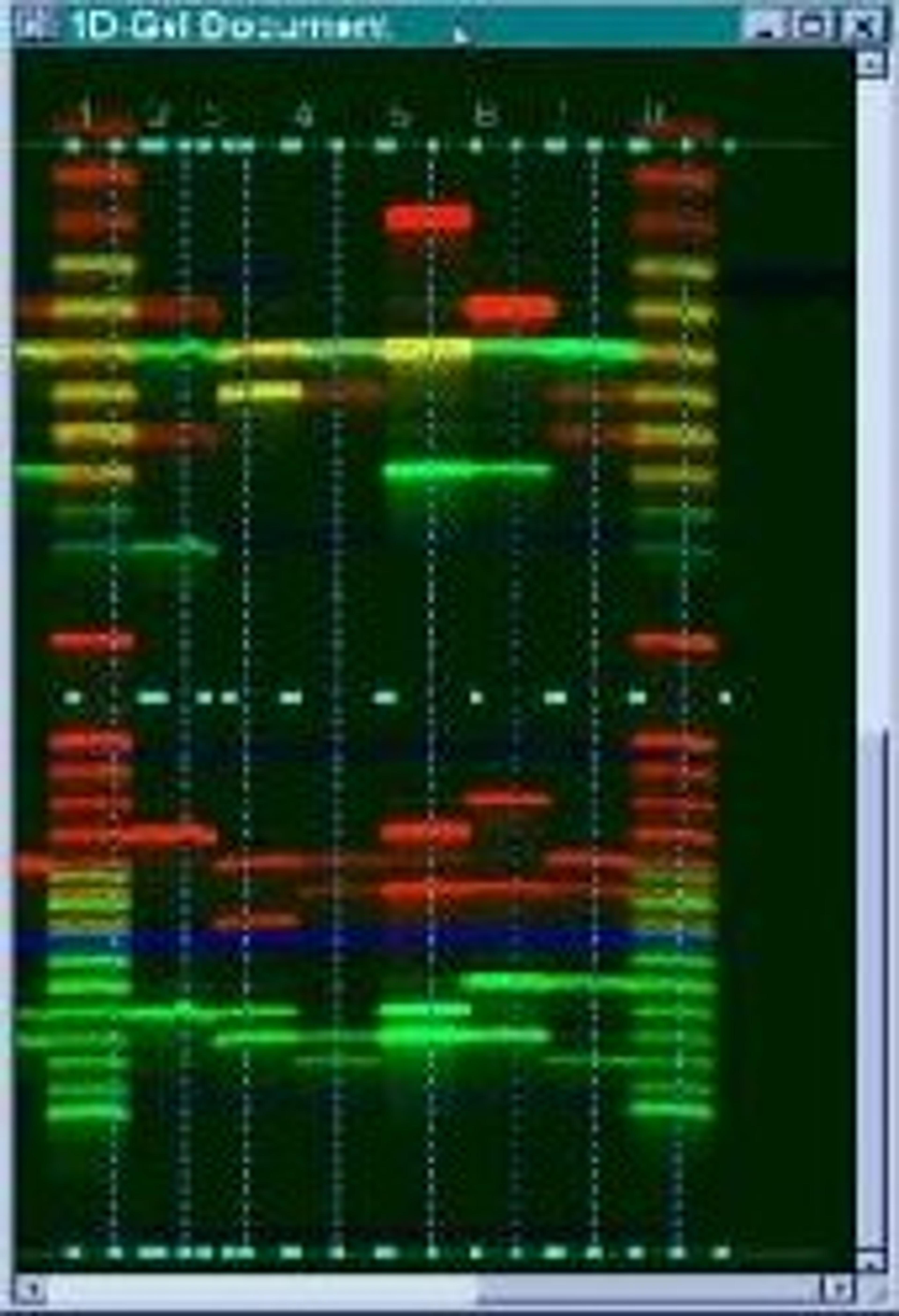
The Image Analysis is a software program for the analysis and digital storage of data from 1-D, 2-D, 96 well, slot/dot blot and DNA sequencing electrophoretic gel images. Image Analysis combines the convenience and economy of a flatbed scanner with the ease and sophistication of Hitachi Software's programming capabilities. Image Analysis will analyze almost any TIFF gel image from a wide range of sources including, fluoresce…
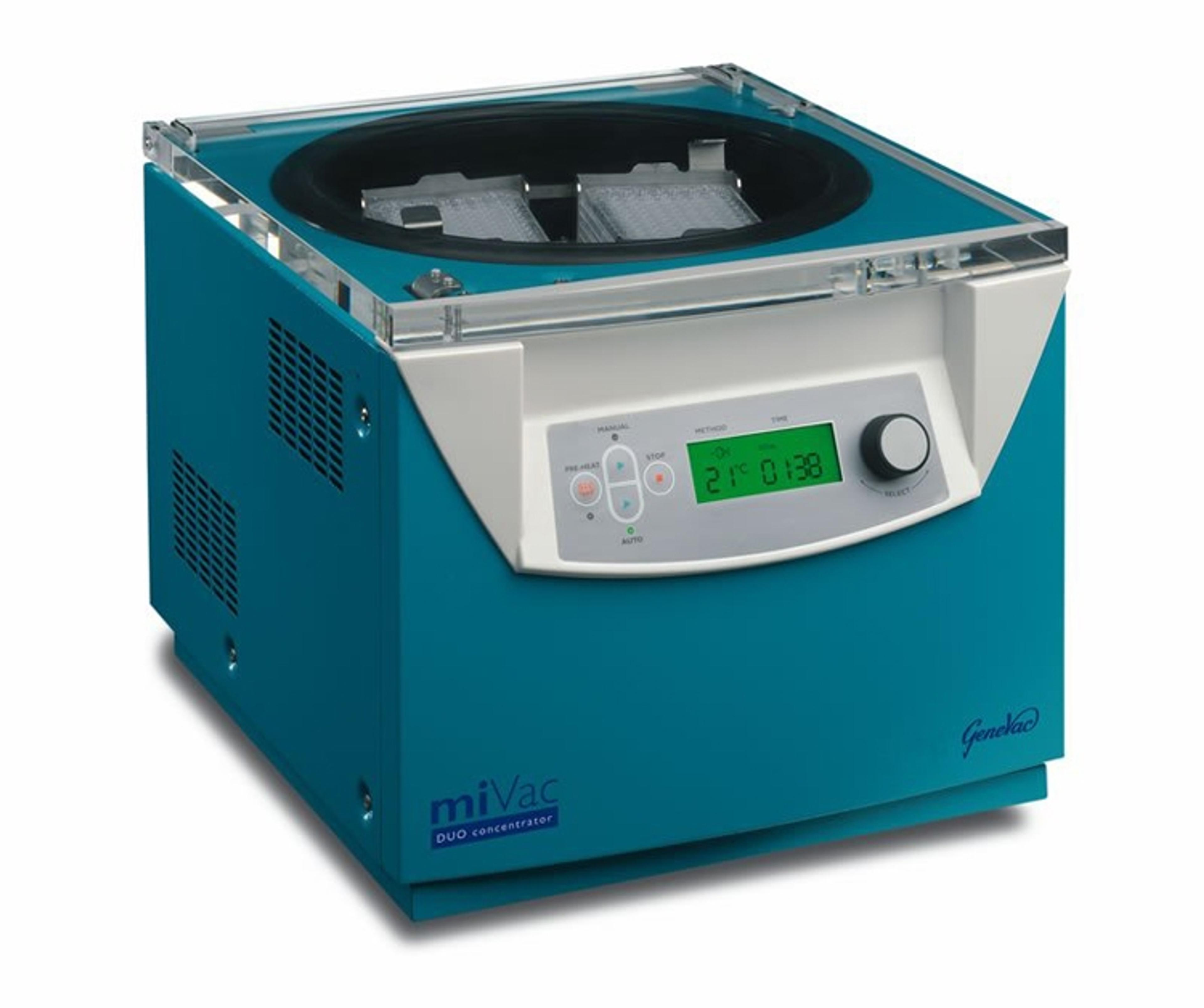
The miVac Duo Concentrator is suitable for use with a wide range of solvents, from volatile organic solvents through to water and some of the higher boiling point solvents. The Duo is a centrifugal vacuum concentrator compatable for use with variety of sample formats including tubes, microplates, vials and round bottom flasks. Making it an ideal "work-horse" concentrator for the busy lab. Combined with a SpeedTrap and JetRo…
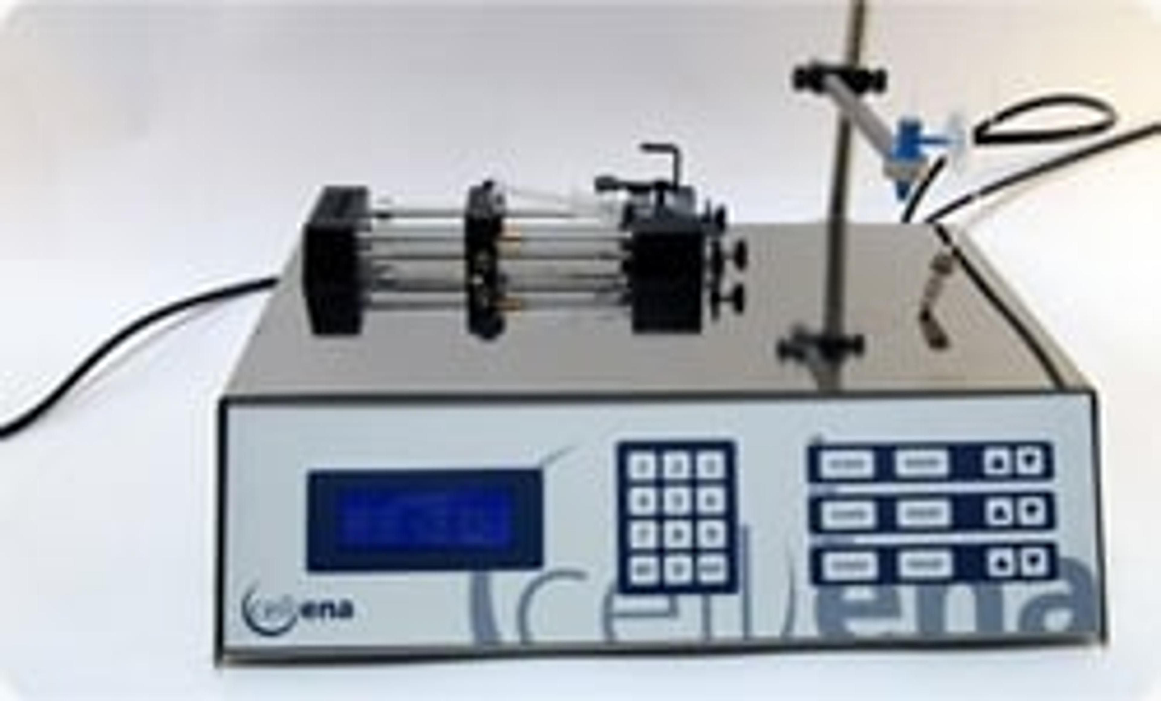
Cellena® is a bioencapsulation portable platform able to encapsulate high molecular weight compounds, microorganisms and cells in homogeneus particles of predictable and controlable size. Designed by Ingeniatrics, it has been specifically developed for biotechnological research. Its performance is based on Flow Focusing® technology. By means of this technology, the elements to be encapsulated are immobilized within a semiperme…
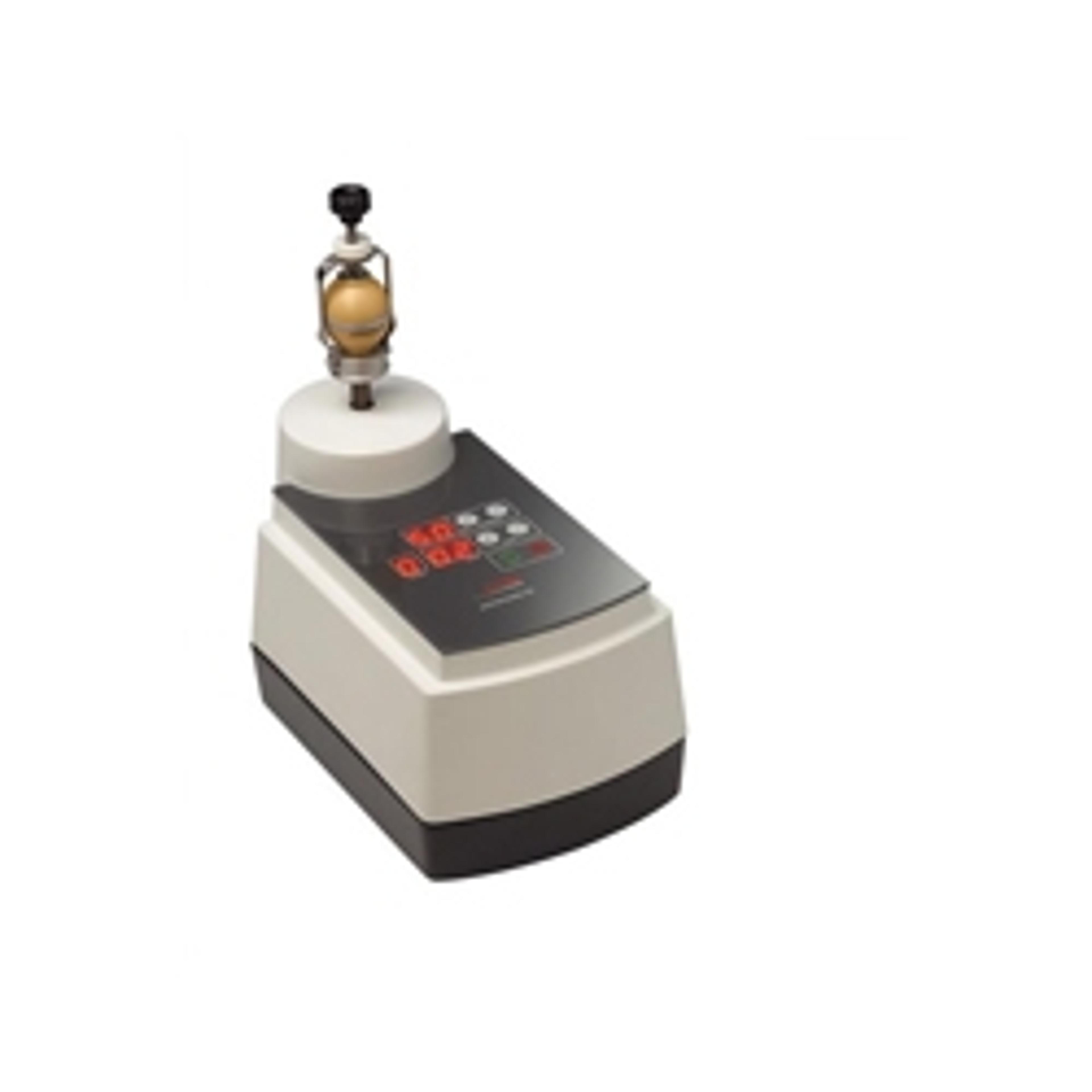
The smallest instrument for small quantities
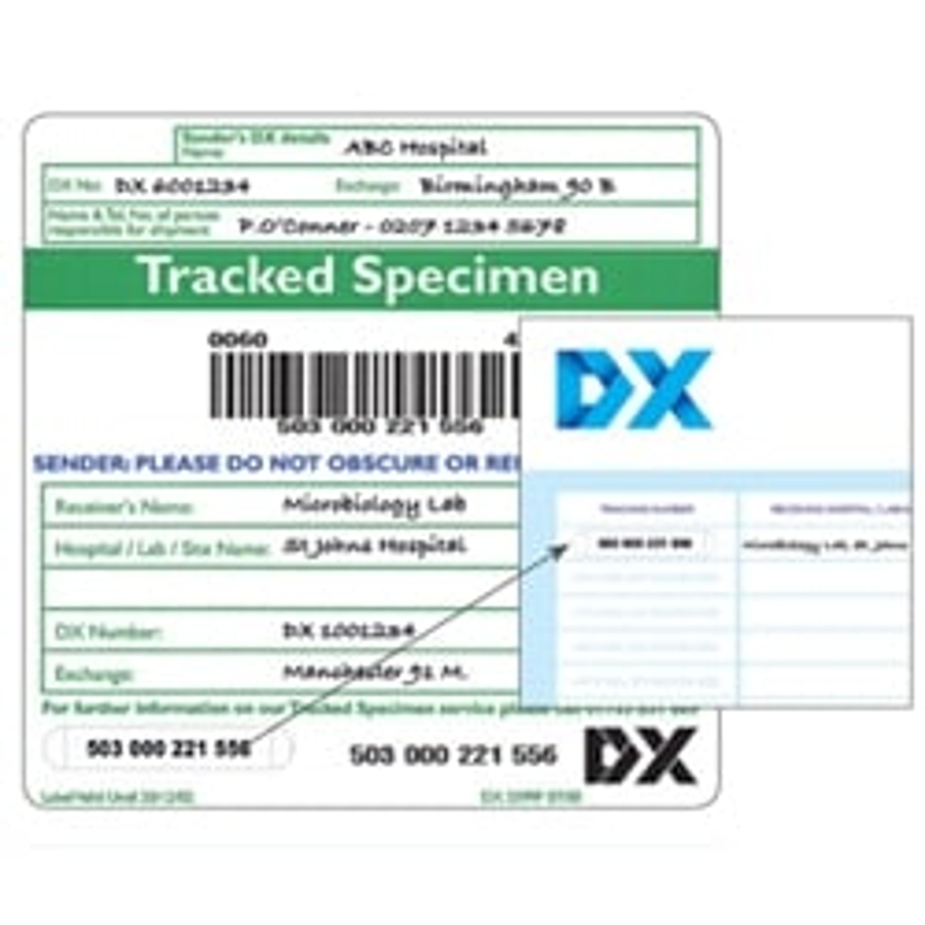
The DX Tracked Specimen Delivery Service CATEGORY B BIOLOGICAL SUBSTANCES Using our Tracked Specimen service, you can send your Category B biological substances through our dedicated pre 9 am delivery network and enjoy the benefits of safe and compliant packaging, bar-code tracking and confirmation that your items are out for delivery. CATEGORY A SPECIMENS – FOR CATEGORY A INFECTIOUS SAMPLES DX does not carry Category A spe…
Created specifically for solid dosage forms, Parteck® excipients are distinguished by outstanding functionalities and a unique particle structure. All Parteck® products are developed under our Functional Particle Engineering concept comprising advanced EMD Millipore technologies for quite extraordinary functionally relevant particle properties. These properties of Parteck® excipients offer decisive advantages to the formulato…
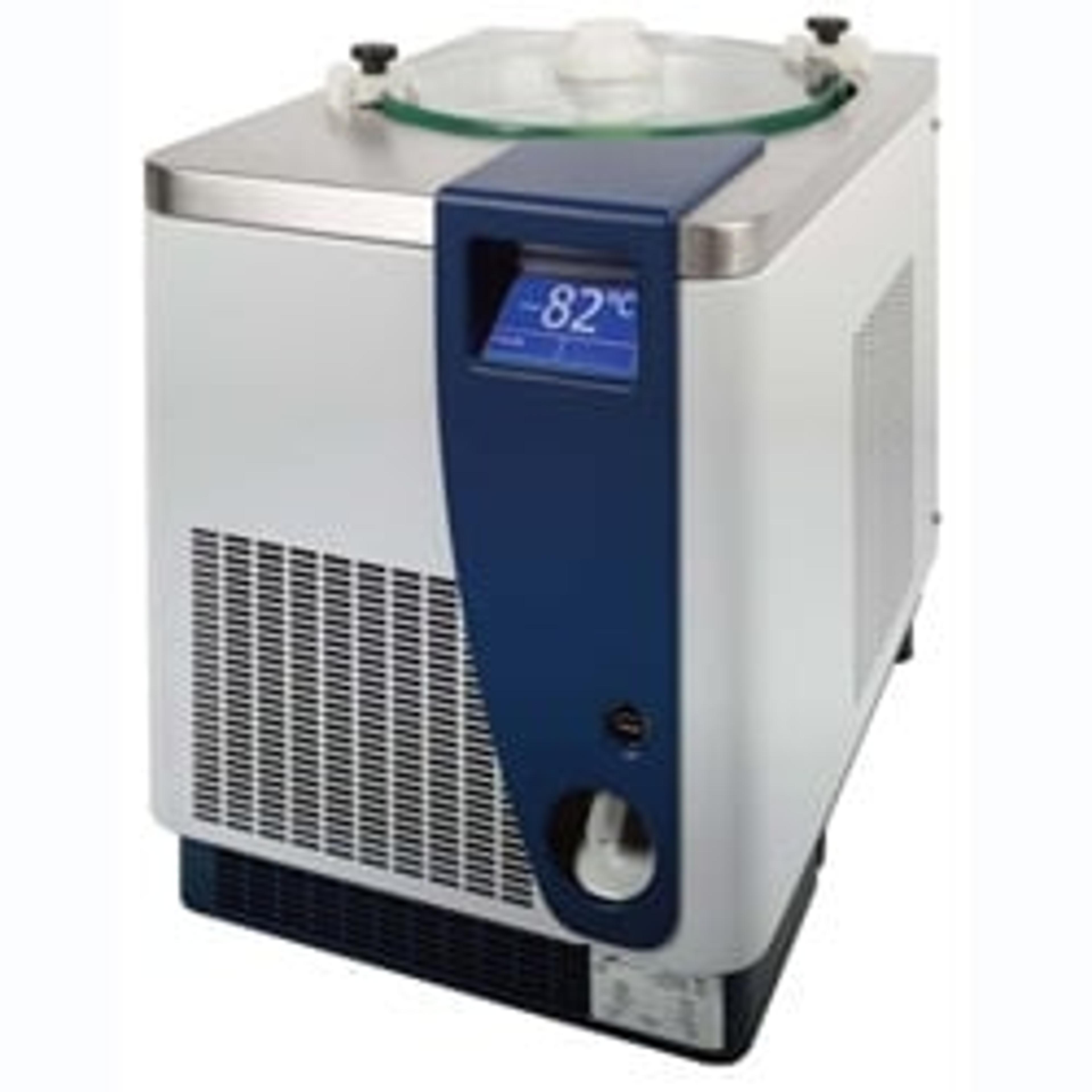
The demand for a more powerful and colder condenser has naturally arisen as the boundaries of evaporation science are pushed further and further. Users increasingly regard as critical the importance of having the right condenser for the job, with the correct power and temperature profile matched to the characteristics of the evaporator. Genevac has developed the super-powerful and super-cold SuperCool 75, in collaboration wit…
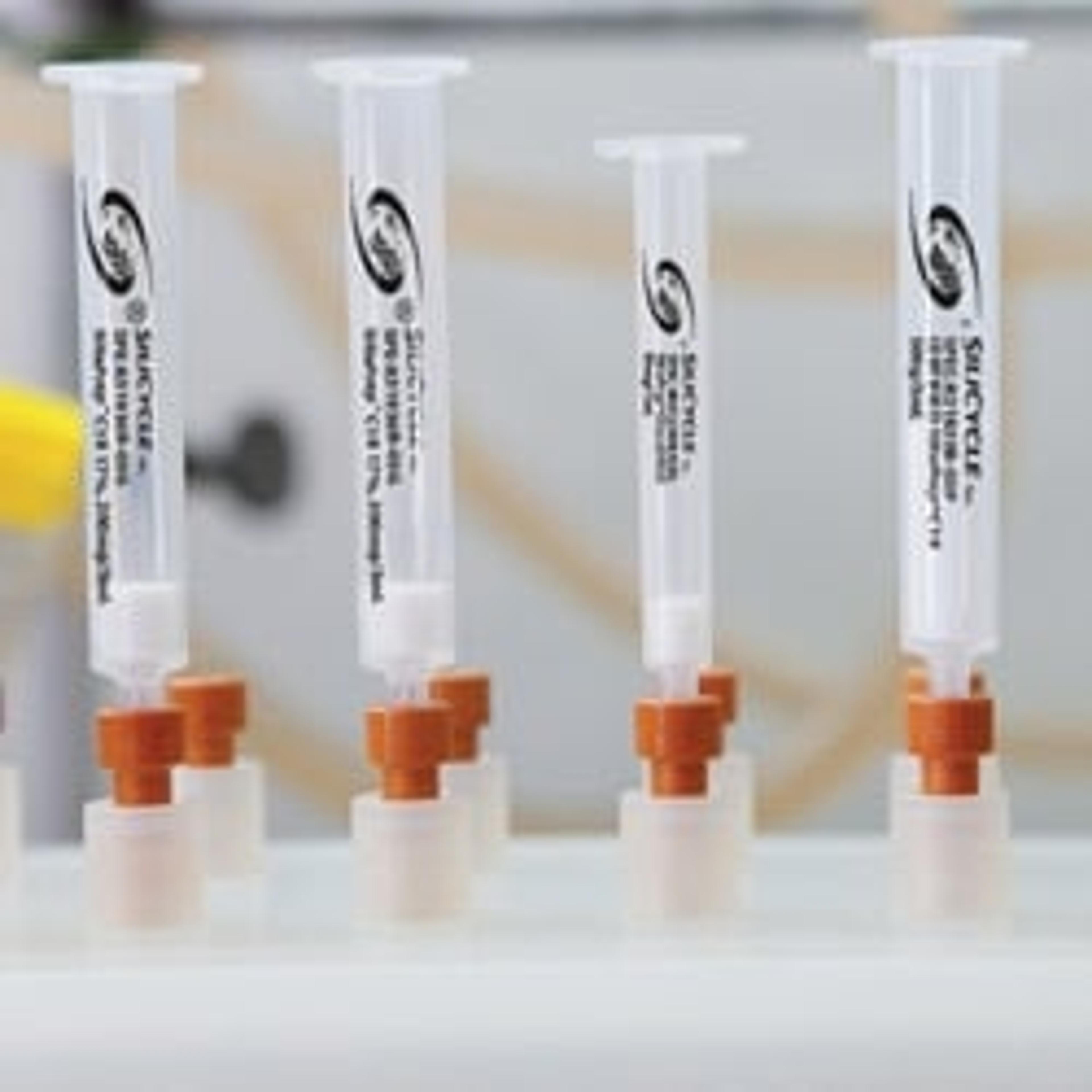
Solid Phase Extraction (SPE) is the basis of analytical laboratories. A crucial step of this process is sample preparation. Therefore we know that a clean sample will yield better results. To achieve this goal SiliCycle has designed SiliaPrep, which is a silica based sorbent exempt of fines, and SiliaPrepX, polymeric based sorbent, that can be found in cartridges, well plates and SPME (Solid Phase Micro-Extraction) cartridges…
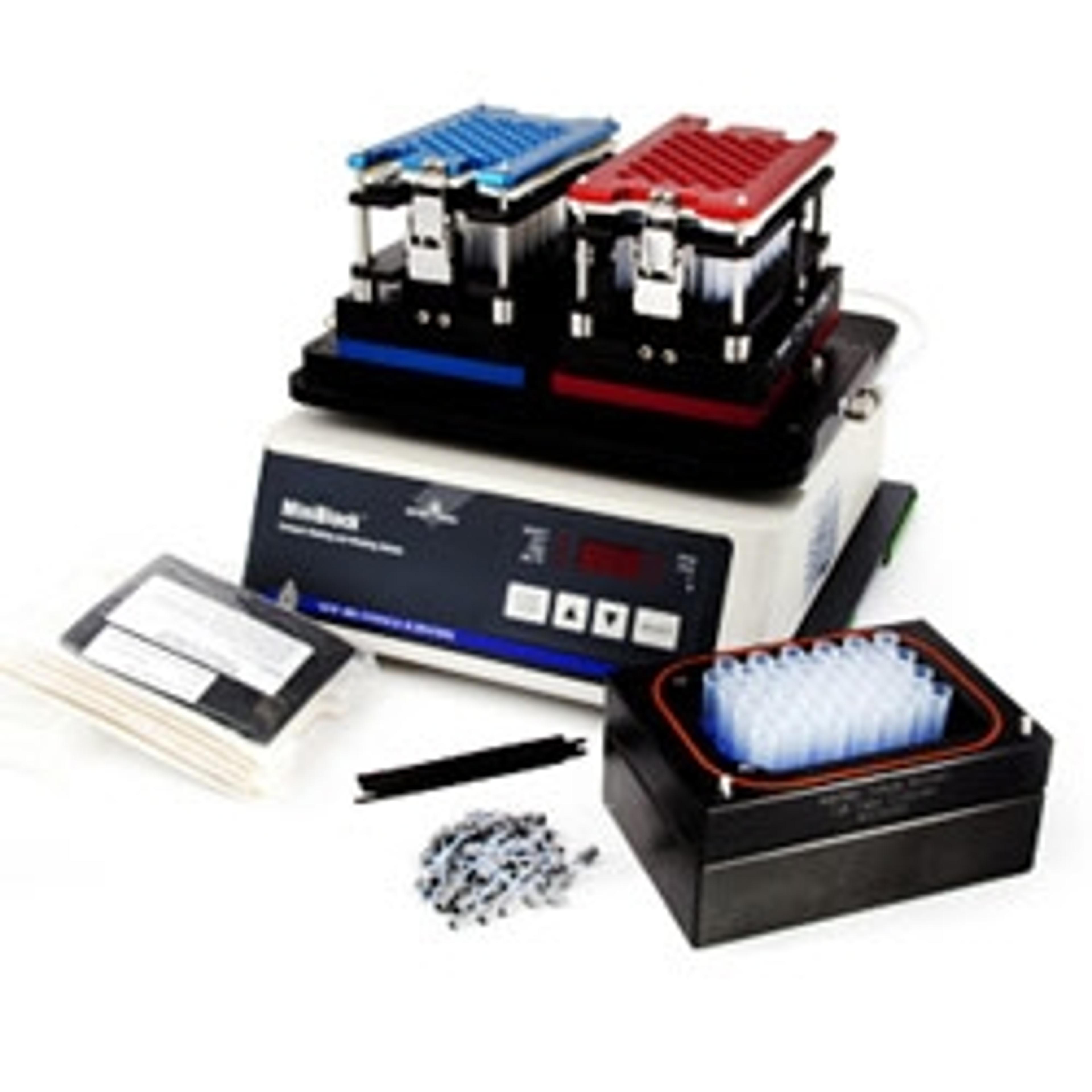
The MiniBlock® is an easy-to-use reaction block designed for parallel synthesis and screening. Its unique valve body design enables processes where filtration is critical, including solid-phase organic synthesis, use of scavenger resins with solution phase synthesis and parallel purification via Solid-Phase Extraction (SPE).Make the choice many chemists have made to increase productivity, more so than any other similar tool.…
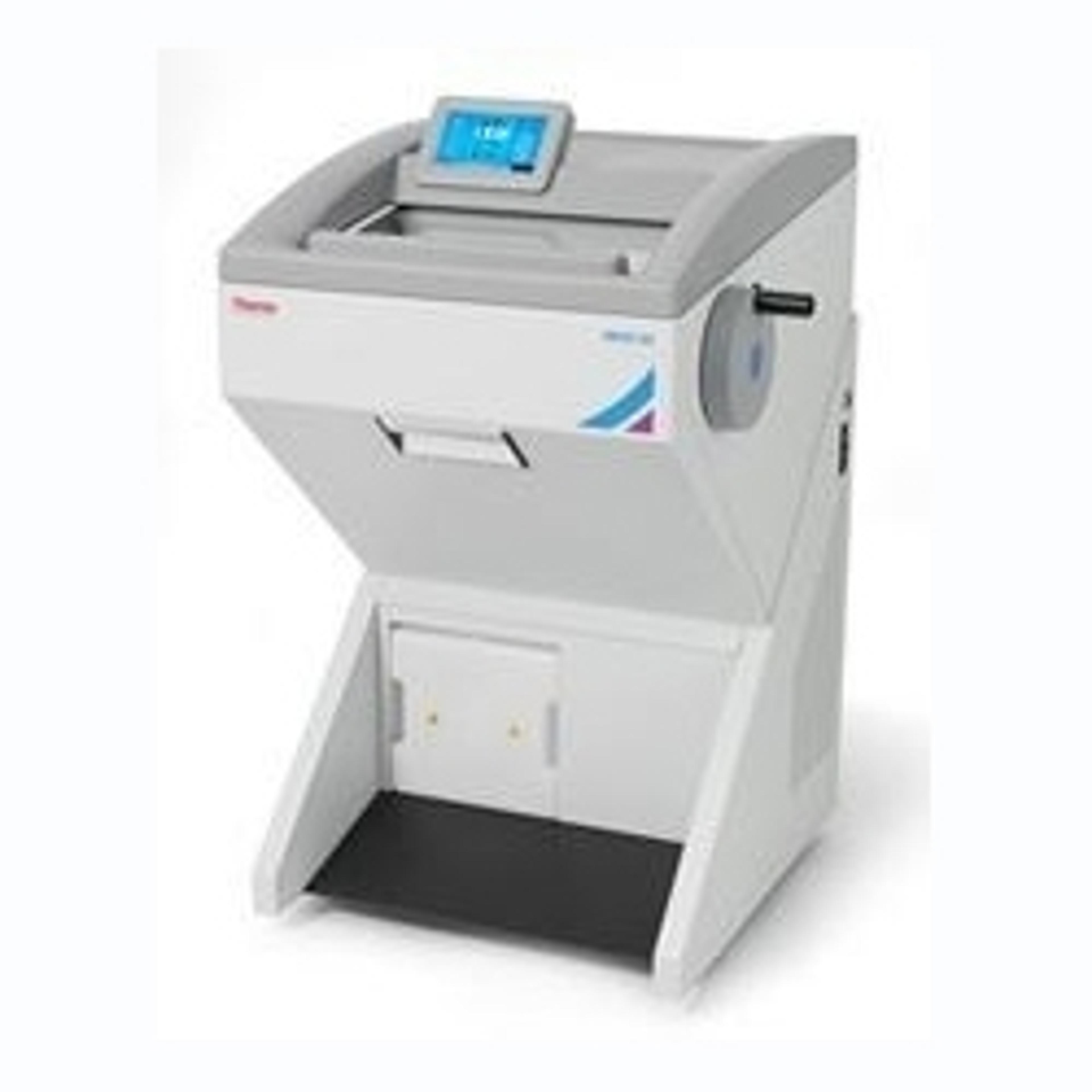
Achieve superior consistency and reliability of sectioning results with the The Thermo Scientific™ Microm™ HM 525 Cryostat. This semi-electronic cryostat with stepper motor advance allows for fast ,stress-free production of high-quality samples. Ergonomically designed, all settings can be set outside of the cryostat chamber.Microm™ HM 525 Cryostat Features: 7 total freezing stations Peltier with four freezing stations…
WIN IN EFFICIENCY, AUTOMATION, SPEED & SAFETY WITH OUR NEW SAMPLE PREPARATION DEVICES HORIBA Scientific offers a great range of possible solutions for our analyzers, to save time and ensure that the end results are as accurate as possible! Whatever your analytical solutions: ICP-OES, ICP-MS, AA, Fluorescence X, elemental analyzers, etc.… HORIBA Scientific can provide you with many sample preparation devices, such as: • Grinder…
Registering an API is complex and thus very time-consuming. That’s why we’ve offer EMPROVE® api: high-quality raw materials for use as active pharmaceutical ingredients. EMPROVE® api raw materials are produced according to cGMP guideline ICH Q7. What’s more, all of our EMPROVE® API production plants are located in Western Europe and follow current environmental standards. And in order to support your products’ compliance with…
Raw material qualification is a key factor in assuring the quality and safety of biopharmaceuticals. Even still, performing tests and compiling required documentation on your own is costly and time-consuming. Enter EMPROVE® bio raw materials – your solution for getting products to market faster. EMPROVE® bio raw materials for use in biopharmaceutical production offer specifications adapted for use in upstream or downstream pr…
EMPROVE® exp for use as excipient As drug approval procedures grow stricter, regulatory requirements are becoming more demanding for excipients. Our high-quality EMPROVE® exp raw materials for use as excipients not only comply with relevant regulations, but also come with detailed documentation, facilitating lower costs of qualification, so you can get to market faster. EMPROVE® exp dossiers comprise information on the manuf…